U
NIVERSIDADE DEL
ISBOAF
ACULDADE DEM
OTRICIDADEH
UMANAStrength, Water Compartments and Phase Angle in
Breast Cancer Survivors
Dissertação elaborada com vista à obtenção do Grau de Mestre em
Exercício e Saúde
Orientador: Professora Doutora Analiza Mónica Lopes de Almeida Silva
Co-orientadora: Professora Doutora Catarina Nunes Matias
Júri:
Presidente
Professor Doutor José Henrique Fuentes Gomes Pereira
Vogais
Professora Doutora Analiza Mónica Lopes de Almeida Silva
Professora Doutora Diana de Aguiar Santos
Mafalda Maria Travado Reis
Acknowledgments
I would like to express my gratitude to the following:
Professor Analiza Mónica Silva she was very enthusiastic from the beginning, helping me organizing this project with her knowledge and very rare work ability. I truly appreciate her support in this journey and her total availability to answer my e-mails or to meet me to discuss some doubts. She gave me wings to fly, she let me manage my project as I wish and gave me full support whenever I needed it, making me more confident.
Professor Catarina Matias who made possible all the practical component of my thesis. She helped me to learn the procedures of bioelectrical impedance and Jamar dynamometer, she was always available to teach me over and over and to meet with me to explain it at any time. Without her help, patience and availability I would not have finish this thesis in time. Thank you for always being there, even during her vacations when my SPSS was crashing and I did not know what to do, she gave me opening to be in contact with her almost 24/7 and I truly appreciate it. She is an unbelievable person with an amazing heart.
Viva Mulher Viva Associação especially Dra. Cidália, Dr. Araújo and Sofia. They were amazing, Dra Cidália was always worried if everything was ok, if the room where I was doing the measurements was good enough, if the mattress was the one I needed, if I had enough alcohol and compresses, if the number of participants was ok and so on, she was really amazing! Dr. Araújo helped me with information regarding the patients and Sofia made herself responsible for the phone calls and finding the participants for the study. Without Sofia availability to call to all the survivors of the Association I would not have had enough participants.
Thank you Viva Mulher Viva Associação for making it possible.
I also want to thank to Rita Pinto, former colleague, for all her assistance with endnote and SPSS.
I would like to thank to all my friends for their support.
Table of Contents
Tables and figures ... 7
Abbreviations ... 9
Abstract ... 11
Resumo ... 12
Introduction ... 13
1.1 Background ... 14
1.1.1. What is Breast Cancer ... 14
1.1.2. Breast Cancer Staging ... 15
1.1.3. Breast Cancer treatment ... 16
1.1.4. Follow up care... 17
1.2. Muscular Strength and Breast Cancer... 18
1.2.1. Measures of Strength ... 20
1.2.2. Handgrip Test... 21
1.2.3. Muscle Dysfunction and Obesity ... 23
1.3. Physical Activity ... 26
1.4. Water Compartments and Breast Cancer ... 29
1.4.1. Bioelectrical impedance ... 31
1.4.1.1. Single Frequency Bioelectrical Impedance Analysis ... 34
1.4.1.2. Multiple Frequency Bioelectrical Impedance Analysis ... 34
1.4.2. Validation in Clinical Populations ... 36
1.4.3. Phase Angle ... 38
2. Relevance of the Study ... 43
3. Objectives ... 45
4. Methodology ... 45
4.2. Body composition measurements ... 46
4.2.1. Preparation ... 46
4.2.2. Anthropometry ... 46
4.3. Bioelectrical Impedance Spectroscopy (BIS) ... 47
4.4. Water Compartments ... 48
4.5. Phase Angle (PhA) ... 48
4.6. Handgrip Strength ... 49
4.7. Physical Activity ... 49
4.8. Statistical Analysis ... 50
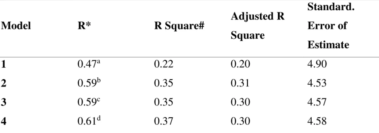
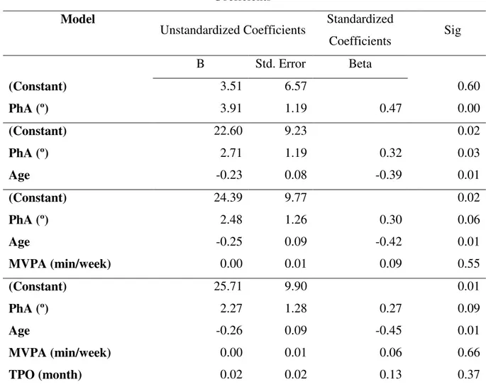
5. Results ... 53
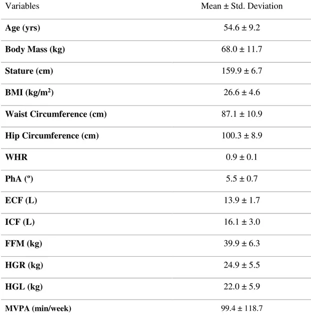
5.1. Characteristics of the Sample ... 53
5.2. Preliminary Tests ... 53
5.3. Multiple Regression Analysis ... 53
6. Discussion ... 57
7. Limitations ... 63
8. Conclusions ... 65
9. Future work ... 65
Tables and figures
Figures List
Figure 1: on the left side - lymph nodes distribution; on the right side - inside of a breast (http://www.cancer.gov/types/breast) ... 14
Figure 2: Diagram of the graphical derivation of the phase angle and its relationship with R, Xc and Z.136 ... 39
Figure 3: Example of how the electrodes must be placed in the right hand and foot for BIS assessment... 47
Tables List
Table 1: Participants characteristics ... 54 Table 2: Predictive power of phase angle in explaining muscular strength ... 55 Table 3: Adjusted and unadjusted coefficients for the phase angle in determining muscular strength ... 56
Chart List
Abbreviations
ASHT American Society of Hand Therapists
BC breast cancer
BCS breast cancer survivors
BCM body cell mass
BI bioelectric impedance
BIA bioelectrical impedance analysis
BIS bioelectrical impedance spectroscopy
BMI body mass index
CFR cancer related fatigue
ECW extracellular water
FFM fat free mass
HC hip circumference
HGdom handgrip dominant side
HGL handgrip left side
HGR handgrip right side
HIV human immunodeficiency virus
ICW intracellular water
IPAQ International Physical Activity Questionnaire
IPAQ-SF international physical activity questionnaire short form
MF-BIA multiple-frequency bioelectrical impedance analysis
MVPA moderate-vigorous phisical activity
PA physical activity
QOL quality of life
R resistance
SF-BIA single frequency bioelectrical impedance analysis
SM skeletal muscle
TBW total body water
TNM tumor-node-metastasis
TPO time post operation
WC waist circumference
WHR wait-to-hip ratio
Xc reactance
Abstract
Background: Accurate prognostic tools are determinant for decision-making in cancer care planning. Objective measures such as bioelectrical impedance spectroscopy (BIS) may improve the accuracy of prognostic. In this cross-sectional study the goal was to determine if the water compartments and the phase angle were predictors of muscular strength in breast cancer survivors (BCS).
Methods: A total of 41 BCS (age 54.6 ± 9.2) were evaluated. Water compartments and phase angle were assessed with BIS and muscular strength was assessed with handgrip dynamometer. Moderate-to-vigorous physical activity (MVPA) was assessed using the International Physical Activity Questionnaire (IPAQ). Measurements were performed in the morning after an overnight feast.
Results: Linear regression analysis showed that phase angle explained 22% (r2 = 0.216)
of the variance of the handgrip. Independently of MVPA and time post-operation, phase angle remained a significant predictor (B=2.269, p=0.085). No associations were found between water compartments and handgrip strength (p>0.05).
Conclusions: The findings of this study suggest that phase angle is an important predictor of muscular strength in breast cancer survivors.
Key words: breast cancer, phase angle, muscular strength, water compartments,
Resumo
Introdução: É necessário definir medidas de prognóstico precisas para que haja uma melhor tomada de decisão relativamente ao planeamento do tratamento de cancro da mama. Medidas objetivas como a bioimpedância elétrica multiespectral (BIS) podem melhorar a precisão de prognóstico. Neste estudo transversal o objetivo será determinar se os compartimentos hídricos e o ângulo de fase são preditores da força muscular em sobreviventes de cancro da mama.
Métodos: A amostra consistiu em 41 sobreviventes de cancro da mama (idade 54.6 ± 9.2 anos). Os compartimentos hídricos e o ângulo de fase foram medidos com a BIS e a força muscular com um dinamómetro. A atividade física moderada a vigorosa (MVPA) foi avaliada através do Questionário Internacional de Atividade Física (IPAQ). As medições foram realizadas durante a manhã com os participantes em jejum.
Resultados: A análise da regressão linear mostra que o ângulo de fase explica 22% (r2
= 0.216) da variação da força muscular. Independentemente da MVPA e tempo pós-operatório, o ângulo de fase manteve-se um preditor significativo (B=2.269, p=0.085). Não foram encontradas associações entre os compartimentos hídricos e força muscular (p>0.05).
Conclusão: Os resultados deste estudo sugerem que o ângulo de fase é um importante preditor da força muscular em sobreviventes de cancro da mama.
Palavras-chave: cancro da mama, ângulo de fase, força muscular, compartimentos
Introduction
Humans and animals have had cancer throughout recorded history. Some of the earliest evidence of cancer was found in ancient Egyptian manuscripts describing cases of tumor, or ulcers, of the breast that were removed by cauterization that date back to 3000BC. At this time, they were describing this disease as not treatable.1
Cancer is a term used for cells that start to grow out of control in a certain part of the body, because of damaged DNA. These cells are different from normal cells, instead of dying or repair the damage, they continue to grow and form new abnormal cells that the body does not need. Most DNA damage is caused by mistakes that happen while the normal cell is reproducing or by something in our environment.2 Cancer cells can
also invade other tissues, throughout the bloodstream or lymph vessels of our body,
where they begin to grow and form new tumors that replace normal tissue. This is a process called metastasis.2
Breast cancer (BC) is the most common cancer among women in developed countries.3 In recent years, the BC incidence rates have been increasing throughout the
world.3 With respect to Portugal, data from the report of the International Agency for
Research on Cancer (IARC) from 20124 show that the number of new cancer cases per
year (incidence) was 49,200 and the number who died (mortality) was 24,100. In terms of incidence and mortality, colorectal cancer is both the leading cause of cancer in our country, with 7,129 new cases diagnosed per year, as well as the leading cause of cancer mortality, with 3,797 reported deaths. This is followed by prostate cancer with 6,622 incident cases and 1,582 deaths and breast cancer with respectively 6,088 (incident cases) and 1,570 (deaths).
In Portugal, BC ranks in first among cancers affecting women, with an age-standardized incidence rate of 60.0/100 000 females.3Due to early detection and more advanced treatment options, the mortality rates of breast cancer worldwide have decreased in the last decade, with an estimated annual percentage charge of -2% year in Portugal.3,5
However, despite the decrease in mortality rates, BC incidence continues to rise, with a concerning increase of 20% newly diagnosed cases since 2008.6 This increase
dietary choices and lack of physical activity, both been considered major risk factors for the development of the disease.6
1.1 Background
1.1.1. What is Breast Cancer
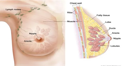
A woman's breast is made up of lobules (glands that can make milk), ducts (small tubes that carry milk from the lobules to the nipple), fatty and connective tissue, blood vessels, and lymph vessels (Figure 1). Most breast cancers begin in the cells that line the ducts.7
The lymph system is one of the main ways breast cancer spreads. Normally, lymph nodes, which are filters connected by vessels that carry a clear fluid called lymph, are small, bean-shaped tissues that contain a certain kind of immune system cells that try to catch and trap cancer cells before they reach other part of the body.8
Most of the lymph vessels of the breast drain into lymph nodes under the arm (axillary nodes); lymph nodes around the collarbone (supraclavicular and infraclavicular lymph nodes); and lymph nodes inside the chest near the breastbone (internal mammary lymph nodes).
Figure 1: on the left side - lymph nodes distribution; on the right side - inside of a breast (http://www.cancer.gov/types/breast)
(bimanual palpation of the breasts and locoregional lymph nodes assessment) and an imaging test (bilateral mammography, ultrasound of the breast and regional lymph nodes and MRI) are performed to look more closely to the chest and to help finding abnormalities.7,9
After the diagnosis of breast cancer, disease stage should be assessed to help organize the different factors and some of the personality features of the cancer into categories, in order to best understand prognosis, to guide treatment decisions and to provide a common way to describe the breast cancer so that results of the treatment can be compared and understood.8,10 The tumor-node-metastasis (TNM) staging system is
the most common system used to describe the different stages and it was first implemented by Pierre Denoix in 1942.10 It is based on whether the cancer is invasive
(cancer has grown into normal tissues and cancer cells have spread to other parts of the body through the blood or lymph system) or non-invasive (cancer that stay within the milk ducts or milk lobules in the breast), the size of the tumor (T), how many lymph nodes (N) are involved, and whether it has spread to other parts of the body (M for metastasis).7
1.1.2. Breast Cancer Staging
So, accordingly with TNM system BC is divided into Stage 0 used to describe non-invasive breast cancers. There is no evidence of cancer cells or non-cancerous abnormal cells breaking out of the part of the breast, in which they started, or getting through to or invading neighboring normal tissue.
Stage I the tumor has <2cm and it can be found in small groups of cancer cells in the lymph nodes. It is divided into subcategories known as IA and IB.
Stage II the tumor has <2cm and it has spread to the axillary lymph nodes or the tumor has between 2-5cm but it hasn’t spread to the axillary lymph nodes. It is divided into subcategories known as IIA and IIB.
Stage IV the tumor has spread beyond the breast to other organs of the body, such as bones, distant lymph nodes or skin, lungs, liver and brain. The words used to describe stage IV breast cancer are advanced and metastatic.
It is not known what exactly causes BC but it is known that some risk factors such as diet (associated with obesity), alcohol, age, genetic predisposition and physical activity contribute to the rising incidence of the disease. However, the presence of a risk factor, or even several, does not mean that a woman will get breast cancer. Although many risk factors may increase the chance of having breast cancer, it is not yet known just how some of these risk factors cause cells to become cancer.7,9
1.1.3. Breast Cancer treatment
The most common treatments for BC are surgery, radiation therapy, chemotherapy, hormonal therapy and biological therapy.
When developing a treatment plan there are factors to consider such as the type of BC, age, menopausal status, overall health and personal preferences or situation. Other factors, regarding the tumor, need to be taken into consideration such as the stage of cancer (size of tumor and if it has spread); grade of cancer (how cancer cells look and behave); hormone receptor status (if cancer cells have receptors for estrogen and progesterone); HER2 status (HER2 is a protein on the surface of the breast cells that promotes growth).11
reduces the levels of these hormones in the body or blocks their effect on cancer cells. This therapy is often given after surgery, radiation and chemotherapy to reduce the risk of cancer coming back; e) biological therapy works with the immune system to help protect the body from the disease.11
1.1.4. Follow up care
The transition from active treatment to post treatment care is critical to long-term health. When treatment ends some women feel lost, anxious and worried that cancer might come back. Some women may also have low self-esteem due to loss of breast, hair, scars and lack of mood for sex. The inclusion in support groups for women who survived is of extreme importance to help regain confidence, improve response to treatment, speed recovery, reduce risk of recurrence and improve quality of life.11,12
Cancer related fatigue (CFR) is a highly prevalent and multifactorial symptom classically defined as ‘a persistent, subjective sense of tiredness related to cancer or cancer treatment that interferes with usual functioning’. This symptom does not get better with rest and is very common in breast cancer survivors (BCS).13
A survivor is anyone who has been diagnosed with cancer, from the time of diagnosis through the rest of life.12 For some survivors, this kind of fatigue lasts a long
time after treatment and can make it hard for them to exercise. A study conducted by Winters-Stone et al14 reported that women with lower extremity muscle weakness, high
body fat and lower physical activity levels had greater fatigue. The specific etiology of CRF is still unknown but it is frequently associated with a wide variety of psychosocial factors (e.g. clinical depression), and exacerbating symptoms (e.g. chronic pain, nausea) as well as treatment side effects.13,15 Studies have shown that patients who
follow an exercise program tailored to their needs feel better physical and emotionally and it helps reduce fatigue.7,16
The post-surgery period is also crucial due to the possibility of developing complications, such as lymphedema, reduction in muscle function, decrease in upper limb functionality and muscular strength.17
Although adjuvant therapy combinations and advances in early detection have improved survival rates (5 year survival rates of approximately 90%)17-21, BCS often
exhibit reduction in muscle strength (impaired shoulder function with decrements in shoulder muscular strength, shoulder mobility and functional capacity) associated with cancer related symptoms.17,20 Limitations in upper body strength such as pushing,
lifting, reaching were reported as especially problematic in this population.22 Such
limitations may be due to surgical trauma and activity avoidance.20
It is important to introduce BCS in exercise programs to help them recovering their strength and reduce their weight so they can recover their self-esteem and quality of life.
1.2. Muscular Strength and Breast Cancer
The skeletal muscle (SM) is the largest organ in the human body, constituting 40-50% of total body mass in healthy non-obese humans.23 It is responsible for
performing muscular contractions, generating external mechanical force, which enables the realization of daily activities and exercise and plays an important role in primary and secondary disease prevention as an essential regulator of metabolic and inflammatory homeostasis.23,24 It is also an influential organ in hormonal, immune and
metabolic function.25
Cancer treatments have acute and chronic effects on the muscle system. The loss of lean muscle mass results in muscle weakness, decreased functional work capacity, decreased flexibility and reduced mobility. These series of events occur due to a decline in protein synthesis in conjunction with enhanced protein catabolism caused by cancer treatment and consequent deconditioning, leading to a diminished quality of life.17,19 The contractile and metabolic proteins that are lost are responsible for muscle
contraction, force generation, extensibility and the production of energy (ATP).19
The muscle has a very important role in whole body protein metabolism in the response to stress and therefore prevention of many pathologic conditions and chronic diseases.24 Maintenance of the protein content of certain tissues and organs such as the
breakdown. Physiologic responses necessary for recovery lead to the accelerated synthesis of protein due to the greater demand of amino acids. Studies suggest that a protein intake >3g.kg.d is required to provide the necessary precursors for the synthesis of protein for normal healing of, for example, a burn injury in 50% of whole body. This means that individuals with limited reserves of muscle mass respond poorly to stress.24
Despite the efficacy of cancer treatments in improving survival, BCS usually suffer from substantial impairments that affect their level of physical activity such as increased body fat, reduced aerobic exercise capacity and muscle weakness.26 These
are well known risk factors responsible for loss of muscular strength, which leads to a poor quality of life.14,25,27-31 Also, deconditioning during active treatment may
contribute to declines in upper and lower extremities strength,13,18,32 regardless of
disease stage.18,23,30
The role of muscular strength in the performance of activities of daily living and exercise, as well as in the prevention of chronic diseases, is increasingly being recognized. Important findings suggest that poor muscle strength is a predictor of death from all causes, cardiovascular disease and cancer.27,33-36
Reduced muscle strength is associated with loss of physical functionality and with negative impact on recovery after surgery or illness, which partly explains the high predictive power of muscle function tests.35 In addition, muscular strength has been
recognized as an important health related component of fitness and has been shown to be positively related to cardiorespiratory fitness.36,37
The loss of muscle mass is detrimental for cancer survival. A significant portion of patients return for rehabilitation treatment after several months to even years after discharge, with several complaints of upper limb dysfunction that incapacitate performance of daily tasks.38 Evidence suggests that the ability to perform physical
tasks in daily life is determined by a threshold level of muscular strength.31 Increasing
the physical activity level during and after the treatment may improve the muscle strength and control the atrophy as well as improving quality of life outcomes.30,31 A
study performed with BC patients by Vardar-Yagli et al30 found a positive association
between physical activity levels and peripheral muscle strength. Another study31
with changes in physical functioning. In addition to this studies, Harrington et al.26
found that all strength measures assessed in his study were decreased in the BCS when compared to healthy subjects.39
Skeletal muscle has a greater adaptability when given appropriate training stimuli, even in cases of severe muscle atrophy and fatigue. Enhancing lean body mass size and function will improve survival after cancer treatment.25,31
In order to determine the severity of SM loss, muscle strength needs to be assessed. The exertion of muscle contraction is measured as muscle strength.23
1.2.1. Measures of Strength
There are several methods for the measurement of voluntary and involuntary muscle function. One method used to determine involuntary muscle contraction is electrical stimulation at various frequencies but constant isometric length. Although this method is considered the superior procedure in regard to objectivity, it is not suitable for clinical routine. Regarding measurements of voluntary strength (handgrip, knee extension and hip flexion strength) the handgrip is the most common measure of muscle function and functional capacity for clinical purposes due to its easy use, reliability, validity, feasibility, generalization of overall strength, since it correlates with elbow flexion strength, knee extension strength and trunk extension strength and because it is a significant predictor of health related quality of life in cancer patients.17,18,27,32,35,36,40
Grip strength has been shown to be a predictor of postoperative complications, functional limitations, functional decline, disability and mortality.18,27,36,41 However, as
recently shown by Norman et al.42 mortality prediction of handgrip strength is
dependent on which further parameters are included in the regression models. Handgrip strength predicted 6 months mortality in cancer patients but lost its significance when bioelectrical phase angle was introduced in the model.35 Also, several factors are
negatively associated with handgrip strength in BC such as mood, fatigue, pain, hypersensitivity and neck shoulder mobility.17 Depressed mood is a potential
voluntary handgrip contraction, meaning that women with lower strength have more fatigue compared with women with higher strength.14,32
Several studies have shown that having low muscular strength is a predictor of all-cause mortality, as well as mortality due to cardiovascular diseases and cancer in healthy and diseased people23,32-34,36,44 and so it seems important to assess handgrip
strength and to determine whether and to what extent the measured values are abnormally low.37 Handgrip strength is strongly correlated with post-operative
complications and has been reported to be predictive of length of hospital stay, loss of functional status, body cell mass depletion, post-surgery complication, short-term survival in hospitalized patients as well as associated with probability of premature mortality and earlier onset of disability.32,35,45,46
In conclusion, higher levels of strength will have a protective effect from disability and mortality.18,36,47
1.2.2. Handgrip Test
The gold standard for assessment of muscle strength is the force exerted in a maximum voluntary contraction with force output measured by a dynamometer.23,36,40
The strongest evidence in relation to mortality has been reported for the handgrip strength test.36,37 This test reflects the maximum strength derived from combined
contraction of extrinsic and intrinsic hand muscles, which leads to the flexion of hand joints. It was originally developed for hand surgery but quickly become the focus of interest in numerous studies due to its feasibility and prognostic relevance.35
Therefore, handgrip strength test has been recommended as an assessment of muscle function for oncology rehabilitation and it is an important indicator of health-related quality of life in BCS.18,32,36,48 Grip strength test is commonly used to evaluate
the integrated performances of muscles by determining maximal grip force produced in one muscular contraction.40 It likely reflects the combined influences of genetic
predisposition, acquired modifications of physical constitution, aging processes and chronic diseases.47 It has been used in a variety of clinical areas and for multiple
dystrophy, determining the efficacy of different treatments for people with a range of disabilities, part of an overall fitness assessment and determining the level of effort exerted.49
A wide range of instruments is available to measure grip strength. There are four basic categories for the measurement of handgrip strength: a) hydraulic which is a sealed system that records grip strength in kg or pounds of force. This includes the Jamar dynamometer, a measure with static grip with handles that can be adjusted to 5 different positions (2.5, 3.8, 5.1, 6.4 and 7.6 cm apart). This is the most widely used instrument; b) pneumatic instruments which uses the compression of an air-filled bulb or bag to determined grip pressure. Commonly used by individuals who have painful hands or fragile skin, as they are viewed as being more comfortable to grasp and softer. A criticism is that this method can only measure the pressure of grip and not its force;50
c) mechanical instruments that record grip strength based on the amount of tension produced in a steel spring; d) strain gauges: commonly measure grip strength in Newtons of force.49
The California Medical Association Committee studied some dynamometers and found that the Jamar dynamometer was “perfect to the extent that its sealed hydraulic system is a nearly leak proof as any mechanical appliance can be made”, recommending it as the best measure of grip strength.50 This Committee summarized
three basic factors of grip measurements: grip is a force, grip is not a pressure and the measurement of grip must be in force units such as pounds or grams.50
Therefore, the Jamar dynamometer has been reported to be the most reliable, valid, fast and easy to perform instrument with the highest calibration accuracy for the measurement of grip strength, as well as better repeatability than other equipment and has been recently validated in advanced cancer patients.32,37,48-51
Although there is not any consensus in measurement protocols, consistency is crucial since posture, arm side and handle position of the dynamometer easily alter maximum grip strength.35,50 The American Society of Hand Therapists (ASHT)
be used with the Jamar dynamometer.49 Most of the studies follow the ASHT protocol
in an attempt to control known risk of errors48 however, the handgrip strength values
depend on many other factors such as the model of the dynamometer in use, inadequate calibration, sample characteristics (physical activity level, etc.) and probably ethnical differences.46
Regarding the handle position, every individual should use the most comfortable, however, it seems advantageous to standardize the method as much as possible and reports suggest that the second position is the most comfortable and has the higher grip strengths.48-50,52 It was also reported that a 3 second time of contraction
should be used as it is sufficient to obtain a maximum reading without exposing the patient to adverse effects such as increased blood pressure or heart rate, which accompanies prolonged isometric contraction.32,49,51
This technique has allowed for large cohorts of handgrip strength data to be obtained and to make predictions concerning key health indicators, especially in aging and clinical populations where malnutrition and cachexia are prevalent.32
A study held by Kilgour et al.32 was the first to link handgrip strength with
survival in advanced cancer. They also found that an overall reduction in handgrip strength was related with the appearance of sarcopenia.32
A systematic review conducted by Neil-Sztramko et al18 pooled grip strength
data from 26 studies in BC population and reported that in women off treatment the mean value was 22.8 kg (95% CI 20.6 to 25.1).18
Normative values are essential if informed decisions are to be made about the individual’s status relative to the general population.
Even though the distribution of a given measure for a specific population does not necessarily mean the health state of the population, it is reasonable to assume that handgrip strength values in the lower end of the distribution may be indicative of several outcomes.45,46
1.2.3. Muscle Dysfunction and Obesity
skeletal muscle arises from a fall in protein synthesis and an increase in protein degradation, which is a side effect of cancer treatment.17,23,53,55-57 It is often
misdiagnosed as a condition of weight loss but it is actually a highly complex metabolic disorder and one of the most frequent effects of malignancy, which is why within oncology, interest in muscle function has traditionally been confined to the clinical entity of cancer cachexia.23,58
Cancer cachexia is characterized by severe muscle wasting, systemic inflammation and malnutrition leading to both acute and chronic impairments in various aspects of physical function18,23,58 such as extensive loss of muscle mass,
strength and metabolic function24 and reduced fitness levels17, and contributes to nearly
one third of all cancer deaths.58
The cachectic state is particularly problematic in cancer, typifying poor prognosis and often lowering the responses to chemotherapy or radiation treatment.58
In a study conducted by Wolfe et al24 it was found that the amount of body protein,
which is related to the amount of muscle mass, predicted recurrence in lung cancer patients that were receiving radiation therapy.
Although cancer cachexia is associated with loss of SM, gain of adipose tissue can occur, culminating in the condition of sarcopenic obesity.17,54 Martin et al54 found
that most of the cancer patients of his study were more commonly overweight or obese and often had occult severe pre-existing muscle depletion. Sarcopenia (progressive loss of muscle mass and function)24 itself is an independently prognostic of lower survival
in obese patients with cancer.54 According to Cantarero-Villanueva et al15 the lack of
strength associated with an increased body mass index (BMI) may be related to sarcopenia and a decreased grip strength.
One common side effect that occurs in over 50% of BCS is the gain of weight, which can be related to the development of comorbid conditions that affect survival 15,59-62 as well as risk of cancer recurrence due to increasing endogenous estrogen
production,63 with most explanations focusing on adiposity rather than body weight per
se.61,64,65 Evidence62 suggests that reported weight gain happens during the first two
years after diagnosis, with the weight gained ranging from few grams to several kilograms. In a study conducted by Arpino et al62 it was shown that weight gain is also
circumference and hip circumference. Although weight is the simplest anthropometric index of excess adiposity, it does not distinguish between fat free tissues (comprised primarily of muscle, bone and extracellular water) and adipose tissue.63 Waist and hip
circumference and waist-to-hip ratio (WHR) are additional measures of body fat distribution that provide an index of both subcutaneous and intra-abdominal adipose tissue.62,66-68 The evidence on abdominal obesity on BC survival highlights the need of
using general obesity (body mass index) as well as fat distribution (waist circumference and WHR) to evaluate prognosis.69
It is well established that obesity has an impact on BC occurrence and it is important to distinguish 2 types of obesity: android (WHR>0.8) in which fat is mainly distributed in the upper body (shoulders and abdomen) and gynoid (WHR<0.8) where fat accumulates in the lower part of the body (buttocks, thighs).66 According to
Pacholczak et al66 women with BC present an android type of silhouette with the
distribution of fat tissue present in the central and upper parts of the body. They also mention that this type of obesity cause more pronounced abnormalities in metabolic and hormonal systems66 perhaps because of its relation with visceral adiposity.70
Women with increased WHR have a twofold higher risk of all-cause mortality compared with women with a lower WHR, in models adjusted for physical activity and BMI.69 Therefore, women with central adiposity may be at higher risk of BC than
women whose fat is primarily distributed subcutaneously over hips and buttocks70
because abdominal obesity, is associated with an elevated level of circulating insulin that is mitogenic, anti-apoptotic, and pro-angiogenic, and has been found to be associated with worse BC prognosis.68 Several adipokines produced by adipose tissue
are related to hyperinsulinemia and angiogenesis promotion, which is a major contributor to the aggressive behavior of BC.69
Cancer patients with a body mass index (BMI) > 35kg/m2 have worse disease
free survival than those of normal weight, independent of age, race, treatment and sex.15
BMI is a measure of weight adjusted for height that provides a better approximation of the proportion or total amount of adipose tissue in the body than does weight alone.63
The validity of BMI as a measure of adiposity is further supported by its association with obesity-related risk factors such as total cholesterol and blood glucose.63 It’s
calculated as weight in kilograms (kg) divided by height in meters squared (m2).62 The
24.9), overweight (BMI 25-29.9) and obese (BMI > 30) and is used in adults > 20 years old.71 When the BMI is over 25 kg/m2 that person is at increased risk of developing
health problems due to abnormal or excessive fat accumulation.68,71 Central obesity
promotes a concomitant increase in WHR at the same level of BMI therefore, and with increasing evidence of health risks associated with abdominal fat, WHR and waist circumference have been commonly used in epidemiologic studies as measures of central adiposity.63,68 World Health Organization cutoff points of waist circumference
and WHR for substantial increased risk of metabolic complications in women are >88 cm and 0.85, respectively.69
A sedentary lifestyle combined with an unhealthy BMI increases the risk of cardiorespiratory diseases in BCS, which is among the most frequent concomitant comorbidity in women with BC.15 In addition to this, abdominal obesity is a
predisposing factor67 which means that having a waist circumference superior than 88
cm is associated with and increased risk of this comorbidity. This value of waist circumference is exceeded by almost 100% of the obese BC patients and is linked with increased mortality.15
Due to all these factors it is expected that muscular strength is reduced in obese patients due to a decreased level of fitness.15
All cancer patients are subjected to a wide range of degenerative factors, which are all potent causes of muscle dysfunction including aging, malnutrition, physical inactivity and factors directly related to disease pathophysiology and therapy toxicity.23
Lintermans et al72 reported a negative impact of taking aromatase inhibitors –
medication that suppresses the plasmatic concentration of estrogen, such as Anastrazol - on the SM system with more than half of patients complaining of this with loss of grip strength. The loss of SM explains why patients with cachexia have a reduced mobility and quality of life, together with a shorter life span.57
1.3. Physical Activity
In long-term cancer survivors, physical activity (PA) has gained interest as a modifiable lifestyle factor that may improve mortality.73
According to some authors30,31,64,74-77 exercise interventions in cancer patients
body weight, reduced recurrence risk, mitigating cancer treatment side effects (i.e. fatigue), improvements in quality of life (QOL), psychological well-being, body image as well as improvements in physical functioning (i.e. oxygen capacity, flexibility, strength measures), anthropometric measures (i.e. body weight) and health related biomarkers (i.e. blood pressure, heart rate). Therefore, it should be a priority to have an appropriate weight, a healthful diet and a physically active lifestyle aimed at preventing recurrence, second primary cancers and other chronic conditions.12
A report from Europe’s largest survey assessing outcomes in people living with and beyond cancer show that <25% met the current physical activity guidelines, 43% had trouble with fatigue and 45% experience fear of disease recurrence.77 Another
study78 mention that up to 70% of BCS do not meet recommendations of 150 minutes
per week of moderate to vigorous intensity PA (e.g. brisk walking, jogging, swimming).
The combined high prevalence of inactivity and sedentary time may be particularly concerning for BCS given their already heightened risk for poor health and disabilities.78 Several studies79-82 show that women who engaged in PA after a diagnosis
of BC had a statistically significant 20-50% lower risk of death from BC, especially if this PA was within the guidelines. Hormonal changes induced by exercise, such as a reduction of circulating estrogen concentrations mediated by a reduction in adipose tissue or PA independent increase in the amount of sex hormone-binding globulin and improvement in insulin sensitivity may explain the relationship between PA and survival among BCS.73,80,81 Additionally, PA may also improve the immune response,
possibly by promoting killer cell, macrophage and cytokine activity, as well as up regulating antioxidant enzyme activity, which may protect against DNA damage.73
Studies show that regular exercise after BC diagnosis is significantly associated with improved overall and disease free survival following a dose response pattern81 and
that PA influence extends to women regardless of age at diagnosis, stage of disease at diagnosis and post-diagnosis BMI.80
In a study conducted by Bradshaw et al73 the beneficial effect of PA appear
Monitoring physical function during and after treatment may help health professionals to identify declines in order to increase muscle strength and control the atrophy, which will improve morbidity, mortality and quality of life.17,18,25,30,53
To monitor trends and evaluate public health or individual interventions aiming at increasing levels of PA, reliable and valid measures of habitual PA are essential.
Several routine instruments are available to measure PA, including self-report questionnaires, indirect calorimetry, direct observation, heart rate telemetry and movement sensors.84,85 All these methods have well known limitations and for PA there
is currently no perfect gold standard criterion.86
In large-scale cohort studies questionnaires are more frequently used due to their low cost and easy administration.84 There are at least 85 self-administered PA
questionnaires for adults but the International Physical Activity Questionnaire (IPAQ) is the most widely used.85 The IPAQ was developed in an attempt to standardize
assessment of the prevalence of PA in different countries and cultures around the world.87 Its reliability and validity was tested in 14 centers in 12 countries during the
year 2000 with the support of World Health Organization and centers for Disease Control.84,88 From this study it was suggested that the last 7-day short form of IPAQ
could be used for national and regional prevalence studies.88 IPAQ has two versions
available: 31-item long form (IPAQ-LF) and the 9-item short form (IPAQ-SF). The short form records the activity of 4 intensity levels: 1) vigorous-intensity such as aerobics, 2) moderate-intensity such as leisure cycling, 3) walking and 4) sitting.85 The
short version is specific to the ‘last 7 day recall’ because the burden on participants to report their activity is smaller85 and there is no difference between the reliability and
validity of the short and long form.88
Although IPAQ-SF is recommended and widely used, some studies85,87 found
that this method tends to overestimate the amount of PA reported compared to an objective device. Hence, the evidence to support the use of IPAQ-SF as an indicator of relative or absolute PA is weak. However, a study conducted in BCS assessed PA with the IPAQ-SF and found a positive association between PA levels and peripheral muscle strength, showing the significant protective effect of PA on BC risk.30
capacity to mitigate and/or reverse muscle dysfunction in patients with cancer23,30 as
shown by Christensen et al23 BC patients who performed resistance training during
chemotherapy had an increase of 0.8kg of muscle mass. Another study19 suggested that
moderate-intensity exercise can provide a sufficient physiological stimulus to improve muscular performance in cancer survivors, whether exercise is performed during or after cancer treatment. But it is important to note that, according to Doyle et al12 low to
moderate intensity for healthy population may be of high intensity for cancer survivors.
The ability to perform physical tasks in daily life is determined by a threshold level of muscular strength and that’s why strength training in cancer patients would seem to be a potent physiological intervention for regaining lost muscles and improving QOL.31
1.4. Water Compartments and Breast Cancer
Being the major constituent in the human body, water comprises about 40-70% of the entire body mass and constitutes approximately 72-74% of free fat mass (lean tissues) and approximately 10% of fat mass (relatively nonaqueous).89-91 The total
amount of water in the body referred as total body water (TBW) is divided in two main compartments: intracellular water (ICW) corresponding to the fluid within the cells and
it is 66% (2/3), and extracellular water (ECW) which includes all fluid outside the
cells 33% (1/3). These fluid compartments are separated by plasma membranes that surround the cell.92
When conditions outside the body change, these changes are reflected in the composition of ECW, which surrounds the individual cells of the body. Uncorrected deviations in factors such as oxygen and carbon dioxide exchange, can lead to disease and/or death. Therefore it is crucial to maintain concentration gradients and the movement of solutes and water across barriers to preserve normal body function.92 In
some clinical conditions, alterations in body hydration and fluid distribution cause differences in the ratio ECW to ICW, and with certain drugs, the body can retain or lose significant amounts of water and therefore present different proportions of TBW.92,93
athletes by Silva et al.94, it was found that independently of changes in body weight and
arm lean soft tissue, athletes that decreased the intracellular water (ICW) compartment were more likely to reduce grip strength.94
Additionally, TBW estimations have been used to monitor nutritional status and identify disease states, such as dehydration and chronic kidney disease.95
In healthy individuals the hydration of fat-free mass (FFM) and the ratio between ECW and TBW are tightly regulated. In malnourished patients and under a variety of other diseases there is usually a relative increase in ECW, which often results in an increase of TBW.96
The most significant confounding variable in BCS is edema of the distal extremities, which may result from lymphedema, and will affect impedance measurements.93
Lymphedema is the most problematic and dreaded complication of BC treatment, particularly after surgery or lymph node removal,17,97,98 characterized by the
accumulation of protein-rich extracellular fluid, resulting from damaged or blocked vessels99 leading to a significant increase in the volume of the affected limb, resulting
of an impairment in the ability of the lymphatic system to drain the proteins and macromolecules of the interstitium.17 It is an incurable, progressive, disfiguring and
disabling disorder that is often misdiagnosed and treated too late or not treated at all.100
It may present immediately or years after treatment.97 Incidence may vary from
6-30%.21 Early treatment due to early diagnosis may prevent progression to the chronic
phase.100 Despite the causes of lymphedema, such as stiffness and decreased range of
motion of the affected limb joints, handgrip strength test, mentioned in the previous chapter was unchanged regardless the presence or absence of lymphedema.17
Measurements of TBW and ECW involve the use of invasive tracer-dilution techniques. These techniques are expensive and time consuming for routine procedures. Until recently it has not been feasible to use field body composition methods, such as bioelectrical impedance analysis (BIA), to assess routinely the nutritional status of hospitalized patients.96
lymphedema assessment through BIA detects ECW changes. Increases in the volume of this fluid reflects the contribution of lymph accumulation when the subject being at study is at risk of developing lymphedema.100
Intracellular fluid is unaffected by the advert or progression of the disease. Hence, an ECW/ICW ratio is constructed to help detecting the early onset of lymphedema. This ratio is a validated method and appears to have equal or better sensitivity than other techniques in detecting lymphedema.100,101 However, if assessed
too early, as within 3 months of surgery, normal postoperative swelling could be misconstrued as evidence of lymphedema.97 A participant was classified as having
lymphedema when impedance ratio was more than 3 standard deviations above population mean.97
1.4.1. Bioelectrical impedance
The principles of bioelectric impedance (BI) have been established for more than 40 years but methods for estimating components of body composition, specifically total body water, intra and extracellular water, and fat-free mass, are comparatively recent.102
The use of BI to estimate body composition is based on the principle that an electrical current flows at different rates through the human body and is resisted by body tissues and water.93 It is also based on the greater electrolyte content and
conductivity of free fat mass (FFM) compared to that of adipose tissue or bone and upon the geometrical relationship between impedance and volume of the conductor.102,103
Therefore, impedance (Z) is the frequency dependent opposition of a conductor to the flow of an alternating current and is composed by two vectors: resistance (R) and reactance (Xc).93,102
The R is described as pure opposition of the conductor (intracellular fluids, body
fluids and electrolytes) to the flow of an electric current and is related to the amount of
water present in the tissues.104 The Xc is the resistive effect produced by tissues
interface and cell membranes, is the inverse of capacitance, where capacitance is the storage of energy in a circuit by a capacitor.61,105 In human body, capacitance occurs
conductivity (i.e. cell membranes).105 Therefore, ECW and ICW act as resistors in
parallel while the cell membrane behaves as an imperfect capacitor and introduces a reactive and hence frequency dependent component to the total impedance.106
At 5kHz signal pathway is only conducted through ECW because there is very little capacitive penetration of the signal into the intracellular volume and it’s assumed that measured impedance is totally resistance.106,107 At midrange frequencies (e.g.
50kHz) a small amount of intracellular penetration occur due to some capacitors being electrically charged. Therefore, at 50 kHz the resistance is lower than at 0 kHz because there is a greater cross sectional area and a shorter path to destination electrode.105 At
high frequencies (>500kHz), the current is able to penetrate the cell membrane of the intracellular compartment107, taking a more direct pathway through the body, therefore
including both water compartments105,106 assuming resistance index is linearly
correlated with TBW.107
Therefore, the 0/ kHz parallel model (Cole-Cole) is the most accurate model for the analysis of water compartmentalization. The success of this method can be attributed to the fact that ECW and ICW are the major electrical conductors in the body and they reside adjacent to each other, with ICW being isolated from ECW by low conductivity membranes.108 This model is very useful for the assessment of body water
in diseased populations in which the ratio ECW to TBW is altered.107
BI depends on static assumptions and dynamic relationships regarding electrical properties of the body. Anthropometric measurements (e.g. height, weight, age, sex,
race, etc.) are often used in many of the BI prediction equations.106,109 These parameters
are included to reduce the effects of interindividual variance in R and Z values, related to differences in body size and in shape and to increase the prediction accuracy of body
composition estimates.109 It is also important to control some aspects to guarantee the
validity, reproducibility, and precision of the measurement in similar populations. Therefore, some standardizations have been suggested in order to replicate results, such as the subject body position, length of time in supine position before measurement, correct stature measurement, skin preparation with alcohol before electrode placement, electrode position, hydration and menstrual cycle, consumption of food and beverages before test, ambient air and skin temperature, recent physical activity and conductance
BI is considered to be a feasible filed method as it is an easy, portable, inexpensive, noninvasive, and less time consuming method,61,93,105,112-114 especially if
compared with laboratorial methods such as deuterium dilution, tritiated water dilution, bromide dilution, and even dual energy x-ray absorptiometry.104 BI results are available
immediately and measurements can be repeated as often as desired without side effects for the participant.
Malnutrition is a frequent manifestation in patients with advanced cancer and is a major contributor to morbidity and mortality. Malnutrition is characterized by changes in cellular membrane integrity and alterations in fluid balance with an increase in ECW, which often results in an increase in TBW.96 As a result, measurement of body
composition is an important component of overall nutritional evaluation in cancer patients.61
At 50 kHz the current does not completely penetrate the cell membranes but in normal subjects this is not particularly important because the intracellular volume represents a relatively constant proportion of the TBW. In patients, there may be significant variations in the proportions of ICW and ECW and it is more important that the frequency is high enough to allow the current to completely penetrate the intracellular space.115 Clinical advantage to monitor both ECW and ICW makes BI the
preferred technique for future clinical research.109
A decrease in ECW/ICW ratio would result in a relatively lower resistance per unit of body water and higher TBW.114,116
To estimate a certain body compartment based on bioelectrical impedance methods it is important to choose the type of device that will be used. The single frequency bioelectrical impedance analysis (SF-BIA) device and multiple-frequency bioelectrical impedance analysis (MF-BIA) have the measured resistance and/or reactance incorporated into empirical prediction equations, developed by regression analysis.104 The bioelectrical impedance spectroscopy approach (BIS), estimates body
composition through a mathematical modeling technique108,117 on a range of resistance
1.4.1.1. Single Frequency Bioelectrical Impedance Analysis
Single-frequency bioelectrical impedance analysis is by far the most widely available impedance methodology, which involves the application of an electrical current for the measurement of impedance at a single frequency, typically 50 kHz.118
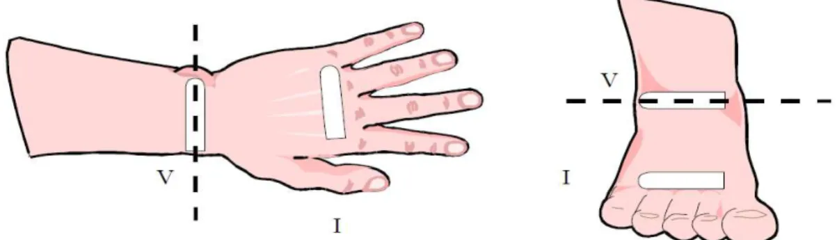
The current passes between surface electrodes placed on hand and/or foot. Some instruments use foot-to-foot or hand-to-hand electrodes.110
SF-BIA strictly measures a weighted sum of ECW and ICW resistivity. Impedance data is entered into predictive equations derived through statistical regression in order to determine TBW, from which FFM is calculated according to the assumption that FFM is constantly hydrated at 0.73.118 However, SF-BIA cannot
determine differences in ICW. Therefore, a problem of using a single-frequency measurement to predict TBW is that the sensitivity of a single high-frequency measurement to changes in ECW and ICW is different due to their different resistivities, a simple change in the ratio ECW/ICW will alter TBW resistivity and cause error.119
As so, the SF-BIA is not valid under conditions of significantly altered hydration, but this does not negate its use to predict absolute FFM or TBW in normally hydrated subjects.110,120 In malnourished patients or under a variety of other diseases such as
edema, there is usually an increase in ECW, which often results in and increase of TBW,96,110,118 when this occurs, Cole-Cole model should be used.107
In SF-BIA the assumption that FFM is constantly hydrated is a major limitation of this technique in the assessment of body composition, as FFM hydration may not be constant for all populations.105
1.4.1.2. Multiple Frequency Bioelectrical Impedance Analysis
At 50kHz, the electrical pathway is primarily extracellular because there is very little capacitive penetration of the signal into the intracellular volume, which means, the cell membrane acts as an insulator, and it is assumed that the impedance is principally a function of ECW, responsible for the measured R at R0.105,107,110,119 Thus,one can conclude that SF-BIA is limited in the ability to distinguish the distribution of body water into its intra- and extracellular compartments.121
the total body R (R) is a function of both ICW and ECW (TBW), which is caused by
cell membrane capacitance.106,110,119
Therefore, TBW assessment by SF-BIA was replaced by MF-BIA devices that apply the current at limited and defined frequencies (e.g. 5, 50, 100, 200 or 500kHz)
and offer the potential of measuring TBW, ECW and ICW separately.103,118
Due to its efficacy in accurately predict TBW, MF-BIA was used to monitor the efficacy of treatment for lymphedema in patients following surgery for BC. This method was shown to be significantly more sensitive than others to detect small differences in the extracellular volumes between the arms of any individual. ECW was elevated after clinical diagnosis of lymphedema. This index does not require normalization to another body segment and can be used to detect all types of peripheral edema including both uni- and bilateral lymphedema.101
There are two approaches to the use of MF-BIA data:
1. Multiple Frequency Bioelectrical Impedance Analysis
MF-BIA first introduced by Thomasset el al.103 used impedance data at two
frequencies: one at very low frequencies (usually 5kHz) and the other at very high
frequencies (typically 50, 100, 200 to 500kHz). The impedance data are applied to
regression-derived equations in order to predict TBW, ECW, and ICW.118
Unlike the SF-BIA, this model correctly assumes that the specific resistivities of intra- and extracellular fluid are different. It assumes that R at low frequencies is the resistance of the extracellular fluid (Re) because virtually no conduction occurs due to high cell membrane capacitance. On the other hand, R at high frequencies is the resistance of whole fluid (Rt) because there is total conduction through the cell membrane.118 The resistance of the intracellular fluid (Ri), is a function of both low and
high frequency.105
However, the use of more than one frequency gives a variability of results and no conclusions can be made regarding the validity of one frequency over another, in the prediction of body fluid compartments.105
2. Bioelectrical Impedance Spectroscopy
modeling and mixture equations (e.g. Cole-Cole and Hanai formula)108,117 to generate
relationships between R and body fluid compartments, instead of regression equations.110 BIS measures the impedance across a spectrum of frequencies and can
accommodate interindividual variation due to the mathematical modeling generated factors. As so, BIS provides a more direct, individualized measurement of ECW and ICW than other impedance approaches.118
In commercial BIS instruments using the Cole-Cole model108, multifrequency
impedance data are mathematically modeled to reduce the influence of artifacts at low and high frequencies. In this model, the body is viewed as an electrical circuit with intracellular and extracellular pathways in parallel and having cell membranes serve as capacitors for the intracellular pathway.105
Cole model is computed by using nonlinear curve fitting to extrapolate data to the low and high frequency limits. This procedure generates Cole model terms, including Re (resistance associated with the ECW); Ri (resistance associated with the ICW); Cm (cell membrane capacitance); and exponent . Cole model terms are then applied to equations derived from the Hanai mixture theory, which is essentially based on the notion that the body is a conducting medium of water, electrolyte-rich tissues (e.g. blood and muscle) in addition to nonconducting material within it (eg, bone, fat and air filled spaces).120 This theory was applied to improve the Cole-Cole linear model
as (1) it accounts for the effects of non-conducting substances in the body water, (2) removes the apparent population-specificity found with Cole-Cole linear equations, and (3) improves sensitivity to body water changes 122.
The TBW is assumed to be the sum of ECW and ICW 118,123,124, and the FFM is
calculated based on the mean density of the intra- and extracellular water and its associated material.
Therefore, Cole-Cole model and Hanai theory108 will be useful for the
assessment of body water compartmentalization in diseased populations in which ratio ECW to TBW is altered.107,119
1.4.2. Validation in Clinical Populations
variety of patients, including cancer.61,96,105,112,125-127 The existent data is, so far,
inconclusive, nevertheless, BIS seem to present itself as a valid alternative to assess body composition and body water compartments, in clinical populations.
In Netherlands, BIA is routinely used in surgical and oncological patients, where quick measurements of body compartments are needed.104 According with
Earthman et al.,118 BIS is the only field technology available that has the potential to
measure body water volumes and body cell mass (BCM) in the clinical setting. The BCM has clinical relevance because it has been defined as the total mass of metabolically active, living, functioning cells. A loss of BCM can cause a decrease in physical strength and immune function, with an increase in susceptibility to infections, as observed in human immunodeficiency virus (HIV) patients.118 BIA also predicts
short-term survival in HIV patients.109
Therefore, from a clinical perspective, the ability of BIS to accurately estimate BCM by quantifying ICW, as well as the ability to monitor fluid distribution between the ICW and ECW compartments, would greatly enhance nutrition assessment, as well as the overall clinical care of the patient.118
However, according with the review of 11 studies in oncological and surgical patients made by Haverkort et al.,104 BIA measurements underestimated TBW and FFM
irrespective of the equation or device used. The results of these studies indicate that the application of the Heitmann equation contributes, to some extent, to a valid estimation of TBW in patients with incurable cancer. They concluded that BIA estimations in the individual patient care with regard to oncological and surgical patients can be useful when performed longitudinally and under strict conditions.104
Fredix et al.,128 also concluded that BI is a promising method for the assessment
of body composition in clinical practice provided that population specific prediction formula are used.128
According to Moon et al.,120 an increase in ECW, which can be due to a high
1.4.3. Phase Angle
In healthy subjects, age, sex and BMI are the major determinants of phase angle (PhA). Aging is associated with a decrease in tissue mass (reduced Xc) and in TBW (increased R) resulting in decreased PhA. A higher PhA is observed in persons with an increased BMI because PhA is directly related to cell membranes (amount and functional status) and persons with increased BMI have more fat cells resulting in higher PhA values. For BMI > 40 kg/m2 an inverse relationship is observed, this has
been attributed to a higher tissue hydration or a pathological fluid overload.129-131
Patients often exceed normal BMI range and a differentiation between PhA as an indicator of body composition and cellular function altered by disease may require BMI-specific reference values to exclude an influence of body composition,132 since
increased BMI is a common feature of BC patients.17,30,57
As mentioned before, measurement of body composition is an important component of overall nutritional evaluation in cancer patients and in association with cell membrane functional analysis it is able to reflect cell function and integrity.133
Changes in cell integrity are characterized by malnutrition, which is a major contributor to morbidity and mortality and so, a predictor of shortened survival.42,134
Nutritional status has been evaluated through objective measures such as anthropometric and laboratory methods. This methods are not ideal in the clinical setting because they are time consuming and require well-trained staff.61 The use of
impedance methods as a measurement of assessment of body composition and nutritional status overcomes the aforementioned issue and other challenges that might appear with the assessment in the clinical setting.131
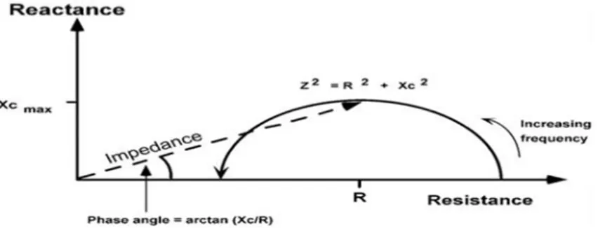
We now know that cell membranes produce capacitance (Xc) by storing parts of the charge as a capacitor. The storage of the current creates a phase shift that can be regarded as the ratio of R and Xc and is expressed as phase angle .135 Bioelectrical PhA
has consistently been shown to have great prognostic relevance with regard to morbidity and mortality in several conditions such as HIV/AIDS, liver cirrhosis, sepsis, hemodialysis, lung, colorectal and pancreatic cancer.42,131-134 PhA is the angle the
Phase Angle = (Resistance/Reactance)*(180/π).102
Although the biological meaning of PhA is not well understood, it is considered a novel marker of cellular function,44 as reflects in one hand, the capacitance behavior
of tissues (reactance) and is associated with cellularity, cell size and integrity of the cell membrane and, on the other hand, on its pure resistive behavior (resistance), which is dependent on lean tissue mass and tissue hydration between intra- and extracellular spaces.42,61,113,129,131,132,134-136 PhA is positively associated with Xc and negatively
associated with R.61
Figure 2:Diagram of the graphical derivation of the phase angle and its relationship with R,
Xc and Z.136
Changes in the extracellular to body cell mass ratio are probably associated with changes of the PhA. This ratio is a known sensitive marker of malnutrition, characterized by both increased ECW and decreased BCM (mainly muscle mass), typical features of systemic illness, and PhA appears to reflect its prognostic significance.42,129,136,137 Castanho et al.133 discovered a direct association between
ECW/BCM ratio and tumor volume in lung cancer patients.
A low PhA is associated with cell death or decreased cell integrity (reduced Xc)135, a loss of ICW reflects BCM loss, which is frequently accompanied by an
increase in ECW in mainly clinical populations characterizing edema/extracellular accumulation and poor health42,118,138 while a higher PhA is an indicator of wellness,
low ECW:ICW ratio,137 associated with large quantities of intact cell membranes of
skeletal mass and BCM reflecting stronger cell function.42,61,113,130,136,139,140 Due to this
feature, PhA is positively correlated with muscle mass as well as with muscle strength (assessed by handgrip strength test) in many diseases such as cancer,42,112,113,135,136,139