ISSN 0103-8478
Valéria Maria LaraI Sueli Akemi TaniwakiII João Pessoa Araújo JúniorII
Occurrence of feline immunodeficiency virus infection in cats
Ocorrência da infecção pelo vírus da imunodeficiência felina em gatos
ABSTRACT
The occurrence of feline immunodeficiency virus (FIV) in Brazil has been previously described. This study aimed to investigate the frequency of FIV infection in 454 blood samples from healthy and sick domestic cats from 13 cities of São Paulo State, Brazil as well as to evaluate the risk factors associated with the infection. The results showed that 14.7% (67/454) of the cats were infected with FIV. The clinical evaluation showed that 29.2% of the FIV-positive animals were sick, while 7.3% did not show any type of clinical manifestation. In addition, the vast majority (23.1%) of positive cases corresponded to free-roaming owned cats. The incidence of FIV infection was higher in males (20.3%) than in females (9.7%). The results suggest that certain characteristics such as gender, health status and lifestyle may be associated with the risk of being infected with FIV in the population of cats studied.
Key words: feline immunodeficiency virus, epidemiology, cats, FIV, risk factors.
RESUMO
No Brasil, a ocorrência da infecção pelo vírus da imunodeficiência felina (FIV) já foi descrita. Neste estudo, objetivou-se investigar a freqüência da infecção pelo FIV em 454 amostras de sangue de gatos domésticos doentes e sadios, oriundos de 13 cidades do Estado de São Paulo, assim como avaliar os fatores de risco associados à infecção pelo FIV. Os resultados demonstraram que 14,7% (67/454) dos gatos estavam infectados pelo FIV. A avaliação clínica dos animais investigados mostrou que 29,2% dos animais soropositivos para FIV estavam doentes, enquanto 7,3% não apresentavam nenhuma manifestação clínica. Além disso, a vasta maioria dos animais positivos (23,1%) vivia em residências e tinha livre acesso à rua. A incidência da infecção pelo FIV foi maior nos gatos machos (20,3%) do que nas fêmeas (9,7%). Os
resultados sugerem que certas características como sexo, estilo de vida e estado de saúde podem estar associadas ao risco de contrair a infecção pelo FIV na população de gatos estudada.
Palavras-chave:vírus da imunodeficiência felina, FIV, gatos, fatores de risco.
INTRODUCTION
Feline immunodeficiency virus (FIV) was originally isolated in 1986 from a feline leukemia virus (FeLV)-negative cat with chronic opportunistic infections (PEDERSEN et al., 1987). FIV is a typical lentivirus with ultrastructural morphology, structural protein profile, and reverse transcriptase requirement resembling those of human (HIV) and simian (SIV) immunodeficiency viruses (VAN REGENMORTEL et al., 2000). FIV isolates have been classified into five subtypes (A to E) based on the sequence diversity in variable regions (V3 to V5) of the envelope gene (SODORA et al., 1994; KAKINUMA et al., 1995; PECORARO et al., 1996). In domestic cats, FIV infection results in an AIDS-like pathology characterized by a period of latency followed by a gradual depletion of CD4+ lymphocytes, which ultimately results in
immunossuppression (WILLET et al., 1997). Following a prolonged asymptomatic period, some infected cats suffer from secondary or opportunistic infections, severe weight loss, and sometimes show neurological signs and neoplasia (BENDINELLI et al., 1995).
IDepartamento de Microbiologia e Parasitologia (DeMIP), Universidade Federal de Santa Maria (UFSM), 97105-900, Santa Maria,
RS, Brasil. E-mail: [email protected]. Autor para correspondência.
FIV infection has been identified in different countries hitherto (GENTILE et al., 1996; PECORARO et al., 1996; HARTMANN, 1998; HOHDATSU et al., 1998; STEINRIGL & KLEIN, 2003; DUARTE & TAVARES, 2005; LURIA et al., 2004; LEVY et al., 2006). Worldwide epidemiologic studies show different values for its prevalence, which range from 2.5% to 44% (HOHDATSU et al., 1998; LURIA et al., 2004; LEVY et al., 2006). These differences in prevalence have been shown to be related to age, gender, lifestyle, physical condition, and geographic location (HARTMANN, 1998; LEVY et al., 2006). Since bites are the most effective mode of transmission of the virus, higher prevalence is found in regions where cats are allowed to freely roam outdoors (HARTMANN, 1998).
The prevalence of FIV infection has been investigated in some Brazilian regions (RECHE JR et al., 1997; CALDAS et al., 2000; SOUZA et al., 2002). One of these studies, a feline immunodeficiency syndrome clinical trial involving 401 domestic cats in São Paulo city, found that 11.7% of the animals were positive for FIV infection, and that 75% of these were males. This is in agreement with other studies showing high prevalence of FIV infection in males (LURIA et al., 2004; LEVY et al., 2006). In 83% of the positive cases, the animals presented with clinical signs associated with immunossuppression (RECHE JR et al., 1997).
CALDAS et al. (2000) have evaluated the occurrence of FIV infection in Rio Grande do Sul State, Brazil, and their analysis demonstrated that the prevalence of FIV was 37.5% in 40 sick cats. Furthermore, a study involving 126 healthy or sick cats performed in Rio de Janeiro city during 1998 and 1999 showed that 20.2% of the animals were FIV-positive. In 3.7% of the positive cases, no clinical signs were observed (SOUZA et al., 2002). In the same study, the lifestyle of FIV-positive animals was evaluated. The occurrence of the infection in cats from animal shelters and/or animal care associations was shown to be higher than in free-roaming domestic cats. However, this difference was not confirmed in a similar study performed in Turkey (YILMAZ et al., 2000). The aim of the present work was to estimate the occurrence of FIV infection in healthy and sick cats from several cities of São Paulo State, Brazil, and to identify possible risk factors associated with the infection.
MATERIALS AND METHODS
This study included 454 cats from 13 cities of São Paulo State, Brazil, and was performed from October 2000 to October 2002. All animals were
clinically examined by a veterinarian and the data collected included age, gender, origin, lifestyle and clinical history prior to inclusion in the study. None of the animals had previous history of vaccination against FIV.
Cats were initially classified into two groups. The first group consisted of 300 animals with no clinical signs of FIV that were taken by their owners to a veterinary practice for vaccination or routine checkup, and cats from animal shelters. Cats were considered asymptomatic based on information provided by their caretakers, handlers and veterinarians. The second group consisted of 154 animals showing as anorexia, depression, lymphomegaly, stomatitis, diarrhea, generalized skin infections or tumors, as these clinical signs could be related to infection with FIV. Samples from these animals were obtained from private veterinary clinics or from the Veterinary Hospital of College of Veterinary Medicine, São Paulo State University upon Botucatu, São Paulo State, Brazil.
The cats were also classified into two populations according to their lifestyle. Cats belonging to the first population were kept in their owners’ houses but were allowed to roam free (access to the street). This population is referred to as ‘domestic’ cat group in this study. The second population included cats that were kept in animal shelters with high population density and had also been free-roaming cats, and is herein referred to as ‘sheltered’ group. Data concerning owned cats were obtained from their owners.
Blood samples were obtained by jugular venipuncture and were placed in sterile tubes containing sodium citrate (pH 7.2). Whole blood was stored at 8oC until time use. A nested PCR technique
was employed to detect FIV-positive animals. The DNA was directly extracted from 300μL of whole blood using GFX™ Genomic Blood DNA Purification Kit (Amersham Pharmacia Biotech, USA), according to the manufacturer’s instructions.
GTT CTT GAG TT 3’. The first PCR reaction amplifies a 658bp DNA fragment and the second reaction results in a 329bp amplicon. The primers were chosen after carefully comparing at least 10 sequences of the different subtypes of FIV published in GenBank (GenBank accession numbers: FIV-A [M25381, D37820, M36968], FIV-B [M59418, D37821], FIV-C [AY369384, U02397], FIV-D [D37818, D37822] and FIV-E [AJ304961], using MEGA v.3.1 software for Windows (Molecular Evolutionary Genetics Analysis) as previously described (KUMAR et al., 2004). The PCR reaction was performed in a final volume of 25µl, and contained 5µl of template DNA, primers at 10pmol/µl, 1 U of Tth DNA Polymerase (Biotools, Spain), 2.5µl of PCR 10X buffer (Biotools, Spain), MgCl2 at 50mmol/L of (Biotools, Spain) and dNTPs (Gibco-BRL,USA) at10mM. The PCR conditions used were: initial denaturation at 96oC for 4 min, followed by 30 cycles of
1 min at 94oC for DNA denaturation, 58oC for 1 min for
primer annealing, 72oC for 90 s for primer extension,
and a final extension step at 72oC for 5 minutes. PCR
products were analyzed under UV light after electrophoresis in 1.5% agarose gel containing ethidium bromide (0.5µg.mL-1). Total DNA extracted
from the blood of a cat experimentally infected with FIV was used as positive control for each reaction; DNA extracted from the blood of a noninfected cat was used as negative control. The reaction specificity was verified by nucleotide sequencing of 10 randomly chosen PCR products in an ABI 377 automatic sequencer using DYEnamic® ET Terminator Cycle
Sequencing Kit (Amersham Biosciences) according to the manufacturer’s instructions. The sequencing was carried out in both directions using the external primers described above.
Statistical analysis was performed with EPIINFO software version 6.04/2001. In addition, the results were analyzed using prevalence ratios (PR) and 95% confidence intervals (95% CI). Being a male cat, being a sick patient, and having a “domestic” lifestyle were considered risk factors. A two-side Chi-square test (χ2) was performed to analyze the categories under
each risk factor. Significant differences observed in the tests are presented in table 1. P values lower than 0.05 were considered significant.
RESULTS
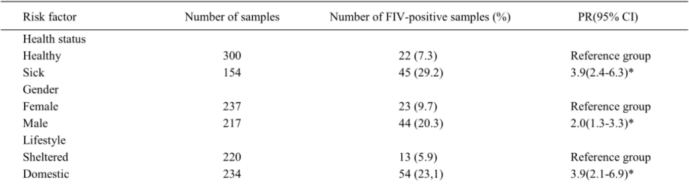
Of the total number of blood samples analyzed by nested PCR, 14.7% (67/454) were positive for FIV. Additionally, the 10 samples randomly selected to verify the specificity of the test were confirmed to be FIV-specific. Table 1 shows the data from FIV-positive
animals organized by health status, gender and lifestyle. The clinical evaluation showed that 29.2% of the FIV-positive animals were sick during the sample collection period, while 7.3% did not present any type of clinical sign.
Of the 454 total samples collected, 220 were obtained from animals living in shelters with a high population density (‘sheltered’ group). Thirteen animals (5.9%) in this group were FIV-positive. In the ‘domestic’ cats group, the percentage of FIV-positive individuals was 23.1%. Moreover, the percentage of positive cases was higher in males (20.3%, or 65.7% from the total) than in females (9.7%, or 34.3% from the total). It was not possible to precisely estimate the age of the FIV-positive animals because most of them had come from places with no records concerning their birth dates.
The risk of being infected with FIV was higher for males (P<0.01, PR=2.0, 95% CI=1.3-3.3). In addition, the likelihood of being FIV-positive was found to be associated with lifestyle, being higher in the “domestic” group (P<0.01, PR=3.9, 95% CI=2.1-6.9), and with a bad health status (P<0.01, PR=3.9, 95% CI=2.4-6.3).
DISCUSSION
There are few studies on the prevalence of FIV infection in São Paulo State. To date, only a single study has demonstrated the occurrence of FIV infection in the population of cats in São Paulo city (RECHE JR et al., 1997). The present study shows the occurrence of FIV infection in the population of cats from several cities of São Paulo State.
The percentage of FIV-positive cats found here was similar to that observed in other countries (PERI et al., 1994; GENTILE et al., 1996). However, it was higher than that described for São Paulo city (RECHE JR et al., 1997) and the United States (LURIA et al., 2004; LEVY et al., 2006), and lower than that described for sick animals in Rio Grande do Sul State (CALDAS et al., 2000). Moreover, when the group comprising sick animals was analyzed separately, the percentage of positive cases was higher to 29.2%. The physiological status of the animals studied could explain these differences in prevalence (HARTMANN, 1998; LEVY et al., 2006). FIV infection prevalence is higher (29.2%) in the group consisting of sick animals as compared to the healthy group (7.3%), as described by others (CALDAS et al., 2000; SOUZA et al., 2002; LEVY et al., 2006).
and hence more easily transmitted in environments with a high density of individuals. However, previous studies have shown that the transmission occurs mainly through bites during fights for territory, breeding and feeding, rather than through direct contact (HARTMANN, 1998). During sample collection, we observed that the ‘sheltered’ animals were not aggressive in crowded environments, and probably for this reason they did not get involved in fights. On the other hand, the owners of ‘domestic’ animals related that their cats usually had bite signs when they returned home. Thus, we suggest that behavioral aspects are more important than origin in determining susceptibility to FIV infection (BRADSHAW & HALL, 1999). Other authors, on the other hand, argue that origin is more important than behavior for FIV transmission (YILMAZ et al., 2000; SOUZA et al., 2002), and only additional studies will elucidate the factors involved in the transmission of this infection.
The behavioral characteristics of male cats observed in previous studies have explained why they are more susceptible to FIV infection than females (BRADSHAW & HALL, 1999). In fact, this study shows that the percentage of positive cases is higher in male than in female cats, and the same result has been found by others (YILMAZ et al., 2000; LURIA et al., 2004; LEVY et al., 2006). However, SOUZA et al. (2002) did not observe the same result in a study performed in Rio de Janeiro city, but in that case most male cats were neutered, which decreases the fights for breeding and, consequently, the risk of FIV transmission.
The results suggest that certain characteristics such as gender, health status and lifestyle may be associated with the likelihood of being FIV-positive in the population of cats studied. The high prevalence of FIV infection observed in São Paulo State urges clinician veterinarians to establish a differential
diagnosis of FIV infection so as not to mistake it for other infectious diseases. Finally, increasing the knowledge of the epidemiologic aspects of FIV is necessary to introduce efficient goals for the management and prevention of FIV infection.
ACKNOWLEDGEMENTS
The authors would like to thank Dr. Adriano Bonfim Carregaro for the collection of biological specimens. This work was supported by grants from Coordenação de Aperfeiçoamento de Pessoal de Nível Superior (CAPES) and Fundação de Amparo à Pesquisa do Estado de São Paulo (FAPESP - Processo 02/02649-0).
REFERENCES
BENDINELLI et al. Feline immunodeficiency virus: an interesting model for AIDS studies and an important cat pathogen. Clin Microbiol Rev, v.8, p.87-112, 1995. BRADSHAW, J.W.S.; HALL, L.S. Affiliative behaviour of related and unrelated pairs of cats in catteries: a preliminary report. Appl Anim Behav Sci, v.63, p.251-255, 1999. CALDAS, A.P.F. et al. Detecção do provírus da imunodeficiência felina em gatos domésticos pela técnica de reação em cadeia da polimerase. Pesq Vet Bras, v.20, p.20-25, 2000.
DUARTE, A.; TAVARES L. Phylogenetic analysis of portuguese feline immunodeficiency virus sequences reveals high genetic diversity. Vet Microbiol, v.114, p.25-33, 2005.
GENTILE, G. et al. Infección por el virus de la inmunodeficiencia felina: estudio seroepidemiológico y clínico em Bologna (Itália). Arch Med Vet, v.28, p.153-156, 1996. HARTMANN, K. Feline Immunodeficiency virus infection: an overview. Vet J, v.155, p.123-127, 1998.
HOHDATSU, T. et al. Genetic subtyping and epidemiological study of feline immunodeficiency virus by nested polymerase chain reaction-restriction fragment lenght polymorphism analysis of the gag gene. J Virol Methods, v.70, p.107-111, 1998. Table 1 – Results of the analyses of potential risk factors for FIV infection in 454 cats from veterinary clinics and animal shelters in São
Paulo State, Brazil.
Risk factor Number of samples Number of FIV-positive samples (%) PR(95% CI) Health status
Healthy 300 22 (7.3) Reference group
Sick 154 45 (29.2) 3.9(2.4-6.3)*
Gender
Female 237 23 (9.7) Reference group
Male 217 44 (20.3) 2.0(1.3-3.3)*
Lifestyle
Sheltered 220 13 (5.9) Reference group
Domestic 234 54 (23,1) 3.9(2.1-6.9)*
KAKINUMA, S. et al. Nucleotide sequence of feline immunodeficiency virus: classification of Japanese isolates into two subtypes which are distinct from non-japanese subtypes. J Virol, v.69, p.3639-3646, 1995.
KUMAR, S. et al. MEGA3: integrated software for molecular evolutionary genetics analysis and sequence alignment. Brief Bioinform, v.5, p.150-163, 2004.
LEVY, J.K. et al. Seroprevalence of feline leukemia virus and feline immunodeficiency virus infection among cats in North America and risk factors for seropositivity. J Am Vet Med Assoc, v.228, p.371-376, 2006.
LURIA, B.J. et al. Pevalence of infectious diseases in feral cats in Northern Florida. J Fel Med Surg, v.6, p.287-296, 2004. PECORARO, M.R. et al. Genetic diversity of Argentina isolates of feline immunodeficiency virus. J Gen Virol, v.77, p.2031-2035, 1996.
PEDERSEN, N.C et al. Isolation of a T-lymphotropic virus from domestic cats with an immunodeficiency-like syndrome.
Science, v.235, p.790-793, 1987.
PERI, E. et al. Seroepidemio-logical and clinical survey of feline immuno-deficiency virus infection in northern Italy. Vet Immunol Immunopathol, v.40, p.285-297, 1994.
RECHE JR, A. et al. Clinical study of acquired immunodeficiency syndrome in domestic cats in São Paulo. Braz J Vet Res Anim Sci, v.34, p.152-155, 1997.
SODORA, D.L. et al. Identification of three feline immunodeficiency vírus (FIV) env gene subtypes and comparison of the FIV and human immunodeficiency virus type evolutionary patterns. J Virol, v.68, p.2230-2238, 1994.
SOUZA, H.J.M. et al. Estudo epidemiológico de infeções pelo vírus da leucemia e/ou imunodeficiência felina, em gatos domésticos do município do Rio de Janeiro. Clin Vet, v.36, p.14-21, 2002.
STEINRIGL, A.; KLEIN D. Phylogenetic analysis of feline immunodeficiency virus in Central Europe: a prerequisite for vaccination and molecular diagnostics. J Gen Virol, v.84, p.1301-1307, 2003.
VAN REGENMORTEL, M.H.V. et al. Virus taxonomy: an
update. In: INTERNATIONAL COMMITTEE ON
TAXONOMY OF VIRUSES. 2000. [Consulted in 21 feb. 2006]. [online]. http:// www.virustaxonomyonline.com/virtax/ lpext.dll?f=templates&fn=main-h.htm
WILLET, B.J. et al. FIV infection of the domestic cat: an animal model for AIDS. I mmunol Today, v.18, p.182-189, 1997.