Hematologic reference values of Vinaceous-breasted Amazon (
Amazona vinacea
)
Valores hematológicos de referência em Papagaio-de-Peito-Roxo (Amazona vinacea)
Vanessa Rafaella Foletto da SilvaI Patricia Pereira SerafiniII Joice Reche PedrosoIII Denise Pereira LemeIV Vanessa Tavares KanaanIII
ISSN 1678-4596
ABSTRACT
Avian hematologic reference intervals are useful tools to evaluate body homeostasis and diagnose diseases. However,
there are few species-specific reference intervals published. The
present study reports Vinaceous-breasted Amazon (Amazona vinacea) hematologic reference values obtained during the health status evaluation of release candidates as part of this species reintroduction efforts at the Araucárias National Park. Parameters reported are erythrogram (erythrocytes, hemoglobin, packed cell volume, mean cell volume, mean corpuscular hemoglobin concentration and mean corpuscular hemoglobin), Red Cell Distribution Width (RDW), white cells total and differential (heterophiles, lymphocytes, basophils, eosinophils and monocytes),
thrombocytes and total plasma protein. For the first time results on
RDW and thrombocytes were described and a larger sample size were provided for all parameters analyzed. Intervals demonstrated
in the present study showed significant differences from those
considered normal in other parrot species and consequently have contributed to bring valuable information to base actions for the conservation of this endangered species of great biological value.
Key words: avian, araucárias national park, hemogram, laboratory medicine, psittacidae, parrot.
RESUMO
Os valores de intervalos de referência para parâmetros hematológicos aviários são ferramentas úteis para avaliação da homeostase corporal e diagnóstico de doenças. No entanto, são escassos os relatos de intervalos de referência
espécie-específico, o que aumenta a probabilidade de um erro na
interpretação de dados laboratoriais. Dessa forma, o presente
estudo relata valores hematológicos de referência específicos
para papagaio-de-peito-roxo (Amazona vinacea). Esses valores foram obtidos e calculados durante um projeto de reintrodução
como importante e adequada forma para avaliar o estado de saúde de candidatos à soltura. Os analíticos observados foram: eritrograma (eritrócitos, hemoglobina, hematócrito, volume celular, concentração de hemoglobina corpuscular e hemoglobina corpuscular média), distribuição das células vermelhas (RDW),
leucograma (basófilos, eosinofilis mielócitos, metamielócitos, heterófilos, linfócitos e monócitos), trombócitos e proteína
plasmática total. Este estudo traz, pela primeira vez, resultados referentes a RDW e a contagem de trombócitos para a espécie, além de fornecer um tamanho amostral maior que estudos anteriores. Os intervalos, demonstrados neste estudo, relatam valores diferentes dos considerados normais para outras espécies de papagaios e, consequentemente, vêm contribuindo para embasar a conservação dessa espécie ameaçada de extinção, de grande valor biológico.
Palavras-chave: aves, Parque Nacional das Araucárias, hemograma, medicina laboratorial, psitacídeos.
InTRODUCTIOn
The Vinaceous-breasted Amazon (Amazona vinacea) is one of the most endangered parrot species of the Atlantic Forest, a world’s top biodiversity hotspot. Two of the main reasons for the rapid and on-going population decline are habitat destruction and illegal nest poaching to satisfy the demand for companionship worldwide.Its estimated population ranges from 1,000-2,500 individuals in an area that includes parts of Brazil, Paraguay and Argentina (NAVARRO; PACHALY, 1994; “The
ICentro de Ciências da Saúde, Universidade Federal de Santa Catarina (UFSC), 88040-400, Florianópolis, SC, Brasil. E-mail: vanessafoletto@uol.com.br. Corresponding author.
IICEMAVE, Instituto Chico Mendes de Conservação da Biodiversidade (ICMBIO), Florianópolis, Santa Catarina, Brasil. IIIInstituto Espaço Silvestre, Itajaí, Santa Catarina, Brasil.
International Union for Conservation of Nature (IUCN)
Red List of Threatened Species”). In 2010, the first
parrot reintroduction project in a Brazilian National Park was initiated with a long-term goal to establish a viable A. vinacea population at the Araucárias National Park (ANP) Santa Catarina, Brazil. A total of 102 parrots (95 were victims of illegal wildlife trade,
6 offspring of confiscated birds born at the Curitiba Zoo and 1 was a fledgling rescued at the ANP) have
been rehabilitated, 83 were released and have been monitored continuously (KANAAN & RECHE, 2012; KANAAN, 2016). In order to be evaluated for release in the wild, each bird went through a four to six-month rehabilitation process, in which they underwent all laboratory and clinical exams listed in the Ordinance 179 (IBAMA 2008), including a complete hemogram.
Hematologic values are widely used for several purposes, such as illness diagnosis, evaluation of body condition and reproduction, as an indicator of physiological responses in adaptation to new surroundings, among others (MEHREN, 1999; MMA, 2003; OTS et al., 1998; WEISS & WARDROP, 2010). However, the literature describing hematologic values of free wild birds and those in captivity is very scarce (GOULART, 2006; MASELLO & QUILLFELDT, 2004). Thus, reference range data from different species are often used to perform blood examinations interpretation (VIGGERS et al., 1993). The lack of standardized data that can be used to evaluate health may limit adequate diagnosis (FUDGE, 2000; VALLE et al., 2008). There are few published articles describing
A. vinacea hematologic and serum biochemical values (POLO et al., 1998; SCHMIDT et al., 2009). Sample sizes of these studies are small with few analytes
concentrations quantified. Therefore, professionals may not find enough information in the literature and may have difficulties diagnosing and managing
patients until results from avian research efforts begin to satisfy the demand for data (OTS et al., 1998). Due to gaps in literature, the goal of this study was to provide data regarding hematological values of A. vinacea. All efforts were made to consider the described intervals as part of a reference population using biological and clinical criteria. The subjects of this study were healthy individuals according to statements and special reports developed by the American Society for Veterinary Clinical Pathology (ASVCP),
MATERIALS AnD METHODS
Animals
Subjects of this study were 102 release candidates of the project “Reintroduction of A.
vinacea at the Araucárias National Park, Santa Catarina, Brazil”, divided temporally in four main bird groups since 2010 (KANAAN, 2016). The research team prospectively selected individuals that were available within the ex-situ population in Southern Brazil institutions and one youngster rescued at the release area. Only animals considered representative of the healthy population kept in captivity, based on physical examinations and with no clinical disease were selected. To allow diversity within the target population studied, all sexes and body conditions were included even though the precise origins of the illegal trade apprehension were not always available (they were limited to the Southern Region of Brasil),
which made difficult to identify each bird’s exact
age; however, all of them were full grown with average weight: 365g. The sex of majority of birds was determined through DNA analyses (48 Males, 36 Females and 18 undetermined).
Birds received adequate nutrition with
seasonal fruits, leaves, flowers and seeds in its wild
state, as well as psittacidae seed mix. Fresh dietary items and water were provided ad libitum and changed daily.
As a condition for release, pathogen screening testing were also performed during the time they had their blood sample collected for:
Aspergillus sp., Coronavirus, Cryptococcus sp.,
Influenza Type A, Escherichia coli, Mycoplasma
sp., Newcastle Disease, Herpesvirus, Circovirus and Polyomavirus by PCR Real Time; Candida
sp. by fungal culture; Salmonella spp. by bacterial culture; hemoparasites investigation by the panoptic staining method visualized in an optical microscope; and parasitological feces examination by fecal
sedimentation and floatation tests.
Sample collections and laboratory techniques In order to collect blood samples, birds were physically restrained. Individuals were removed from the enclosure using a bird net, transferred to a procedure room and restrained with a towel. A veterinarian physically examined all birds. Next, each bird was held upright on a table giving the veterinarian free access to the ventral surface of the wing, which allowed for blood collection. If any sign of distress was noted, including respiratory distress, dyspnea, or open-mouthed breathing, blood collection was discontinued and it was concluded at a later time, when the bird showed no sign of distress.
The blood (1ml) was collected through venipuncture of the brachial vein with a syringe
UltraFine®, Becton Dickinson ind., Curitiba, Paraná,
80030-000, Brazil) containing 1% of heparin as anticoagulant (Hepamax®, Blau Farmacêutica,
Cotia, São Paulo, 06705-030, Brazil). The collected blood was transferred into another tube for the hemogram analysis. Blood smears were immediately carried after this blood collection, using the sample obtained with heparinized syringes. All samples were refrigerated, for up to 12 hours, and sent to a private laboratory. All analysis of samples were performed using methodology validated for avian species, taking into consideration the differentiation between red and white cells with a continuous quality control process (GEFFRÉ et al., 2011; FRIEDRICHS et al., 2012).
The hematologic parameters analyzed were erythrogram including red blood cell count (RCB), hemoglobin (Hb), packed cell volume (PCV), mean cell volume (MCV), mean corpuscular hemoglobin concentration (MCHC), mean corpuscular hemoglobin (MCH), red cell distribution width (RDW). Leucogram included white cells total count (white blood cells-WBC), white blood differential counts (heterophils, lymphocytes, basophils, eosinophils and monocytes) and thrombocytes.
In order to analyze the hemogram the methodology proposed by ELLIS & CAMPBELL,
(2013) was followed. Briefly, the PCV was
measured with a microhematocrit centrifuge (Jorvet
J503®, Jorgensen Labs, Loveland, Colorado,
80538, USA) and a PCV card reader. The total concentration of hemoglobin was determined by the cyanmethemoglobin method (Bioclin®, Quibasa
Química Básica, Belo Horizonte, Minas Gerais, 30130-000 Brazil) with centrifuged sample to remove nuclear content before analyzed in a spectrophotometer (EMP 168 VET®, Shenzhen Emperor Electronic
Technology, Nanshan, Shenzhen, 518108, China). Total erythrocytes concentration (RBC) was obtained by automated reading impedance within hematologic counter veterinary (Exigo®, Boule Diagnostics AB,
Plantation, Florida, 33311, USA). The RBC indices were obtained after determining PCV, RBC and Hb sample concentrations, using the following formulas: MCV (f/L) = PCV (%) x 10 / RBC; MCH (pg) = Hb (g dL-1) x 10 / RBC and MCHC (%) = Hb (g dL-1)
x 100 / PCV (%) (ELLIS & CAMPBELL, 2013). Total plasma protein was obtained by a photometric colorimetric test using the Biuret method (ELLIS & CAMPBELL, 2013).
To determine total leukocyte count, whole blood was diluted in a solution of Natt & Herrick and the concentration of total leukocytes was obtained
by counting the leukocyte cells in nine fields in a
Neubauer chamber (Zuzi®, Auxilab, Beriain, Navarra,
31191, Spain). The differential count of leukocytes and morphological evaluation were obtained in Diff Quik stained smears under light microscopy (1000x, Premiere®, Microscopes America, Cumming,
Georgia, 30041-4095, USA). The estimated number of thrombocytes was obtained by reading the blood smears using the following formula: Estimated number of thrombocytes = average of thrombocytes
in 5 oil-immersion fields (1000x)/1000 x 3.500.000.
To calculate the interval values of packed cell volume (35-55%) the following formula was used: Corrected number of thrombocytes = Estimated number of thrombocytes found x PCV / 45 (ELLIS & CAMPBELL, 2013).
Data presentation and statistical analysis
Statistical analysis was carried out
using Microsoft Office Excel® software (Microsoft
Corporation, Redmond, Washington, 98052-7329, USA) with the macroinstructions Reference Value Advisor (GEFFRÉ et al., 2011). Intervals were calculated in accordance with the recommendations of the American Society for Veterinary Clinical Pathology reference interval guidelines (FRIEDRICHS et al., 2012).The normality was tested using the Anderson–Darling test and by
histograms, while ‘‘outliers’’ were identified by
Tukey and Dixon Tests. Following the International Federation of Clinical Chemistry (IFCC) – Clinical and Laboratory Standards Institute (CLSI) recommendations (GEFFRÉ et al., 2011), unless outliers were known to be aberrant observations, emphasis was put on retaining rather than deleting these data points (CLSI, 2008). Thus, after evaluating the assumptions regarding symmetrical distribution and removal of extreme outliers, intervals were calculated for each analyze, which resulted in different sample sizes. Because, of non Gaussian distribution in majority data group the value limits were obtained using a nonparametric method with untransformed data for all.
RESULTS
and tested negative for several diseases, such as Poxvirus, Aspergillus sp., Candida sp., Coronavirus,
Cryptococcus sp., Escherichia coli, Influenza Type
A, Mycoplasma sp., Newcastle Disease, Alpha-Herpesvirus, Circovirus, Polyomavirus, Salmonella
spp. and parasitological feces examination.
DISCUSSIOn
There are very few published articles describing hematologic values for A. vinacea and others species. Therefore, this A. vinacea hematology
report is a significant contribution to the literature,
reinforcing values of parameters already described
for this species (POLO et al., 1998; SCHMIDT et al., 2009), and also describing novel parameters, such as RDW and trombocytes. Moreover, the sample size (n = 102 birds) was considerably larger than those reported in other studies, n = 6 by POLO et al. (1998), and n = 5 by SCHMIDT et al. (2009).
Although hematologic reference intervals and ranges have been established for some avian species, determined values such as total erythrocyte concentration, packed cell volume (PCV) and
hemoglobin concentration may be influenced by
age, gender, hormones, and other factors. The normal PCV for many bird species ranges approximately between 35% and 55% (MITCHELL & JOHNS,
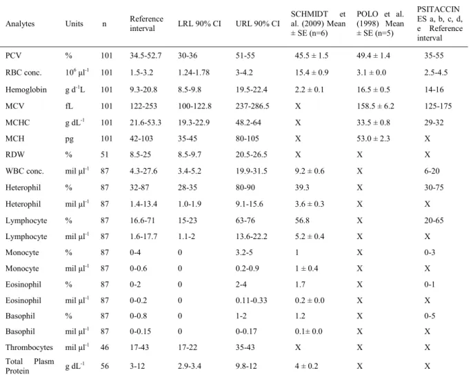
Table 1 - Hemotology reference intervals of Vinaceous Amazon Parrots(Amazona vinacea)and comparison of the hematological intervals of POLO et al. (1998), SCHMIDT et al. (2009) and values for psittacines in general.
Analytes Units n Referenceinterval LRL 90% CI URL 90% CI
SCHMIDT et al. (2009) Mean ± SE (n=6)
POLO et al. (1998) Mean ± SE (n=5)
PSITACCIN ES a, b, c, d, e Reference interval
PCV % 101 34.5-52.7 30-36 51-55 45.5 ± 1.5 49.4 ± 1.4 35-55
RBC conc. 106μl-1 101 1.5-3.2 1.24-1.78 3-4.2 15.4 ± 0.9 3.1 ± 0.0 2.5-4.5
Hemoglobin g d-1L 101 9.3-20.8 8.5-9.8 19.5-22.4 2.2 ± 0.1 16.5 ± 0.5 14-16
MCV fL 101 122-253 100-122.8 237-286.5 X 158.5 ± 6.2 125-175
MCHC g dL-1 101 21.6-53.3 19.3-22.9 48.2-64 X 33.5 ± 0.8 29-32
MCH pg 101 42-103 35-45 80-105 X 53.0 ± 2.3 X
RDW % 51 8.5-25 8.5-9.7 20.5-26.5 X X X
WBC conc. mil μl-1 87 4.3-27.6 3.4-5.2 19.9-31.5 9.2 ± 0.6 X 6-20
Heterophil % 87 32-87 28-35 80-90 39.3 X 30-75
Heterophil mil μl-1 87 1.4-13.4 1.0-1.9 9.1-15.6 3.6 ± 0.3 X X
Lymphocyte % 87 16.6-71 15-23 63-76 56.8 X 20-65
Lymphocyte mil μl-1 87 1.6-17.7 1.1-2 13.6-22.2 5.2 ± 0.4 X X
Monocyte % 87 0-4 0 3.2-5 1 X 0-3
Monocyte mil μl-1 87 0-0.6 0 0.2-0.9 1 ± 0.4 X X
Eosinophil % 87 0-2 0 2-4 1.7 X 0-1
Eosinophil mil μl-1
87 0-0.2 0 0.11-0.33 0.2 ± 0.0 X X
Basophil % 87 0-0.8 0 1-2 1.2 X 0-5
Basophil mil μl-1 87 0-0.15 0 0-0.17 0.1± 0.0 X X
Thrombocytes mil μl-1 46 17-43 17-22 35-43 X X X
Total Plasm Protein g dL
-1 56 3-12 2.9-3.4 9.8-12 4 ± 0.2 X X
“x” symbolizes the parameter not described in the study. LRL, lower reference limit; URL, upper reference limit; PCV, packed cell volume; MCHC, mean cell hemoglobin concentration; WBC, white blood cell; conc., concentration.
2008), similar to the results of this study, despite of the limitations brought up by the lack of knowledge of each individual bird´s history and age, corroborating those previously described for others parrots species (HARRIS, 1991; ELLIS & CAMPBELL, 2013).
Variation in size of avian erythrocytes is seen in peripheral blood smears (ELLIS & CAMPBELL, 2013). Slight anisocytosis is considered
an insignificant finding in birds (MITCHELL &
JOHNS, 2008) and could be calculate using RDW (%), which measures variation in red blood cell size (FUDGE, 2000). RDW% may vary depending on the age or even between laboratories (JONES, 2015).
Interval values of WBC reported are within the expected values for psittacines. However, the white blood cell differential was different from other reports (HARRIS, 1991; HARRISON, 2008; CARPENTER, 2013; ELLIS & CAMPBELL, 2013;). Considerable variability may be found in leukocyte reference values in a wide variety of clinically normal avian species (MAXWELL & TREJO, 1970).
Results showed that the heterophil cells were the highest values of leukocytes, followed by lymphocytes. This pattern was described as acceptable for psittacines by NAVARRO & PACHALY (1994). However, in the majority of species, percentage of lymphocytes is higher than any other types of leukocytes, with values ranging between 40-70% of the total, and heterophils being the second highest group (STURKIE & GRIMINGER, 1986).
Avian thrombocytes play a primary role in hemostasis in a manner similar to mammalian platelets, which may also have a phagocytic function and participate in removing foreign material from the blood (HARRISON, 2008). Thrombocytopenia can be seen in some viral diseases or may appear as an artefact caused by distress. Clumping might also be a serious problem for thrombocyte counts when drawing blood from the bird was a challenge or if the smear had a problem in preparation, resulting in lower thrombocyte numbers. Exactly because of clumping, thrombocytes counts are not routinely performed in avian hematology and reference values are scarce. Interval of thrombocytes obtained in this studyis in agreement with the values described in other studies, but it is important to note that number of these cells varied greatly among individuals, with values that range from 10 to 132 x 103 mm-1 (STURKIE & GRIMINGER, 1986;
HARRISON, 2008;). According to STURKIE & GRIMINGER (1986), the number of thrombocytes of different avian species range from 20 to 30 x 103/mm³, and it is approximately the same as the
number of total leukocytes.However, in this study, no similarity between the values of thrombocytes and leucocytes was observed. The evaluation for
Amazona vinacea showed that the lower limit was
found representative of only five individuals with
values less than 20 x 103 mm-1. Those animals had
no clinical signs of disease and the coagulation time after blood collection was homogeneous within the group, suggesting that a coagulopathy was absent. No viruses were isolated or detected in those birds. Probably those results were caused by
increased susceptibility of those five individuals to
stress during physical restraint for blood sampling. Thrombocytosis has not been documented.
This study did not identified or reported significant leukocyte changes as toxic cells or
polychromatic avian erythrocytes, even though regular appearance of polychromatophilic erythrocytes (1%-5% of the total erythrocyte count) in peripheral blood (FUDGE, 2000). Polychromatic erythrocytes, along with reticulocytes, are indicative of bone marrow activity. There are no reports of any blood cells
morphological characteristic specific to A. vinacea. Further research is necessary to increase the number of publications describing all the hematologic and biochemical values for the A. vinacea. Adequate published references for hematologic values are a powerful tool to evaluate health status and provide adequate diagnosis, especially for animals being considered for release, and consequently contribute to the conservation of this endangered species of great biological value.
COnCLUSIOn
This study has demonstrated the hematology of A. vinacea, corroborating with avian medicine and to efforts to evaluated health of individual birds in order to provide a chance to this species to play their ecological roles, contributing to conservation.
ACKnOWLEDGMEnTS
Laboratório de Etologia Aplicada (LETA), the Pós-Graduação em Agroecossistemas (PGA), the Pós-Graduação em Ecologia (POSECO) of the Universidade Federal de Santa Catarina for technical support, and the Coordenação de Aperfeiçoamento de Pessoal de Nível Superior (CAPES). Finally, we thank the Fundação Grupo O Boticário de Conservação à Natureza, Politrade, Biofaces, Taroii Investiment Group and Zoological Society for the Conservation of Species and Populations (ZGAP)
for their financial support.
BIOETHICS AnD BIOSSECURITY COMMITTEE APPROVAL
All procedures were approved by the Chico Mendes Institute for Biodiversity Conservation (ICMBio- protocol number SISBIO 25133 and 41776), Instituto Brasileiro do Meio Ambiente e dos Recursos Naturais Renováveis, FATMA and Universidade Federal de Santa Catarina ethics committee for animal research (CEUA-UFSC protocol number PP00589).
REFEREnCES
CARPENTER, J.W. Exotic animal formulary. Philadelphia: Saunders, 2013. 564p.
ELLIS, C.K.; CAMPBELL, T.W. Avian and Exotic Animal Hematology and Cytology. New Jersey: Wiley-Blackwell, 2013. 2047p.
CLSI. Defining, establishing, and verifying reference intervals
in the clinical laboratory: approved guideline EP28-A3c. Wayne: Clinical and Laboratory Standards Institute, 2008. Available from: <http://shop.clsi.org/site/Sample_pdf/EP28A3C_sample. pdOnline>. Accessed: May 19, 2016.
FRIEDRICHS, K.R. et al. ASVCP reference interval guidelines: determination of de novo reference intervals in veterinary species and other related topics. Veterinary Clinical Pathology, v.41, n.4, p.441-453, Dec. 2012. Available from: <http://onlinelibrary. wiley.com/doi/10.1111/vcp.12006/full>. Accessed: May 19, 2016. doi:10.1111/vcp.12006.
FUDGE, A.M. Biochemistry. In: FUDGE, A.M. (Ed.).
Laboratory medicine: avian and exotic pets. Philadelphia: Saunders, 2000. p.35-46.
GEFFRÉ, A. et al. Reference value advisor: a new freeware set of macroinstructions to calculate reference intervals with Microsoft Excel. Veterinary Clinical Pathology, v.40, n.1, p.107-112, Mar. 2011. Available from: <http://onlinelibrary.wiley.com/ doi/10.1111/j.1939-165X.2011.00287.x/full>. Accessed: May 19, 2016. doi: 10.1111/j.1939-165X.2011.00287.x.
GOULART, C.E.S. Valores hematológicos de referência para papagaios- verdadeiros (Amazona aestiva - Psittacidae) mantidos em cativeiro. 2006. 80f. Dissertação (Mestrado em Medicina Veterinária Preventiva) – Curso de Pós-graduação em Medicina Veterinária Preventiva, Universidade Federal de Minas Gerais, MG.
HARRIS, D.J. Laboratory testing in pet avian medicine.
Veterinary clinics of north America. Small Animal Practice, v.21, n.6, p.1147-1156, Nov. 1991. Available from: <http://www. sciencedirect.com/science/article/pii/S0195561691501297>.
Accessed: May 19, 2016. doi: 10.1016/S0195-5616(91)50129-7. HARRISON, L. et al. Exotic companion medicine handbook for veterinarians. Florida: Zoological Education Network, 2008. 400p.
IBAMA. Instrução normativa no 179, DE 25 DE JUNHO DE
2008. Brasil, 2008. Available from: <http://www.icmbio.gov.br>. Accessed: Nov. 13, 2015.
JONES, M.P. Avian hematology. Veterinary Clinics of north America: Exotic Animal Practice, v.18, n.1, p.51-61, Jan. 2015. Available from: <http://www.sciencedirect.com/science/article/pii/ S0272271215000554>. Accessed: May 19, 2016. doi: 0.1016/j. cll.2015.05.013.
KANAAN, V.T. Re-introduction of the vinaceous-breasted Amazon at the Araucárias National Park, Santa Catarina, Brazil. In: SOORAE, P. S. (ed.) (2016). Global Re-introduction Perspectives: 2016. Case-studies from around the globe. Gland, Switzerland: IUCN/SSC Reintroduction Specialist Group and Abu Dhabi, UAE: Environment Agency Abu Dhabi. xiv + 276 pp. pp. 106-110. Available from: <https://portals.iucn.org/
library/sites/library/files/documents/2016-006.pdf>. Accessed:
August 22, 2016.
KANAAN, V.T.; RECHE, J. Resumo das atividades do projeto piloto de reintrodução do papagaio-de-peito-roxo (Amazona vinacea) no Parque Nacional das Araucárias, SC. 4.ed. In: REVISTA CETAS e ASMs no Estado de São Paulo– Relatório de Atividades 2012. São Paulo: Superintendência do IBAMA no Estado de São Paulo, Ministério do Meio Ambiente, 2012. p.59-63.
MASELLO, J.F.; QUILLFELDT, P. Are haematological parameters related to body condition, ornamentation and breeding success in wild burrowing parrots Cyanoliseus patagonus?
Journal of Avian Biology, v.35, n.5, p.445-454, Sept. 2004. Available from: <http://onlinelibrary.wiley.com/doi/10.1111/ j.0908-8857.2004.03278.x/full>. Accessed: May 19, 2016. doi: 10.1111/j.0908-8857.2004.03278.x.
MAXWELL, M.H.; TREJO, F. The ultrastructure of white blood cells and thrombocytes of the domestic fowl. British Veterinary Journal, v.126, n.11, p.583-592, Nov. 1970.
MEHREN, K.G. Exotic companion medicine handbook for veterinarians. Lake Woorth: Zoological Education Network, 1999. 400p.
MMA. Instrução normativa n. 03, de 26 de maio de 2003. Available from: <http://www.mma.gov.br>. Accessed: Nov. 13, 2015. NAVARRO, C.; PACHALY, J. Manual de hematologia veterinária. São Paulo: Varela, 1994. 206p.
POLO, F.J. et al. Hematologic and plasma chemistry values in captive psittacine birds. Avian Diseases, v.42, n.3, p.523, July, 1998. Available from: <http://www. jstor.org/stable/1592679>. Accessed: May 19, 2016. doi: 10.2307/1592679.
SCHMIDT, E.M. et al. Hematology of the red-capped parrot (Pionopsitta pileata) and Vinaceous Amazon parrot (Amazona vinacea) in captivity. Journal of Zoo and Wildlife Medicine, v.40, n.1, p.15-17, Mar. 2009. Available from: <http://dx.doi. org/10.1638/2007-0054.1>. Accessed: May 19, 2016. doi: 10.1638/2007-0054.1.
STURKIE, P.D.; GRIMINGER, P. Body fluids: blood. In:
STURKIE, P.D. (Ed.). Avian physiology. New York: Springer New York, 1986. p.102-128.
THE IUCN RED LIST OF THREATENED SPECIES. Available from: <http://www.iucnredlist.org>. Accessed: Nov. 13, 2015. VALLE, S. de F. et al. Parâmetros de bioquímica sérica de machos,