Braz. J. of Develop.,Curitiba, v. 6, n. 1,p. 2209-2215 jan. 2020. ISSN 2525-8761
Homeopathic treatment in dog dermatopathy
Tratamento homeopático em dermatopatia em cão
DOI:10.34117/bjdv6n1-160
Recebimento dos originais: 30/11/2019 Aceitação para publicação: 15/01/2020
Franscinne Brait Narita
Pós Graduada em Homeopatia Clínica e Tecnologia das Altas diluições na High Dilution Science.
Rua João Vilhema, 76, Planalto Paulista, São Paulo/SP [email protected]
Bruna Scardoeli
Pós Graduada em Homeopatia Clínica e Tecnologia das Altas diluições na High Dilution Science.
Rua Oagy Kalile 415, Morumbi, São Paulo/ SP [email protected]
Adalberto do Carmo Braga von Ancken
Mestre em Bem-estar e Medicina Veterinária pela UNISA UNIP/ High Dilution Science
End. Rua Antônio de Barros, 1705 Tatuapé - São Paulo/ SP [email protected]
Cideli de Paula Coelho Doutor em Ciências pela FMVZ- USP Universidade Santo Amaro / High Dilution Science
Rua Amazonas, 1245, São Caetano do Sul -SP [email protected]
ABSTRACT
Between 20% and 75% of veterinary consultations of dogs and cats in clinics and hospitals are believed to be directly related to skin problems. The main complaints reported by tutors during consultations are the symptoms of alopecia, hypotrichosis, pruritus, ulceration, scaling, vesicle and scabs formation, and the presence of skin masses, in which one dermatopathy is often associated with another. Chronic lesions are mainly observed in places where there is intense itching, which is the most desconfortable symptom to tutors, but which, on the other hand, is minimized by several drugs. The most commonly treatments used are highly effective immunomodulations such as glucocorticoids, but it brings by numerous adverse reactions such as polyuria, polyphagia, polydipsia, alopecia, obesity, pancreatitis, gastrointestinal ulcers, diabetes, and predisposition to infections. The aim of this paper is to show the effectiveness of treatment through ultra-diluted medications in chronic dog dermatopathy. After repertorization, the administration of Pulsatilla nigricans 30cH was recommended, and four
Braz. J. of Develop.,Curitiba, v. 6, n. 1,p. 2209-2215 jan. 2020. ISSN 2525-8761
drops were prescribed orally twice a day for 20 consecutive days, associated with Hystaminium 30cH, prescribed the same way, but for 30 days. One week after the beginning of homeopathies, the patient showed considerable improvement in the skin and after 30 days, 80% improvement in the itchy conditions, skin texture, local hyperemia and odor, as well as behavior and rostral physiognomy. suggesting good disposition of the patient.
Keywords: Dermatology; Atopy; Dogs; Ultra dilution. RESUMO
Acredita-se que entre 20% e 75% das consultas veterinárias de cães e gatos em clínicas e hospitais estejam diretamente relacionadas a problemas de pele. As principais queixas relatadas pelos tutores durante as consultas são os sintomas de alopecia, hipotricose, prurido, ulceração, descamação, formação de vesículas e crostas e presença de massas de pele, nas quais uma dermatopatia está frequentemente associada a outra. As lesões crônicas são vistas principalmente em locais onde há prurido intenso, que é o sintoma mais desconfortável para os tutores, mas que, de outra forma, é minimizado por vários medicamentos. Os tratamentos mais comumente usados são imunomodulações altamente eficazes, como glicocorticóides, mas apresentam inúmeras reações adversas, como poliúria, polifagia, polidipsia, alopecia, obesidade, pancreatite, úlceras gastrointestinais, diabetes e predisposição a infecções. O objetivo deste artigo é mostrar a eficácia do tratamento ultra-diluído na dermatopatia crônica em cães. Após repertorização, foi recomendada a administração de Pulsatilla nigricans 30cH e quatro gotas foram prescritas por via oral duas vezes ao dia por 20 dias consecutivos, associadas ao Hystaminium 30cH, prescrito da mesma maneira, mas por 30 dias. Uma semana após o início das homeopatias, o paciente apresentou melhora considerável na pele e, após 30 dias, melhora de 80% nas condições de prurido, textura da pele, hiperemia e odor local, além de comportamento e fisionomia rostral. sugerindo boa disposição do paciente.
Palavras-chave: Dermatologia; Atopia; Cães; Ultra diluição.
Dermatological resources in dogs and cats prevail from 20% to 75% in clinics and hospitals, being most prominent in patients between 2 and 5 years, without racial predisposition and being also present licking dermatitis in up to 68% of atopic dogs, with 12% of these animals show concomitant seborrhea1.
Skin scraping and mycological examination are imperative for any patient with dermatological issue, including an indication of cytology and bacterial culture2. Blood test and biochemical exam are important complements for the differential diagnosis of endocrinopathies with cutaneous reflexes3, as well as behavioral alterations of separation anxiety4.
The initial symptomatology of dermatopathies, local or generalized itching, is more evident and reported by tutors and can be immediately minimized by administration of glucocorticoids, which unfortunately can provoke various side effects, like diarrhea, anorexia and constipation5, in addition to polyuria, polydipsia, alopecia, polyphagia, obesity, urinary
Braz. J. of Develop.,Curitiba, v. 6, n. 1,p. 2209-2215 jan. 2020. ISSN 2525-8761
tract infections, pancreatitis, ulceration and gastrointestinal perforation6, often associated with limb licking and axillary lesions7,8.
In addition, opportunistically, secondary infections such as superficial and deep bacterial pyodermatite, fungical infections, often included in the patient's clinical symptoms, are treated with antibiotic therapy or systemic antifungicals associated with glucocorticoids and antihistamines, respectively9.
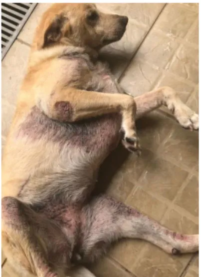
This case report the treatment in a 10 years old dog in March 2018, whose primary complaint was chronic dermatitis (Figure 1), which has been treated by clinical colleagues, dermatology and endocrinology for almost 4 years, but without goods results in all convencional previous treatments.
Figure 1: The patient reported.
Source: Personal Archives, March 31, 2018.
In the first visit, the tutor reports that the symptoms began with the change of residence, where physical examination revealed seborrhea, fetid odor, allopecic areas, erythema, hyperpigmentation and hyperkeratosis, especially in the limb, ventral, cervical regions (Figure 2, 3 and 4), in addition to intense and widespread itching. However, there wasn't notable alterations in blood test, renal function, liver function, triglycerides, cholesterol, TSH, free T4 and skin scraping.
Braz. J. of Develop.,Curitiba, v. 6, n. 1,p. 2209-2215 jan. 2020. ISSN 2525-8761 Figures 2, 3 and 4: Ventral region; member region; and cervical region, respectively.
Source: Personal Archives, March 31, 2018.
Remembering that most dermatopathies are due to behavioral or mental changes, the predisposition of atopic dermatitis increases when the animal separates from the tutor or when there have changes in the life of these individuals10, being an attempt by the body to rebalance itself, thus, physically manifesting the result of this biological process against factors such as heat, food, microorganisms, jealousy, longing, fear, stress, among others11.
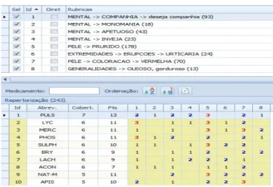
Therefore, we opted for homeopathic treatment after repertorization (Figure 5), in order to stimulate the whole organism, preventing side effects to the patient, based on the Similar Law, described by the ability of a disease to be cured through the administration of a substance that produces the same symptoms in a healthy person12.
The pharmacodynamic action of homeopathic medicines provides small amounts of information to regulatory centers, such as receptors and DNA, perceived as reactive disorders to promote the body's vital harmony, which manifests symptoms due to pathogenic factors that will, then, used to identify the correct drug12. In the traditional homeopathic repertoire model, the pathogenetic symptoms described in the Homeopathic Medical Matter receive the grouping of several drugs, maintaining a hierarchy of those more or less common13.
Braz. J. of Develop.,Curitiba, v. 6, n. 1,p. 2209-2215 jan. 2020. ISSN 2525-8761 Figure 5: Digital repertorization of symptoms.
Source: Personal Archives, March 31, 2018.
Thus, Pulsatilla nigricans, which acts on relapsing urticaria, was administered to docile but suspicious, lewd patients with emotional arousal disorders and the need for affection and companionship, while Histaminum was administered to inhibit histamine production, minimizing pruritus14,15, due to allergen-induced mast cell degranulation7, in which the mechanism of action causes acute inflammation, allergy and urticaria, but which at homeopathic dose responds to these proinflammatory and irritant properties, reducing the manifestations, because at high dilutions there is activity at the level of cellular receptors, thus blocking the effects of histamine16,17.
Thus, 4 drops were prescribed Pulsatilla nigricans 30cH via oral, 2 It was prescribed 4 drops of Pulsatilla nigricans 30cH orally twice a day for 20 days and 2 drops of Hystaminium 30CH orally twice a day for 30 days, already showing significant improvement in skin in just 1 week. After 30 days of treatment (Figures 6, 7 and 8), there was absence of fetid odor, less intense hyperemia, smaller areas of alopecia and seborrhea, as well as minimized itching, resulting in an 80% improvement in treated dermatopathy, always maintaining the well-being of the patient during treatment.
Braz. J. of Develop.,Curitiba, v. 6, n. 1,p. 2209-2215 jan. 2020. ISSN 2525-8761 Figures 6, 7 e 8: Ventral region; member region; and cervical region, respectively.
Source: Personal Archives, March 31, 2018.
However, relying on the key pillars of homeopathy, it was possible through the Similarity Law, promote the path to cure quickly and less harmful, by balancing the patient's vital force18, demonstrating the performance of homeopathic treatment as therapeutic support in the home in prognosis of hypersensitivity and atopic diseases and acute and chronic eczema.
REFERENCES
1. Scott DW, Miller WH, Griffin CE. Small animal dermatology. 6 ed. Philadelphia: WB Sauders Company; 2001.
2. Hillier A. Symposium on atopic dermatits. Veterinary Medicine, Lenexa, KS; 2002. 3. Salzo PS. Entendendo as piodermites: diagnóstico e tratamento. Nosso clínico n. 48; p. 14-18.
4. Appleby D, Pluijmakers J. Separation anxiety in dogs: the function of homeostasis in its development and treatment. Vet. Clin. N. Am. Small Anim. Pract. 33(2); 2004, 321-344. 5. Hiller A. Symposium on atopic dermatits. Veterinary Medicine, Lenexa, KS, v. 97, n.3, p. 196-222, 2002.
6. Scott DW, Miller WH, Griffin CE. Dermatologia de pequenos animais. 5ª ed. Rio de Janeiro: Interlivros, 1996.
7. Thompson JP. Moléstias imunológicas. In: Ettinger SJ; Feldman EC. Tratado de medicina interna veterinária. 4.ed. São Paulo: Manole; 1997.
8. Virga V. Self-directed behaviors & behavioral dermatoses. Atlantic Coast Veterinary Conference; 2007. Acessado eletronicamente em 22/04/2019 disponível em
Braz. J. of Develop.,Curitiba, v. 6, n. 1,p. 2209-2215 jan. 2020. ISSN 2525-8761 http://www.vin.com/Members/Proceedings/Proceedings.plx?CID=ACVC2007&Category=3 101&PID=19119&O=VIN
9. Noli C. Staphylococcal pyoderma. In A. Foster & C. Foil (Eds.) BSAVA manual of small animal dermatology, Gloucester, UK: British small animal veterinary association, 2003. 10. Markolin C. German New Medicine. Explore, volume 6, n 3.Vancouver; 2007, 1-2. 11. Lobão AO. A Homeopatia e os animais de companhia. Jornal de Piracicaba, Piracicaba/SP; 1994, 26-29.
12. Bellavite P. As evidências a favor da homeopatia e as ciências básicas. Revista de Homeopatia; 2018; 81(3/4): 1-15.
13. Teixeira, MZ. Novos medicamentos homeopáticos: Uso dos fármacos modernos segundo o princípio da similitude. Revista de Homeopatia; 2012; 75(1):39-53
14. Torro AR. Homeopatia veterinária: semiologia, materia médica e psicossomática. São Paulo, 1999.
15. Demarque D. Homeopatia: medicina de base experimental. 2ª ed. Ribeirão Preto: Museu de Homeopatia Abrahão Brickmann, 2002.
16. Potevin B, Davenas E, benveniste J. In vitro immunological degranulation of human basophils is modulated by lung histamine and Apis mellifica. Br j Clin Pharmacol; 1988; 25(4): 439-444.
17. Sainte-Laudy J, Belon Ph. Inhibition of basophil activation by histamine: a sensitive and reproducible model for the study of the biological activity of high dilutions. Homeopathy; 2009;98: 186–197.
18. Pustiglione M. Organon da Artes de Curar de Samuel Hahnemann para o século XXI. 1ª Edição. São Paulo: Organon. 2010.