CLINICAL SCIENCE
Heart Institute (InCor) and Department of Medicine-LIM 13, Faculdade de Medicina da Universidade de São Paulo – São Paulo/SP, Brazil.
Phone: 55 11 3069.5068
Emails: ayumi.miyakawa@ incor.usp.br / [email protected] Received for publication on May 09, 2008
Accepted for publication on July 04, 2008
HUMAN SAPHENOUS VEIN ORGAN CULTURE
UNDER CONTROLLED HEMODYNAMIC CONDITIONS
Ayumi Aurea Miyakawa, Luis Alberto Oliveira Dallan, Silvia Lacchini, Thaiz Ferraz Borin, Jose Eduardo Krieger
doi: 10.1590/S1807-59322008000500018
Miyakawa AA, Dallan LAO, Lacchini S, Borin TF, Krieger JE. Human saphenous vein organ culture under controlled hemodynamic conditions. Clinics. 2008:63(5):683-8.
INTRODUCTION: Saphenous vein grafting is still widely used to revascularize ischemic myocardium. The effectiveness of this procedure is limited by neointima formation and accelerated atherosclerosis, which frequently leads to graft occlusion. A better understanding of this process is important to clarify the mechanisms of vein graft disease and to aid in the formulation of strategies for prevention and/or therapeutics.
OBJECTIVE: To develop an ex vivo low system that allows for controlled hemodynamics in order to mimic arterial and venous conditions.
METHODS: Human saphenous veins were cultured either under venous (low: 5 ml/min) or arterial hemodynamic conditions (low: 50 ml/min, pressure: 80 mmHg) for 1-, 2- and 4-day periods. Cell viability, cell density and apoptosis were compared before and after these intervals using MTT, Hoeschst 33258 stain, and TUNEL assays, respectively.
RESULTS: Fresh excised tissue segments were well preserved prior to the study. Hoechst 33258 and MTT stains showed progres-sive losses in cell density and cell viability in veins cultured under arterial hemodynamic conditions from 1 to 4 days, while no alterations were observed in veins cultured under venous conditions. Although the cell density from 1-day cultured veins under arterial conditions was similar to that of freshly excised veins, the TUNEL assay indicated that most of these cells were undergo-ing apoptosis.
CONCLUSION: The results observed resemble the events taking place during early in vivo arterial-vein grafting and provide evidence that an ex vivo perfusion system may be useful for the identiication of new therapeutic targets that ameliorate vein graft remodeling and increase graft patency over time.
KEYWORDS: Saphenous vein graft; Ex vivo organ culture; Vascular biology.
INTRODUCTION
Coronary artery bypass graft (CABG) is the standard procedure for the revascularization of ischemic heart tissue. Saphenous vein grafts were the first vascular conduits used in CABG, but their effectiveness has been limited by accelerated atherosclerosis that develops within the vein conduit.1,2 In contrast, arterial conduits display better
long-term patency and have been increasingly used instead
of venous grafts. Despite the increased usage of arterial conduits, full arterial revascularizations are carried out in only a small minority of eligible patients; currently,CABG surgery utilizes a combination of arterial and venous grafts.3,4
However, a better understanding of the molecular events associated with the early arterialization of vein grafts may give rise to alternative approaches to overcoming the poor long-term patency of saphenous vein grafts.
stresses are increased.5-10 Ex vivo organ culture has also
been used in vascular biology studies; most of the protocols make use of vessel rings that may be maintained up to 8 days in culture media without tissue degeneration.11,12
More recently, perfusion organ culture systems have been developed for the study of vessels under well-controlled hemodynamic conditions. Arteries kept in organ culture systems have been shown to be viable and with active vasomotion tone for up to 7 days.13,14 In contrast, vein
culture studies have been limited to no more than 24 hours of culture.15,16
In the present investigation, we developed an ex vivo perfusion system, which mimics in vivo hemodynamic conditions, allowing for the study of viable vein grafts under venous or arterial hemodynamic conditions for up to 4 days. We demonstrated that this ex vivo system reproduces early events observed in in vivo animal models during the arterialization of vein grafts.
MATERIALS AND METHODS
Saphenous vein preparation
Saphenous veins were obtained from patients undergoing aortocoronary bypass surgery at the Heart Institute (InCor), University of São Paulo Medical School. The vein segments were kept in physiological solution until mounting in the culture system. This study protocol was approved by the local ethical committee (SDC – 1834/01/22, CAPPesq – 299/01).
Organ culture system
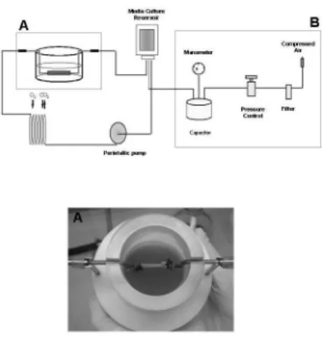
The organ culture system is schematically represented in Figure 1. The vessel chamber was connected to a perfusion
system where the media from the reservoir was pumped at a set low rate. This circuit was adapted from the CELLMAX Artiicial Capillary System (Spectrum Laboratories, Inc. Rancho Dominguez, CA) according to previously described methods.13 Vessel chambers and a pressure device were
developed in-house to properly place the vessel segments and to enable pressure and low to be controlled independently. The flow circuit was maintained in a humidified CO2 incubator at 37°C.
Saphenous vein segments (approximately 2 cm each) were placed in the chamber illed with Dulbecco’s modiied Eagle’s medium containing 10% fetal bovine serum, 100 U/ ml penicillin, and 100 µg/ml streptomycin. Vein segments from the same patient were connected to the perfusion system and cultured either under venous conditions (low: 5 ml/min) or arterial conditions (low: 50 ml/min, pressure: 80 mmHg) for 1, 2, or 4 days. Fresh, non-mounted segments were also collected for analysis.
All culture solutions were purchased from GIBCOTM
(Invitrogen Corporation, Carlsbad, CA).
Histological analysis
Tissue samples were ixed in 4% phosphate-buffered formalin and embedded in paraplast (Oxford, St. Louis, MO). Sections of 3-µm thickness were stained with haematoxylin-eosin (HE) and Verhoeff’s-Van Gieson (VVG) for elastin. The sections were also used for Hoechst staining and TUNEL assays as described below.
Hoechst staining
Cell nuclei were labeled with Hoechst 33258 (Sigma Chemical Co., St. Louis, MO) as described previously.17
Briefly, tissue sections were deparaffinized in citrisolv (Fisher Scientiic Company, Pittsburgh, PA), rehydrated in serial alcohol dilutions, and treated with 0.5% Triton X-100 for 15 min at 20 °C. The staining reaction was performed using Hoechst 33258 20 µg/ml diluted in assay buffer (mmol/L): NaCl 137, KCl 5, Na2HPO4 0.4, NaHCO3 4, glucose 5.5, MgCl2 2, EGTA 2, PIPES, pH 6.1. The sections were visualized under luorescence microscopy (Carl Zeiss Inc., Thornwood, NY) and quantiication was performed by an observer who was blinded to the hemodynamic conditions and duration of the cultures. Each section was measured at 10 different randomly selected points and positive nuclei stains were quantiied. Tissue area was quantiied using Leica Qwin software (version 2.2) calibrated as per the manufacturer’s instructions (40X magniication, 1 pixel = 0.228 µm). The cellular density was expressed as the total cell number per vessel area.
Terminal transferase-mediated dUTP nick end labeling (TUNEL) assay
DNA fragmentation was detected using the In Situ Cell Death Detection kit, AP (Roche Molecular Biochemicals, Mannheim, Germany) according to the manufacturer’s instructions. Briely, tissue sections were deparafinized in citrisolv, rehydrated in serial alcohol dilutions, and permeabilized with 0.5% Triton X-100 in 0.1% sodium citrate. The reaction with terminal deoxynucleotidyl transferase and alkaline phosphatase conversion was performed and the cross-sections were examined by light microscopy. Quantification was performed using Leica QWin software (version 2.2). TUNEL-positive cells and total cells were quantiied by an observer who was blinded to the hemodynamic conditions and the duration of culture. The results were expressed as a percentage of TUNEL-positive cells among all cells evaluated.
MTT stain
To verify tissue viability, the vein segments were stained with methylthiazol tetrazolium (MTT - Sigma Chemical Co., St. Louis, MO) as described elsewhere.14 When reduced by
active mitochondria and other cellular enzymes, the MTT dye gives rise to a dark purpleprecipitate only in viable areas within the tissue fragments, making them readily visible. Briely, the vein was incubated at 37°C for 1 hour with MTT 0.5 mg/ml in PBS solution; then, pictures were taken from the samples submitted to both hemodynamic conditions.
Statistical Analyses
Data analyses were performed by two-way ANOVA, followed by Tukey’s post hoc test, as appropriate. Values for p < 0.05 were considered statistically signiicant.
RESULTS
The human saphenous vein segments were cultured under venous and arterial hemodynamic conditions for 1, 2 and 4 days. Before and after these periods, analysis of cell viability, cell density, and apoptosis were performed. In the fresh excised segment of human saphenous vein, tissue morphology and integrity were veriied by HE and VVG staining, and also by Hoechst 33258 nuclei stain and TUNEL assay (Figure 2). Furthermore, there was no evidence for apoptotic process indicating that the vessels used were well preserved (Figure 2).
The HE staining was also performed in vein segments cultured under venous and arterial conditions, and the
number of nuclei stained by hematoxylin gradually diminished from 1 to 4 days when cultured in the arterial condition. No alteration was observed in veins cultured under venous conditions (data not shown). To further conirm the HE staining results, Hoechst 33258 nuclei staining was used to verify cell density in the vessel segments cultured under venous and arterial stimuli. As noted before, cellular density tended to decrease in specimens cultured under arterial conditions for 4 days, whereas no modiication was observed in the veins cultured under venous conditions (Figures 3 and 4).
Tissue viability of the segments was examined by MTT staining (Figure 5). A control experiment was performed
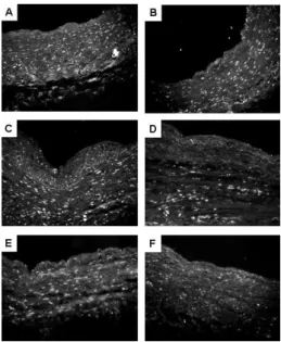
Figure 2 - Morphological and integrity analyses of fresh excised human saphenous vein. HE stain (A) and VVG stain (B) demonstrating the tissue structure. Hoechst 33258-labeled cell nuclei (C) and TUNEL assay (D)
showing tissue cellular density with no presence of apoptotic processes. 100X magniication
Figure 3 - Hoechst 33258 nuclei stain of human saphenous vein cultured in the ex vivo perfusion system. The segments were cultured under venous (A,
C, E) and arterial (B, D, F) hemodynamic conditions for 1 (A, B), 2 (C, D)
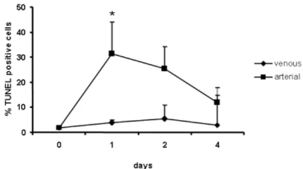
using a freshly isolated saphenous segment and a dead, refrigerated vessel. Since MTT stains only living cells, the fresh cross-section was completely stained, while no staining was observed in the dead vein segment (Figures 5Aand 5B). The segments cultured under venous conditions from 1 to 4 days contained viable cells throughout the vessel wall, but few viable cells were veriied in segments cultured under arterial conditions for 4 days (Figure 5H). To examine if the decreased cell density and viability were due to apoptotic events, a TUNEL assay was performed to detect in situ DNA fragmentation (Figures 6 and 7). Almost no apoptotic cells were observed in the saphenous vein cultured under venous
conditions for up to 4 days. In contrast, cultured segments maintained for 1 and 2 days under arterial hemodynamic stimuli showed portions with several TUNEL-positive cells (Figures 6B and 6D), suggesting that the cellular losses observed in the segments cultured for 4 days may have been a result of apoptosis.
DISCUSSION
In this work, we present a perfusion organ culture system developed to mimic the arterialization of human saphenous vein grafts. This ex vivo culture system reproduces the initial events taking place when the grafted vein is submitted to arterial hemodynamic conditions and may be a valuable approach to the identiication of molecular mechanisms underlying the early stages of bypass grafting, which have been validated in our lab using an in situ rat model (data not shown).
Figure 4 - Analysis of cellular density of human saphenous vein cultured in the ex vivo perfusion system. The results are expressed as mean ± standard error of total cell number per vessel area of 3-5 samples stained with Hoechst 33258. () represents human saphenous vein cultured under venous condi-tions and () under arterial conditions
Figure 5 - MTT stain of human saphenous vein cultured in the ex vivo perfusion system. The segments were cultured under venous (C, E, G) and arterial (D, F, H) hemodynamic conditions for 1 (C, D), 2 (E, F) or 4 (G, H) days. The freshly excised tissue (A) and dead samples (B) were used as positive and negative controls for the visualization of tissue viability. 1.5X magniication
Figure 6 - TUNEL assay of human saphenous vein cultured in the ex vivo perfusion system. The segments were cultured under venous (A, C, E) and arterial (B, D, F) hemodynamic conditions for 1 (A, B), 2 (C, D) or 4 (E, F) days. 100X magniication
Several in vivo studies have demonstrated that cell apoptosis is induced during the initial period of arterial-vein graft due to increased tensile stress and strain.5,6,18,19 In our
system, the cellular density and tissue viability decreased gradually in the human saphenous vein cultured under arterial conditions, whereas no changes in morphology or viability were observed when the veins were cultured up to 4 days under venous hemodynamic conditions (Figures 3 and 5). It is interesting to note that the cell density in veins cultured for 1 day under arterial conditions was similar to that in freshly excised samples, but a substantial number of these cells were TUNEL-positive, indicating the presence of apoptotic processes (Figures 3 and 6), which may explain the cellular losses observed in saphenous veins cultured under arterial hemodynamic conditions. It has been described that in vivo models of increased tensile stress induce cell death
in vein grafts during the irst 24 hours, and this process seems to be associated with increased caspase 3 activity.5,20
More recently, Goldman et al.16 demonstrated that rat vena
cava cultured under arterial conditions showed an increase in caspase 3 activity at 6 hours of culture and progressive -actin ilament degradation and decreases in smooth muscle cell density over 48 hours, all of which are thought to be mediated by p38 MAPK.
It is generally accepted that vascular remodeling results from an imbalance between cell death and proliferation.6,20 In
arterial-vein grafts, apoptosis occurs in response to increased mechanical stress, which is counterbalanced by subsequent proliferation processes. In rat vein graft models 10 to 30 days after surgery, it has been shown that early decreases in cellular density may be compensated for or may even increase in comparison to control veins.19 Liu et al.20 suggested that the
initial cell death observed in experimental vein grafts possibly
mediates the subsequent cellular proliferation. The absence of proliferation in saphenous veins cultured under arterial hemodynamic conditions in our system is not understood, but it may involve a lack of speciic growth factors in the media culture used. Another possibility is that, under the present conditions, the proliferation process may require additional time beyond the 4-day observation period. However, it is important to emphasize that the lack of proliferation did not preclude the reproduction of other early events that have been observed in in vivo vein grafts, such as apoptotic processes and progressive decreases in cellular density and viability that may be associated with increased tensile stress.
The system presented here reproduced the early events observed in in vivo vein graft models with the advantage of working with human samples in very well controlled hemodynamic conditions. Although other components of blood circulation have an important role in vein graft failure, this ex vivo organ culture system provides the opportunity to study the effects of hemodynamic stimuli alone. We believe that the ex vivo organ culture is a useful model for characterizing the early molecular events taking place in the arterialization of human vein grafts and to provide new approaches to prolonging its lifespan.
ACKNOWLEDGMENTS
AAM, SL and TFB were recipients of fellowships from FAPESP (00/09485-7, 99/11908-4, and 03/01828-0, respectively). This work was funded by grants from FAPESP (01/00009-0), from the Conselho Nacional de Pesquisa e Desenvolvimento – CNPq – (478073/2004-6) and from Fundo Bunka de Pesquisa – Banco Sumitomo (Auxilio à Pesquisa 2003).
REFERENCES
1. Motwani JG, Topol EJ. Aortocoronary saphenous vein graft disease: pathogenesis, predisposition, and prevention. Circulation. 1998;97:916-31. Review.
2. Fitzgibbon GM, Kafka HP, Leach AJ, Keon WJ, Hooper GD, Burton JR. Coronary bypass graft fate and patient outcome: angiographic follow-up of 5,065 grafts related to survival and reoperation in 1,388 patients during 25 years. J Am Coll Cardiol.1996;28: 616-26.
3. Akowuah EF, Sheridan PJ, Cooper GJ, Newman C. Preventing saphenous vein graft failure: does gene therapy have a role? Ann Thorac Surg. 2003;76:959-66.
4. Verma S, Szmitko PE, Weisel RD, Bonneau D, Latter D, Errett L, LeClerc Y, et al.Should radial arteries be used routinely for coronary artery bypass grafting? Circulation.2004;110: e40-6.
5. Rodriguez E, Lambert EH, Magno MG, Mannion JD. Contractile smooth muscle cell apoptosis early after saphenous vein grafting. Ann Thorac Surg.2000;70:1145-53.
6. Liu SQ, Moore MM, Yap C. Prevention of mechanical stretch-induced endothelial and smooth muscle cell injury in experimental vein grafts. J Biomech Eng.2000;122:31-8.
7. Gosling M, Golledge J, Turner RJ, Powell JT. Arterial low conditions downregulate thrombomodulin on saphenous vein endothelium. Circulation.1999;99:1047-53.
9. Wernig F, Mayr M, Xu Q. Mechanical stretch-induced apoptosis in smooth muscle cells is mediated by beta1-integrin signaling pathways. Hypertension.2003;41:903-11.
10. Casey PJ, Dattilo JB, Dai G, Albert JA, Tsukurov OI, Orkin RW, et al. The effect of combined arterial hemodynamics on saphenous venous endothelial nitric oxide production. J Vasc Surg.2001;33:1199-205. 11. Yamawaki H, Sato K, Hori M, Ozaki H, Nakamura S, Nakayama H, et
al. Morphological and functional changes of rabbit mesenteric artery cultured with fetal bovine serum. Life Sci. 2000;67:807-20.
12. Huang B, Dreyer T, Heidt M, Yu JC, Philipp M, Hehrlein FW, et al. Insulin and local growth factor PDGF induce intimal hyperplasia in bypass graft culture models of saphenous vein and internal mammary artery. Eur J Cardiothorac Surg.2002;21:1002-8.
13. Bardy N, Karillon GJ, Merval R, Samuel JL, Tedgui A. Differential effects of pressure and low on DNA and protein synthesis and on ibronectin expression by arteries in a novel organ culture system. Circ Res.1995;77:684-94.
14. Han HC, Ku DN. Contractile responses in arteries subjected to hypertensive pressure in seven-day organ culture. Ann Biomed Eng. 2001;29:467-75.
15. Golledge J, Turner RJ, Harley SL, Springall DR, Powell JT. Circumferential deformation and shear stress induce differential responses in saphenous vein endothelium exposed to arterial low. J Clin Invest.1997;99:2719-26.
16. Goldman J, Zhong L, Liu SQ. Degradation of alpha-actin ilaments in venous smooth muscle cells in response to mechanical stretch. Am J Physio Heart and Circ Physiol.2003;284:H1839-47.
17. Liu SQ, Fung YC. Changes in the organization of the smooth muscle cells in rat vein grafts. Ann Biomed Eng.1998;26:86-95.
18. Mayr M, Li C, Zou Y, Huemer U, Hu Y, Xu Q. Biomechanical stress-induced apoptosis in vein grafts involves p38 mitogen-activated protein. FASEB J. 2000;14:261-70.
19. Moore MM, Goldman J, Patel AR, Chien S, Liu SQ. Role of tensile stress and strain in the induction of cell death in experimental vein grafts. J Biomech.2001;34:289-97.