CLINICAL SCIENCE
A new method to analyze the subjective visual vertical
in patients with bilateral vestibular dysfunction
Martha Funabashi,ITaiza Elaine Grespan Santos-Pontelli,IJose´ Fernando Colafeˆmina,IITheo Zeferino Pavan,III Antonio Adilton Oliveira Carneiro,IIIOsvaldo Massaiti TakayanaguiI
IUniversity of Sa˜o Paulo, School of Medicine at Ribeira˜o Preto, Department of Neurosciences and Behavior, Ribeira˜o Preto/SP, Brazil.IIUniversity of Sa˜o
Paulo, School of Medicine at Ribeira˜o Preto, Department of Ophthalmology, Otorhinolaryngology and Head and Neck Surgery, Ribeira˜o Preto/SP, Brazil. IIIUniversity of Sa˜o Paulo, School of Philosophy, Sciences and Letters at Ribeira˜o Preto, Department of Physics, Ribeira˜o Preto/SP, Brazil.
OBJECTIVE: The aim of this study was to assess the subjective visual vertical in patients with bilateral vestibular dysfunction and to propose a new method to analyze subjective visual vertical data in these patients.
METHODS: Static subjective visual vertical tests were performed in 40 subjects split into two groups. Group A consisted of 20 healthy volunteers, and Group B consisted of 20 patients with bilateral vestibular dysfunction. Each patient performed six measurements of the subjective visual vertical test, and the mean values were calculated and analyzed.
RESULTS: Analyses of the numerical values of subjective visual vertical deviations (the conventional method of analysis) showed that the mean deviation was 0.326¡1.13
˚
in Group A and 0.301¡1.87˚
in Group B. However, by analyzing the absolute values of the subjective visual vertical (the new method of analysis proposed), the mean deviation became 1.35¡0.48˚
in Group A and 2.152¡0.93˚
in Group B. The difference in subjective visual vertical deviations between groups was statistically significant (p,0.05) only when the absolute values and the range of deviations were considered.CONCLUSION:An analysis of the absolute values of the subjective visual vertical more accurately reflected the visual vertical misperception in patients with bilateral vestibular dysfunction.
KEYWORDS: Vestibular Function Tests; Vestibular Labyrinth; Vestibular Diseases; Saccule; Utricule.
Funabashi M, Santos-Pontelli TE, Colafeˆmina JF, Pavan TZ, Carneiro AA, Takayanagui OM. A new method to analyze the subjective visual vertical in patients with bilateral vestibular dysfunction. Clinics. 2012;67(10):1127-1131.
Received for publication onMarch 28, 2012;First review completed onMay 10, 2012;Accepted for publication onMay 20, 2012
E-mail: [email protected]
Tel.: 55 16 3602-1225
INTRODUCTION
The perception of spatial orientation in relation to gravity is crucial for the maintenance of upright posture, gait and most motor activities. Four different sensory systems regulate these complex tasks: the vestibular, visual, inter-oceptive and somatosensory systems (1).
Bilateral vestibular dysfunction (BVD) is the result of a functional impairment of both peripheral labyrinths in the inner ear. BVD leads to impairment of the vestibulo-ocular reflex (VOR) and, as a consequence, the inability to stabilize gaze during rapid cephalic movement (2). BVD is a rare but important cause of imbalance and is both under-recognized and poorly understood. The most common and important
symptoms in BVD are unsteadiness and oscillopsia during locomotion (3). In approximately half of all BVD patients, no specific clinical cause can be identified (2,3), thus making the diagnosis of this important condition very difficult.
The subjective visual vertical (SVV) assessment is a valid clinical exam that evaluates an individual’s capacity to determine if an object is aligned in the vertical position, without any real vertical reference. The test is administered by asking an individual to align a luminous bar with a position that the individual judges to be vertical. The tilts of the individual’s chosen position with respect to the earth’s vertical is measured in degrees (4-6). The ability to judge whether the bar is aligned with the real vertical depends on the integrity of visual (4,7,8) and vestibular otolithic information (9-12). This information codes the static gravitational orientation and cephalic linear acceleration movements, with consequent maintenance of posture and balance (8,13). SVV tilts are a sensitive sign of vestibular dysfunction. The signs can be present in either peripheral or central disorders and can be located at any level of the vestibular pathway from the labyrinth to the vestibular cortex (4,5,10,13).
Copyrightß2012CLINICS– This is an Open Access article distributed under
the terms of the Creative Commons Attribution Non-Commercial License (http:// creativecommons.org/licenses/by-nc/3.0/) which permits unrestricted non-commercial use, distribution, and reproduction in any medium, provided the original work is properly cited.
In patients with unilateral vestibular dysfunction, the SVV tilts usually occur on the same side of the vestibular lesion (5,9). This result suggests that the maintenance of the ocular tilt reaction is ipsilateral to the vestibular disorder, central dysfunction or lesion of the otolithic organs, or changes in afferent graviceptive pathways in the vestibular nerve (4,5,14). In previous studies, the SVV in patients with BVD was indistinguishable from that in healthy volunteers when examined using conventional techniques (15-17). To the best of our knowledge, no studies have investigated the optimal method of estimating SVV in patients with BVD, which represents an important gap, identified in the present study, in the conventional analysis of the SVV in BVD patients. To address this issue, the aim of this study was to assess the SVV in patients with BVD and to propose a new method to analyze SVV data in patients with this condition.
MATERIALS AND METHODS
Subjects
Forty subjects were evaluated in this study. Group A was composed of 20 healthy volunteers, and Group B was composed of 20 patients with BVD. All subjects were aged between 30 and 60 years (mean age of Group A: 42.65¡9.4 years; of Group B: 47.20¡8.8 years). Subjects were selected from the Neurotology Clinic of the University of Sa˜o Paulo School of Medicine at Ribeira˜o Preto between July 1, 2007, and December 31, 2009. In Group B, 14 subjects were diagnosed with bilateral endolymphatic hydrops, according to the guidelines of the American Academy of Otolary-ngology – Head and Neck Surgery (AAO-HNS) (18). The other six subjects were diagnosed with bilateral semicircular canal hypofunction resulting from vestibular neuritis (19,20). This study was approved by the University of Sa˜o Paulo Ethics Committee (Comiteˆ de E´tica em Pesquisa – CEP, protocol number 364/2008). Written informed consent was obtained from all of the subjects.
The diagnosis of BVD and the inclusion criteria in this study was in accordance to diagnostic criteria previously described (3). These criteria included clinical symptoms, results of bedside evaluations, laboratory tests and the absence of other causes of unsteadiness and oscillopsia, such as cerebellar disorders without bilateral vestibular failure, intoxication, phobic postural vertigo, vestibular paroxysmia, perilymph fistula, orthostatic hypotension, hyperventilation syndromes or visual disorders and uni-lateral vestibular loss. Laboratory tests, consisting of audiometry and caloric tests, were performed and docu-mented in all patients in Group B. For the caloric test, the patients’ maximum slow-phase velocity (MSPV) distribu-tions resulting from cool (30
˚
C) and warm (44˚
C) water stimulation were analyzed. This study used the term ‘‘most affected ear’’ when referring to the ear that presented the lowest MSPV and ‘‘least affected ear’’ when referring to the ear that presented a less-reduced MSPV.In both groups, the study exclusion criteria included a previous history of migraines, central or peripheral neuro-logic deficits, metabolic disease, tumors, cancer, psychiatric disorders or traumatic brain injury. The healthy volunteers were assessed for possible vestibular dysfunction and balance disorders. Subjects with unilateral vestibular dys-function were excluded. Patients with BVD and other associated vestibular disorders (such as benign paroxysmal positioning vertigo) were also excluded. The subjects with
endolymphatic hydrops must have been out of the crisis period of the disorder. Those who wore visual corrective lenses performed the exam with the lenses in place.
Equipment
The electro-occulography for the caloric test was carried out with NEUROGRAFF - Eletromedicina - VENG digital, model VECWIN (Jundiaı´, SP/Brazil).
To assess the SVV, a 45-cm-tall seat with a 30-cm-long dark tube was used to isolate the volunteer from external visual references. An HP Pavilion 15.4’’ computer was used to display the visual test. For the static SVV test, an 11-cm highlighted line with a visual angle of 20.14
˚
was projected against a white background (6). The software used in this study presented a sensitivity of 0.1˚
(6). A neck brace was used to minimize cephalic tilts during the exam (21).Procedures
The caloric test was conducted according to previously described stimulation techniques (22). Each ear was alter-natively irrigated with a constant water flow of 5 ml/s for 40 seconds at 30
˚
C (for the cool stimulation) or 44˚
C (for the warm stimulation). The order of the stimulation was: the right ear and left ear with warm stimulation, followed by the left ear and right ear with cool stimulation. We introduced an interval between stimulations so that there would be no cumulative effect. The head position was corrected after each stimulation and was maintained at 60˚
of extension with a vertical Frankfurt line. This allowed the horizontal semicircular canals to remain in a vertical position. During the test, we questioned the patients or asked them to make calculations to prevent cortical inhibition in the vestibular system. All patients were directed to close their eyes during the test to prevent nystagmus inhibition. The subjects had not taken anti-vertigo medication during the two days before the neuro-logical exams.
The detailed procedure of the SVV exam was previously described elsewhere (6). Briefly, the SVV exam consisted in adjusting a virtual line composed by a row of seven red circles in the vertical position using the computer mouse. The right button turned the line into the clockwise (CW) direction, the left one turned into the counterclockwise (CCW) direction. By convention, the angular tilts of the virtual line compared with the real vertical were defined as positive if they were tilted CW and negative if they were tilted CCW. The volunteers remained seated with their trunk in an upright position. Their right hand was positioned on a table to control the computer mouse. Six SVV measurements were performed.
Data Analysis
all tests, the criterion for statistical significance was two-tailed and set at p,0.05. Statistics were performed using Statistical Package for Social Sciences (SPSS Inc., Chicago, IL, U.S.A.).
RESULTS
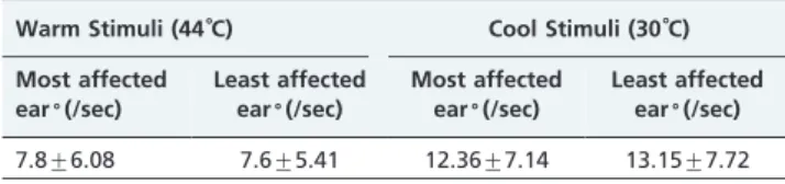
Both ears in all patients in Group B showed a reduced MSPV response to the bithermal caloric test. Despite the bilateral reduced response to the caloric test, the MSPV from one ear was lower than the other ear. Table 1 presents the mean values of the MSPV distributions, resulting from both the cool and warm stimulations, in both ears of all Group B patients.
Our analysis of the numerical values of SVV measure-ments revealed a numerical mean tilt of 0.326¡1.13
˚
in Group A and 0.301˚
¡1.87˚
in Group B. However, this was not a statistically significant difference (Mann-Whitney U test;p= 0.925), as shown in Table 2.Although most of the final SVV tilts values in Group B were tilted toward the patients’ most affected ears (noted in the raw data of each patient in Group B), Group B patients presented larger tilts in both the clockwise (CW) and counter-clockwise (CCW) directions. For this reason, the final arithmetic mean value of 0.301¡1.87
˚
(which is very close to the real vertical of 0˚
) did not correspond to the real variation of tilts exhibited by these patients. Therefore, we analyzed the absolute values of the SVV tilts without taking into consideration the negative or positive sign of the tilts. The absolute value of the difference between the largest CW tilt and the largest CCW tilt was also analyzed. These mean values are shown in Table 2.When comparing the mean absolute SVV tilts between Group A an Group B, a statistically significant difference was observed (Mann-Whitney U test; p= 0.004). We also
observed that Group B presented a significantly higher mean range of SVV tilts compared with Group A (Mann-Whitney U test;p= 0.044).
DISCUSSION
In recent years, new methods of evaluating the vestibular system have been introduced into the clinic. These methods
have verified the otolithic macula as the origin of the VOR (23). Ongoing evaluations of the functions of the otolith organs have resulted in more precise diagnoses and, consequently, improved treatment. Among these evalua-tions, the determination of the SVV is a simple and low-cost assessment of otolith function (23).
The current study aimed to measure the SVV in patients with BVD and to propose a new method to analyze the SVV data in these patients. The vestibular system is a complex system responsible for the detection of linear motion and acceleration of the human head. Information originating in the vestibular system can be influenced by additional sensorial afferent signals, such as proprioception and interoception (24). Due to this complexity, the assessment and identification of vestibular disorders can be difficult and challenging (25). Despite this difficulty, the current study rigorously recruited subjects based on their history, symptoms, physical exam and laboratory tests. To assure the reliability of our results, subjects were included only if they were diagnosed with BVD, as previously described (3). The bithermal caloric test was performed in all subjects in Group B to verify bilateral vestibular involvement. According to a previous study (26), a MSPV of less than 11
˚
/sec under warm water stimulation and less than 6˚
/sec under cool water stimulation indicates a hypoactive semicircular canal. Specifically for BVD, a MSPV of less than 20˚
/sec resulting from the bithermal caloric test is an indicator of decreased VOR function (3). The subjects included in Group B presented a low MSPV in both of their ears. Therefore, considering these values within the pre-viously defined limits for BVD patients (3), BVD in Group B subjects was evident.Traditionally, the SVV is analyzed based on the mean tilt of six trials (8,14,27-29). It is consistently reported in the literature that the upper end of the SVV line tilts toward the affected ear, shifted by several degrees with respect to the gravitational axis (4,9,10,27,30). This finding is in agreement with the present study, where, despite bilateral involvement and large variations in tilts, most of the final mean numerical SVV tilts tilted toward the patients’ most affected ears. It has been previously reported that the ocular tilt reaction is related to otolithic disorders and, consequently, to abnormal SVV tilts (9). Therefore, the SVV tilt toward the side of the most affected ear may be related to the ipsilateral ocular tilt reaction. This reaction can result from reduced neuronal activity in the ipsilateral vestibular nucleus due to a reduction in otolithic inputs (10,14).
SVV mean tilts ranging from -2.0 to+2.0
˚
are considered normal in the healthy population (4,31,32). By convention, positive and negative values indicate that the SVV line is tilted CW and CCW, respectively. In the present study, we noticed that the SVV tilts in Group A were in the normal range (0.326˚
), as described in previous studies (4,6,31,32). The mean SVV tilts in Group B (0.301˚
) were also very close to the real vertical (0˚
), generating a false-negative result.In previous studies, the SVV in patients with BVD was indistinguishable from that in healthy volunteers when examined using the conventional technique (15-17). A previous study (15) found no statistically significant differ-ence between static SVV in patients with BVD, patients with visual vertigo and normal control individuals. Another study (16) noted that the SVV test may be less sensitive in detecting otolith dysfunction in patients with central compensation over time or in patients with bilateral symmetric otolith
Table 1 -Mean values of the maximum slow-phase velocity in both ears of patients in Group B.
Warm Stimuli (44˚C) Cool Stimuli (30˚C)
Most affected ear˚(/sec)
Least affected ear˚(/sec)
Most affected ear˚(/sec)
Least affected ear˚(/sec)
7.8¡6.08 7.6¡5.41 12.36¡7.14 13.15¡7.72
Table 2 -Differential analysis of the mean SVV values: numerical value, absolute value and the range of deviations.
Mean Numerical SVV
Mean Absolute
SVV Mean Range
Control Group
0.326˚¡1.13˚ 1.35˚¡0.48˚ 3.31˚¡1.7˚
BVD Group 0.301˚¡1.87˚ 2.152˚¡0.93˚* 5.15˚¡3.15˚*
SVV = subjective visual vertical; BVD = bilateral vestibular dysfunction. *
dysfunction. In addition, another study (17) evaluated the SVV in patients with unilateral and bilateral vestibular dysfunction with the conventional method. The authors found that the mean SVV tilts in BVD patients did not differ significantly from the tilts in the control subjects (17).
A close observation of the data derived from the current study provides an understandable explanation as to why the SVV in subjects with BVD were previously considered indistinguishable from the SVV in healthy volunteers. We noted that our patients in Group B exhibited larger tilts in both CW (positive) and CCW (negative) directions. Thus, when adding the positive and negative tilts, these arithmetic values cancelled themselves out. The final mean value was 0.301¡1.87
˚
(which is very close to the real vertical of 0˚
) and did not correspond to the true variation of tilts in the subjects. Our results regarding the mean ranges of SVV support our hypothesis and suggest that patients with BVD exhibit a greater range of SVV variance compared with the range exhibited by control subjects (p= 0.044).When we analyzed the absolute values of the tilts (the new method of analysis proposed) in Group B, the positive and negative signs were not taken into consideration, and it was therefore possible to identify a significant difference between the BVD and control subjects (p= 0.004). This
difference was not significant when the tilts in both groups were analyzed using the conventional method (numerical values) (p= 0.925).
The new method of analysis (absolute values and ranges of the absolute SVV values) revealed larger tilts in both the BVD and control subjects when compared to the arithmetic mean. Previous studies analyzing SVV in normal subjects only reported the mean tilts of their subjects, obtained using the conventional method. The tilt in each trial was not available, making it impossible to verify whether normal subjects in previous studies would also present a larger tilt if analyzed using the proposed method. Nevertheless, when analyzing the absolute values of SVV tilts in our control subjects, although they exhibited larger SVV tilts, the mean tilt in Group A remained in the normal range of tilt (4,31,32). This was not observed in Group B.
A description of the physiology underlying the larger absolute SVV values in patients with BVD is beyond the scope of the current study. However, based on our results, we hypothesize that the lesions in BVD patients may cause a significant alteration in the subjects’ perception of verti-cality. This, in turn, results in a large variation in the SVV tilts in both the CW and CCW directions. The pathophysiol-ogy of the verticality misperception in BVD patients is still unclear. However, it may be related to a bilateral reduction in neuronal activity in the vestibular nuclei due to a reduction in bilateral otolithic inputs. Thus, the analysis of the range of tilts and the use of absolute values of SVV tilts proposed in this study more accurately reflect the visual vertical misperception in people with BVD and may prevent false-negative results.
Most prior studies solely used the results of caloric or rotatory chair tests in diagnosing BVD patients (3). However, a recent study reported three cases with absent bilateral vestibular evoked myogenic potentials (VEMP) in the presence of normal caloric responses (33). The authors hypothesized that, while their patients showed normal caloric responses bilaterally, the patients may have had semicircular canal dysfunction in response to stimuli at different frequencies (33). Therefore, the assessment of
VEMP could be an additional test to further investigate the vestibular system in the presence of a normal caloric response (33). Similarly, the SVV test provides different stimuli than the caloric, rotatory and VEMP tests and assesses different paths and elements of the vestibular system. Like the VEMP test, the SVV assessment could be an additional test to assist in the investigation of the complex vestibular system.
Based on the results of the present study, it appears that BVD patients experience a significant misperception of verticality. This finding is clinically important because most human motor activities are based on multisensory integra-tion (34) and spatial orientaintegra-tion (27). Alteraintegra-tions in this orientation may have important consequences for patients’ postural behavior, social activity and quality of life. By identifying a patient’s altered SVV, it may be possible to treat him/her using the strategy suggested for specific verticality misperception. This includes using the intact perception (e.g., subjective postural vertical or subjective haptic vertical) as a reference in the conscious correction of the SVV misperception (35). Thus, by adding the SVV assessment to the clinical exam, clinicians may gain an increased comprehension of the patient’s perception of the world, making it possible to prescribe more specific and efficient treatments with a more detailed characterization of the patient’s symptoms.
This study had some limitations. The small sample size used in this study could have influenced the significance of our findings. However, the limited number of included subjects was due to the use of rigorous inclusion/exclusion criteria to obtain reliable results. In addition, dynamic visual acuity and head impulse tests performed previously (3) were not performed in this study due to the unavailability of appropriate equipment. However, our patients met all of the other criteria defined in the aforementioned study, confirming the presence of bilateral vestibular involvement in our patients.
In conclusion, although the analysis of the absolute values of SVV tilts did not indicate the side of the SVV tilts, it did permit us to obtain a mean value that better corresponded to the real variance of tilts obtained in the exam. Further studies are necessary to more comprehensively investigate the SVV in patients with BVD and other vestibular and neurological disorders.
ACKNOWLEDGMENTS
The authors acknowledge the Coordenac¸a˜o de Aperfeic¸oamento de Pessoal de Nı´vel Superior (CAPES) and the Conselho Nacional de Desenvolvimento Cientı´fico e Tecnolo´gico (CNPq) for providing financial support.
AUTHOR CONTRIBUTIONS
Funabashi M collected data, performed the statistical analysis and drafted the manuscript. Santos-Pontelli TE performed the statistical analysis and drafted the manuscript. Colafeˆmina JF assisted in the development of the study design and critically revised the manuscript. Pavan TZ and Carneiro AA developed the program used in the study and critically revised the manuscript. Takayanagui OM assisted in the development of the study design and critically revised the manuscript.
REFERENCES
2. Jen JC. Bilateral Vestibulopathy: clinical, diagnostic, and genetic considerations. Semin Neurol. 2009;29(5):528-33, http://dx.doi.org/ 10.1055/s-0029-1241035.
3. Kim S, Oh YM, Koo JW, Kim JS. Bilateral Vestibulopathy: Clinical Characteristics and Diagnostic Criteria. Otol Neurotol. 2011;32(5):812-7, http://dx.doi.org/10.1097/MAO.0b013e31821a3b7d.
4. Kanashiro AMK, Pereira CB, Maia FM, Scaff M, Barbosa ER. Subjective visual vertical evaluation in normal Brazilian subjects. Arq Neuropsiquiatr. 2007;65(2B):4472-5.
5. Vibert D, Hausler R, Safran AB. Subjective visual vertical in peripheral unilateral vestibular diseases. Journal of vestibular research : equilibrium & orientation. 1999;9(2):145-52.
6. Pavan TZ, Funabashi M, Carneiro JAO, Santos-Pontelli TEG, Tedeschi W, Colafemina JF, et al. Software for subjective visual vertical assessment: an observational cross-sectional study. Rev Bras Otorrino-laringol. 2012. In press.
7. Bo¨hmer A, Mast F. Assessing otolith function by the subjective visual vertical. Ann N Y Acad Sci. 1999;871:221-31, http://dx.doi.org/10.1111/ j.1749-6632.1999.tb09187.x.
8. Kumagami H, Saino Y, Baba A, Fujiyama D, Takasaki K, Takahashi H. Subjective visual vertical test in patients with chronic dizziness without abnormal findings in routine vestibular function tests. Acta Acta Otolaryngol Suppl. 2009;562:46-9, http://dx.doi.org/10.1080/000164 80902926456.
9. Clarke AH, Scho¨nfeld U, Helling K. Unilateral examination of utricle and saccule. J Vestib Res. 2003;13(4-6):215-25.
10. Kumagami H, Sainoo Y, Fujiyama D, Baba A, Oku R, Takasaki K, et al. Subjective Visual Vertical in acute attacks of Me´nie`re’s disease. Otol Neurotol. 2009;30(2):206-9, http://dx.doi.org/10.1097/MAO.0b013e318192 5010.
11. Pavlou M, Wijnberg N, Faldon ME, Bronstein AM. Effect of semicircular canal stimulation on the perception of the visual vertical. J Neurophysiol. 2003;90(2):622-30, http://dx.doi.org/10.1152/jn.00960.2002.
12. Landau ME, Barner KC. Vestibulocochlear nerve. Semin Neurol 2009;29(1):66-73, http://dx.doi.org/10.1055/s-0028-1124024.
13. Lang EE, Walsh RM. Vestibular function testing. Ir J Med Sci. 2010; 179(2):173-8, http://dx.doi.org/10.1007/s11845-010-0465-7.
14. Faralli M, Ricci G, Molini E, Longari F, Altissimi G, Frenguelli A. Determining Subjective Visual Vertical: Dynamic Versus Static Testing. Otol Neurotol. 2007;28(8):1069-71, http://dx.doi.org/10.1097/ MAO.0b013e31815aea1b.
15. Guerraz M, Yardley L, Berholon P, Pollak K, Rudge P, Gresty MA, et al. Visual vertigo: symptom assessment, spatial orientation and postural control. Brain. 2001;24(Pt 8):1646-56, http://dx.doi.org/10.1093/brain/ 124.8.1646.
16. Kessler P, Tomlinson D, Blakeman A, Rutka J, Ranalli P, Wong A. The high-frequency/acceleration head heave test in detecting otolith dis-eases. Otol Neurotol. 2007;28(7):896-904, http://dx.doi.org/10.1097/ MAO.0b013e3181256543.
17. Tabak S, Collewjin H, Boumans LJ. Deviation of the subjective visual vertical in long-standing unilateral vestibular loss. Acta Otolaryngol. 1997;117(1):1-6, http://dx.doi.org/10.3109/00016489709117982. 18. Committee on Hearing and Equilibrium. Committee on hearing and
equilibrium guidelines for the diagnosis and evaluation of therapy in Meniere’s disease. Otolaryngol Head Neck Surg. 1995;113(3):181-5. 19. Nadol JB. Vestibular neuritis. Otolaryngol Head Neck Surg. 1995;
112(1):162-72, http://dx.doi.org/10.1016/S0194-5998(95)70316-0.
20. Halmagyi GM, Weber KP, Curthoys IS. Vestibular function after acute vestibular neuritis. Restor Neurol Neurosci. 2010;28:37-46.
21. Funabashi M, Silva NNL, Watanabe LM, Santos-Pontelli TEG, Colafemina JF, Carneiro AAO, et al. The use of a neck brace does not influence the visual vertical perception. Arq Neuropsiquiatr. 2011;69(3):509-12, http://dx.doi.org/10.1590/S0004-282X2011000400019. 22. Fitzgerald G, Halpike CS. Studies in human vestibular function: I. Observations on the directional proponderance ("nystagmusber-eitschaft") of caloric nystagmus resulting from cerebral lesions. Brain. 1942;65(2):115-37, http://dx.doi.org/10.1093/brain/65.2.115.
23. Saeys W, Vereeck L, Bedeer A, Lafosse C, Truijen S, Wuyts FL, et al. Suppression of the E-effect during the subjective visual and postural vertical test in healthy subjects. Eur J Appl Physiol. 2010;109(2):297-305, http://dx.doi.org/10.1007/s00421-010-1355-4.
24. Angelaki DE, Cullen KE. Vestibular system: the many facets of a multimodal sense. Annu Rev Neurosci. 2008;31:125-50, http:// dx.doi.org/10.1146/annurev.neuro.31.060407.125555.
25. Paige GD. Otolith Function: basis for modern testing. Ann NY Acad Sci. 2002;956:314-23, http://dx.doi.org/10.1111/j.1749-6632.2002.tb02830.x. 26. Gonc¸alves DU, Felipe L, Lima TMA. Interpretac¸a˜o e utilidade da prova
calo´rica. Rev Bras Otorrinolaringol. 2008;74(3):440-6, http://dx.doi.org/ 10.1590/S0034-72992008000300020.
27. Faralli M, Longari F, Ricci G, Ibba MC, Frenguelli A. Influence of extero-and proprioceptive afferents of the plantar surface in determining subjective visual vertical in patients with unilateral vestibular dysfunc-tion. Acta Otorhinolaryngol Ital. 2009;29(5):245-50.
28. Barbieri G, Gissot AS, Fouque F, Casillas JM, Pozzo T, Perennou D. Does proprioception contribute to the sense of verticality? Exp Brain Res. 2008;185(4):545-52, http://dx.doi.org/10.1007/s00221-007-1177-8. 29. Kobayashi H, Hayashi Y, Higashino K, Saito A, Kunihiro T, Kanzaki J,
et al. Dynamic and static subjective visual vertical with aging. Auris, Nasus, Larynx. 2002;29(4):325-8, http://dx.doi.org/10.1016/S0385-8146(02)00058-5.
30. Mazibrada G, Tariq S, Pe´rennou D, Gresty M, Greenwood R, Bronstein AM. The peripheral nervous system and the perception of verticality. Gait & Posture. 2008;27(2):202-8, http://dx.doi.org/10.1016/j.gaitpost. 2007.03.006.
31. Barra J, Marquer A, Joassin R, Reymond C, Metge L, Chauvineau V, et al. Humans use internal models to construct and update a sense of verticality. Brain. 2010;133(Pt 12):3552-63, http://dx.doi.org/10.1093/ brain/awq311.
32. Perrenou DA, Mazibrada G, Chauvineau V, Greenwood R, Rothwell J, Gresty MA, et al. Lateropulsion, pushing and verticality perception in hemisphere stroke: a causal relationship? Brain. 2008;131(Pt 9):2401-13, http://dx.doi.org/10.1093/brain/awn170.
33. Fujimoto C, Murofushi T, Chihara Y, Suzuki M, Yamasoba T, Iwasaki S. Novel subtype of idiopathic bilateral vestibulopathy: bilateral absence of vestibular evoked myogenic potentials in the presence of normal caloric responses. J Neurol. 2009;256(9):1488-92, http://dx.doi.org/10.1007/ s00415-009-5147-x.
34. Oliveira CB, Medeiros IRT, Greters MG, Frota NAF, Lucato LT, Scaff M, et al. Abnormal sensory integration affects balance control in hemiparetic patients within the first year after stroke. Clinics. 2011;66(12):2043-8, http://dx.doi.org/10.1590/S1807-59322011001200008.