O
riginal
a
r
ticle
RESUMO
Objeivou-se descrever a incidência e as razões de remoção não eleiva do cateter epicutâneo em neonatos, veriicando a associação com o síio de inserção. Es-tudo de coorte prospecivo realizado em unidade de cuidado intensivo neonatal de um hospital privado terciário na cidade de São Paulo. Foram analisadas 266 inserções de cateter epicutâneo. A incidência de remoção não eleiva foi 39,1%. As com-plicações pós-inserção mais frequentes foram suspeita de infecção de corrente sanguínea relacionada ao cateter (25%) e ruptura (23,1%). A maioria dos cateteres foi inserida através do hemisfério corporal direito (65%), membros superiores (77,1%) e veias axilares (31,2%). Os resultados sugerem não haver associação entre a incidência de remoção não eletiva e o síio de inserção do cateter epicutâneo em neonatos. Compete à Enfermagem implementar estratégias para a melhoria da práica assistencial a im de diminuir a frequência de remoções não eleivas do cateter epicutâneo em neonatos.
DESCRITORES Recém-nascido
Cateterismo venoso central Enfermagem neonatal ABSTRACT
This study aimed to describe the incidence and reasons for nonelective removal of epicutaneous catheters in neonates, iden-tifying its association with the catheter inserion site. This was a prospecive cohort study, conducted in a neonatal intensive care unit of a private teriary hospital in the city of São Paulo, Brazil. We analyzed 266 epicutaneous catheter inserions. The incidence of non-elective removal was 39.1%. The most frequent post-inserion complicaions were suspicion of catheter-related bloodstream infecion (25%) and rupture (23.1%). Most catheters were inserted through the right side of the body (65%), in upper limbs (77.1%), and using the axillary veins (31.2%). The indings did not suggest associaion between the incidence of non-elecive removal and the inserion site of the epicutaneous catheter in neo-nates. Nurses should implement strategies to improve care and decrease incidence of non-elecive epicutaneous catheter remov-als among neonates.
DESCRIPTORS Infant, newborn
Catheterizaion central venous Neonatal nursing
RESUMEN
El objeivo fue describir la incidencia y las razones del reiro no elecivo del catéter epicutáneo en neonatos, verificando la asociación con el siio de inserción. Estudio de cohorte prospeciva conducido en una unidad de cuidados intensivos neonatales de un hospital privado terciario, en la ciudad de São Paulo, Brasil. Fueron analizados 266 inserciones de catéteres. La incidencia de reiro no elecivo del catéter epicutáneo fue de 39,1%. Las complicaciones post-in-serción más frecuentes fueron la sospecha de infección del torrente sanguíneo relacio-nada al catéter (25%) y ruptura (23,1%). La mayoría de los catéteres fueron insertados en el hemisferio corporal derecho (65%), en la extremidad superior (77,1%) y en la vena axilar (31,2%). Los resultados sugieren no haber asociación entre el siio de inserción del catéter y la incidencia del retiro no elecivo. Es de competencia de Enfermería implementar estrategias para mejorar la prácica asistencial con el in de disminuir la frecuencia del reiro no elecivo del catéter epicutáneo en neonatos.
DESCRIPTORES Recién-nacido
Cateterismo venoso central Enfermería neonatal
Reasons for non-elective removal of
epicutaneous catheters in neonates
CAUSAS DE REMOÇÃO NÃO ELETIVA DO CATETER EPICUTÂNEO EM NEONATOS
CAUSAS DE RETIRO NO ELECTIVO DEL CATÉTER EPICUTÁNEO EN NEONATOS
Eny Dórea Paiva1, Priscila Costa2, Amélia Fumiko Kimura3,Talita Elci de Castro4
1 Nurse. Doctorate. Professor. Federal Fluminense University. School of Nursing. Rio de Janeiro, Brazil. 2 Nurse. Doctoral student. Teaching specialist.
University of São Paulo. School of Nursing. São Paulo, SP, Brazil. [email protected] 3 Nurse. Doctorate. Professor. University of São Paulo School
INTRODUCTION
High-risk newborns usually require long-term intrave-nously (IV) infused drug treatments. The use of soluions with irritant and vesicant content to peripheral veins is common among IV therapies, such as electrolyte soluions, vasoacive drugs, anibioics and parenteral nutriion. Cen-tral venous access devices have become vital to the recovery and survival of newborns admited to neonatal intensive care units (NICU). In this context, the peripherally inserted central catheter (PICC) or epicutaneous catheter came to address the therapeuic demands of criically ill neonates (1).
Epicutaneous catheters ofer a central venous access route through puncture of a peripheral vein from either the right or let side of body, in the cephalic-cervical region, or in upper or lower limbs. In the upper limbs, PICC can be insert-ed through the following veins: dorsal metacarpal, basilic, cephalic, median cubital, and axillary. In the lower limbs, the most commonly used veins are the great saphenous, the small saphenous and its derivaions, the
plantar venous arch, the medial marginal, the femoral and popliteal. The veins for cen-tral access in the cephalic-cervical region are the temporal, posterior auricular and the external jugular(2-3) veins. However, the most
recommended veins for PICC insertions are in the antecubital fossa in the upper limbs. The basilic vein, due to its favorable anatomy, larger caliber and reduced number of valves is highly recommended, followed by the cephalic vein(3). A study conducted
with 45 newborns in a NICU ideniied the basilic as the vein selected among 22% of the PICC inserions, and the cephalic vein in 20% of the inserions(4).
The incidence of non-elecive removal in PICC-lines may be related to the
cath-eter inserion site. A study of 518 epicutaneous cathcath-eters in neonates compared the incidence of complications between catheters inserted in the femoral vein and in other sites. Its indings showed a signiicant increase in the catheter-related bloodstream infecion rates when the femoral vein was used(5). Regarding the body segment
used at a PICC inserion, a survey with 396 infants with PICC demonstrated lower rates of catheter-related bloodstream infecion among catheters inserted in the lower limbs when compared to those inserted in the upper extremiies(1).
Given the above, the associaion between body side, body segment and vein selected for PICC inserion and the occurrence of complicaions leading to non-elecive removal has not relied on robust evidence to guide clinical pracice of nurses when it comes to the best site of inser-ion in NICU rouine. Despite the relevance of the topic, especially for nurses responsible for the inserion and maintenance of PICC, studies on this topic are insuicient.
Considering the nurse’s role in assessing the newborn venous system for PICC inserion, focusing on the preven-ion of post-inserpreven-ion complicapreven-ions and, consequently, assuring paient safety, we realized the need to determine the incidence and reasons for non-elecive removal of epi-cutaneous catheter in neonates, idenifying its associaion with the inserion site.
METHOD
This was a cohort study with prospecive data collecion. The cohort consisted of neonates who underwent a PICC inserion procedure in a NICU of a large, private hospital in the city of São Paulo, from July 2010 to June 2011. The intensive care unit had 60 beds and the nursing staf con-sisted of 24 nurses and 124 nursing assistants and nursing technicians. Among all nurses, 22 were ceriied in the PICC inserion technique.
The monthly number of births in this insituion was approximately 800, and approximately 30 PICCs per month were inserted in the neo-natal unit. The procedures related to PICC inserion and management followed the insituional guidelines established by the hospital´ intravenous device study group.
The eligible newborns were those born in the hospital maternity, without diagnosis of congenital anomalies or coagulopathy, without loss of skin integrity caused by congenital diseases, and presening data regarding the study variables recorded in the medical charts. The insituion pro-vided single-lumen silicone catheters of 1.9
French (Fr), and polyurethane dual-lumen
catheters of 2.0 Fr.
After bedside PICC insertion using asepic technique by trained nurses, both a neonatologist and nurse evaluated a chest radiograph to check PICC ip posiion, and its use was then allowed or not allowed. The inserion procedure and catheter main-tenance care, as well as removal process, were recorded during every shit.
We considered the following variables for the study pop-ulaion characterizaion: sex, chronological age, weight, cor-rected gestaional age, primary diagnosis, type of catheter used, ip posiion, and PICC indicaion (parenteral nutriion, anibioics, general IV soluion and/or vasoacive drugs).
The indicaion for catheter removal was considered as the main outcome in this study. Elecive removal was con-sidered when the removal occurred at the end of IV therapy. Non-elecive removal was deined as those caused by PICC post-inserion complicaions, such as obstrucion, rupture, catheter-related bloodstream infecion, edema, accidental dislodgement, ip migraion, iniltraion or phlebiis.
...the association between body side,
body segment and vein selected
for peripherally inserted central catheter insertion and the occurrence
of complications leading to non-elective removal has not relied on robust evidence to guide clinical practice
Obstrucion was considered as the impossibility to lush the catheter with a saline soluion, using a 10 ml syringe, and no blood withdraw through the lumen. Catheter rup-ture was considered as the occurrence of a break in the external porion of the catheter. The presence of bacterial or fungal infecion in paients with vascular device, and one or more posiive result of peripheral blood culture, or clini-cal manifestaions of infecion (fever, chills or hypotension), with no other apparent focus of infecion, was considered as suspected of catheter-related bloodstream infecion.
Edema was deined as the ideniicaion of mild to se-vere swelling around the catheter inserion site or in the ex-tremity of a catheterized body part during the permanence of the device. Accidental dislodgement is the inadvertent and accidental removal of the catheter, totally or parially. Migraion is the displacement of the PICC ip, conirmed by radiography. Iniltraion is the invasion of a non-vesicant soluion or drug into the extravascular; extravasaion is the invasion of a vesicant soluion or drug into the extravascu-lar. Phlebiis is a venous inlammaion from a mechanical, chemical or bacterial cause(2-3).
The PICC inserion site was considered to be the indepen-dent variable. We considered the inserion site to be com-posed of: body side (right or let), body segment of venous access (upper limb, lower limb, cephalic-cervical region), and the selected vein (dorsal arch of the hand, dorsal arch of the foot, axillary, basilic, cephalic, median cubital, external jugu-lar, popliteal, posterior auricujugu-lar, saphenous and temporal). The research project was approved by the Ethic Board of the insituion under process number 219/10, following Resoluion 196/96 of the Naional Health Council. Data were collected from medical records by using a speciic instrument from the insituional PICC assessment form. Data were stored in a Microsot Oice Excel 2007 spreadsheet and ana-lyzed using Stata 11.1. Coninuous variables were analyzed using descripive staisics, and categorical variables using absolute and relaive frequency. For categorical variables, the associaion between diferent inserion sites and the occurrence of non-elecive removal was determined by the chi- square test or Fisher’s exact test. The p-value for staisi-cal signiicance was ≤ 0.05 with a 95% conidence interval.
RESULTS
The authors evaluated 309 PICC inserions in neonates regarding their eligibility criteria for inclusion in this study. Ater exclusion of unsuccessful inserions and those without enough data regarding the inserion site (selected vein, body side, segment of inserion), and reason for catheter re-moval, a total number of 266 events remained for analysis. Among the study populaion, most infants were male (163 or 61.2%), appropriate for gestaional age (221 or 83.4%) with a mean gestaional age of 34.1 weeks, weight of 1,888.9 grams and postnatal age of 10.5 days. The most common clinical diagnosis was prematurity in 211 subjects (79.3%),
respiratory distress in 171 (64.3%), twin pregnancy in 83 (31.2%), sepsis in 62 (23.3%), heart disease in 44 (16.5%) and disorders of the gastrointesinal tract in 35 (13.1%) subjects.
The single lumen silicone PICC was used in 187 (70.3%) inserions and the double lumen polyurethane 2.0 Fr cathe-ter in the 79 (29.7%) remaining cathecathe-ters. The epicutaneous catheter ip placement was central in most of the inserions (234 or 88.3%). The main indicaion for catheter inserion was the combinaion of parenteral nutriion and anibioic therapy in 30% of the inserions, followed by electrolyte soluions and anibioic therapy in 20% of catheter usages.
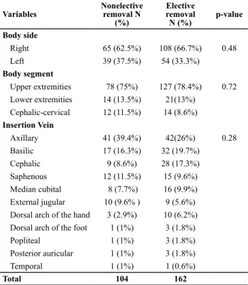
Regarding the PICC placement site, the right side of the body was the most used, in 173 of the inserions (65%). A total of 93 (35%) inserions were made in the let side of the body. The relaive risk was 1.1 with a 95% conidence interval [0.81 to 1.52], therefore, the risk of non-elecive removal of the PICC occurred independent of the body side selected. The most common body segment used was the upper limb, in 205 (77.1%) inserions, followed by the lower limbs in 35 (13.2%), and by the cephalic-cervical region in 26 (9.7%) events. The veins most frequently accessed for PICC inserion were the axillary veins in 83 (31.2%), followed by basilic in 49 (18.4%), cephalic in 37 (13.9%), and saphenous in 27 (10.1%). Elecive removal was ideniied in 162 (60.9%) catheters, and non-elecive removal in 104 (39.1%).
Table 1 –Association between the insertion site and indication for catheter removal – São Paulo, SP, 2011.
Variables
Nonelective removal N
(%)
Elective removal
N (%)
p-value
Body side
Right 65 (62.5%) 108 (66.7%) 0.48
Left 39 (37.5%) 54 (33.3%)
Body segment
Upper extremities 78 (75%) 127 (78.4%) 0.72
Lower extremities 14 (13.5%) 21(13%)
Cephalic-cervical 12 (11.5%) 14 (8.6%)
Insertion Vein
Axillary 41 (39.4%) 42(26%) 0.28
Basilic 17 (16.3%) 32 (19.7%)
Cephalic 9 (8.6%) 28 (17.3%)
Saphenous 12 (11.5%) 15 (9.6%)
Median cubital 8 (7.7%) 16 (9.9%)
External jugular 10 (9.6% ) 9 (5.6%)
Dorsal arch of the hand 3 (2.9%) 10 (6.2%)
Dorsal arch of the foot 1 (1%) 3 (1.8%)
Popliteal 1 (1%) 3 (1.8%)
Posterior auricular 1 (1%) 3 (1.8%)
Temporal 1 (1%) 1 (0.6%)
Total 104 162
However, it was observed that among those epicutaneous catheters that were non-elecively removed, the proporion of the axillary vein as the selected vein was higher: nearly 40% of the catheters. Among those elecively removed, this proporion was only 26%. The opposite occurred in relaion to the cephalic vein: among the elecively removed catheters, this vein was used in approximately 17% of the inserions and among the non-elecively removed, the cephalic was used in 8.6 % of the inserions.
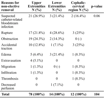
The most frequent complicaions that led to the non-elecive removal of the PICC were: suspicion of catheter-related bloodstream infecion in 26 (25%) removals, rupture of the external PICC hub in 24 (23.1%), obstrucion in 21 (20.2%), and accidental dislodgement in 14 (13.5%). Table 2 shows the associaion between complicaions resuling in non-elecive removal and PICC inserion site.
There was no associaion between the site of PICC inserion and the diferent complicaions ater inserion.
average of corrected gestaional age was of 30.4 weeks, weight of 1,465 grams, and 37.5% of PICC indicaions was due to parenteral nutriion therapy(6).
The prevalence of respiratory distress syndrome was similar to a study in a teriary public hospital in São Paulo that evaluated 37 neonates with PICC(7).
In the present study, although the chronological age average at the ime of PICC inserion was of 10.5 days of life, 37.0% of catheters were inserted in neonates who were less than three days old. The results of a study conducted in a NICU indicated that those PICCs inserted before 5 days of life caused fewer complicaions (15.2%) when compared to catheters inserted later 5 days of life (24.4%)(8). Factors
such as edema, hypotension and dehydraion can inluence the catheter inserion procedure(9). Immediately ater birth,
the neonate loses extracellular luid, reducing the swelling, and consequently making PICC inserion easier.
Regarding the inserion sites ideniied in this study popula-ion, the right side of the body and the upper limbs were the most used. Studies showed a range of 42 to 82.4% of PICC inserions in the right side of the body(10-11). There is similarity
in veins of the upper limb on both right and let side from the hands through to the subclavian veins. From the brachioce-phalic vein, the venous anatomy difers between the right and let side. The let brachiocephalic vein crosses the mediasinum to the right side in a direcion toward the superior vena cava. The professional knowledge of the venous anatomy is essenial for an accurate measurement of the catheter length(12).
Regarding veins, the majority of observaions indicated the use of the axillary vein for PICC inserion. Other studies showed that the veins of the antecubital fossa were the irst choice in 69.5% of atempts. We highlighted the use of the axillary vein in 28.2% of inserions, the basilic in 23.9%, the cephalic in 21.7%, and median cubital in 13%. The arches of the dorsal carpal veins were used in 8.7% of the inserions, and the jugular in 4.4%(6).
The choice of the axillary vein may be related to its larger diameter, which facilitates the puncture and progression of the catheter. Its size allows the use of catheters with larger caliber and a greater number of lumens. The main disadvantage of the axillary vein is the diicult visualizaion in older children caused by larger quaniies of subcutaneous issue. The proximity to the axillary artery increases the risk of arterial puncture.
The basilic was the second most used, followed by the cephalic vein. The basilic caliber is less tortuous than the cephalic, easy to puncture and presents an easy progression into the lumen, requiring less ime to execute the proce-dure. It also allows safer ixaion of catheter dressings and low incidence of phlebiis. The disadvantage is the anatomi-cal proximity to the brachial artery, which increases the risk of accidental arterial puncture. When using the basilic, the PICC ip may migrate to the jugular vein, resuling in poor posiioning of the catheter.
Table 2 – Association between the reasons for non-elective removal of the PICC and the catheter insertion site – São Paulo, SP, 2011.
Reasons for non-elective removal
Upper Extremities
N (%)
Lower Extremities
N (%)
Cephalic-cervical region N (%)
p-value
Suspected catheter-related bloodstream infection
21 (26.9%) 3 (21.4%) 2 (16.4%) 0.06
Rupture 17 (21.8%) 4 (28.6%) 3 (25%)
Obstruction 19 (24.3%) 2 (14.3%) 0 (-)
Accidental traction
10 (12.8%) 1 (7.1%) 3 (25%)
Edema 5 (6.4%) 3 (21.4%) 1 (8.3%)
Extravasation 4 (5.1%) 0 0
Migration 1 (1.3%) 0 (-) 1 (8.3%)
Iniltration 1 (1.3%) 0 1 (8.3%)
Thrombosis 0 0 1 (8.3%)
Decreased perfusion
0 1 (7.1%) 0
Total 78 (100%) 14 (100%) 12 (100%) 104
DISCUSSION
Ensuring safe venous access in neonates is a constant challenge for nurses. However, only in recent decades, technological advances have enabled the development of safe central venous catheters, which are less traumaic and beter tolerated in the venous system. Nonetheless, few studies have explored the inluence of diferent inserion sites and non-elecive PICC removal.
The cephalic vein has a smaller diameter than the basilic and has a smaller angle in it, gathering with the axillary vein, with one part of it going to meet the external jugular vein and the other remaining as the axillary vein. As the cephalic rises along the arm, it become narrow and tortuous, lead-ing to an increased risk of mechanical phlebiis. It can also be more diicult to progress the catheter into the angle of the vein with the shoulder and induce a bad placement of the catheter ip in the axillary vein(3).
However, the veins of the lower limbs were also employed in 13.2% of the inserions, mainly the saphenous vein. The reasons for the elecion of the saphenous may be related to its high caliber and great length, the number of valves (ranging from 7 to 25 valves) and easy visualizaion next to the heel(3).
A retrospecive cohort study conducted in a NICU in Beijing, that analyzed the risk factors for complicaions in 104 catheters inserted in newborns, found that the saphe-nous vein was elected in 11.5% of the inserions and that veins used for catheter inserion were not associated with nonelecive removal (p=0.13). The indings on associaion between the selected vein and the incidence of non-elecive removal are similar to this study’s results(13).
A prospecive, randomized study of post-inserions compli-caions, comparing occurrences in proximal valve polyurethane PICC and distal valve silicone PICC, analyzed 392 catheters and their indings showed no associaion between complicaions and the vein used (p = 0.35) or body side (p = 0.24)(10).
In this study, the incidence of non-elecive removal was similar to other studies conducted in diferent faciliies, which ranged from 31.7%(13) to 47.7%(14). However, the present study
found that the main reasons for non-elecive removal were diferent among the three possible segments of PICC inser-ion. Among the epicutaneous catheters inserted in the upper limbs, the most frequent complicaion was catheter-related bloodstream infecion. It is an essenial part of nursing care to prevent this complicaion, especially in neonates in whom the PICC was inserted in the upper limbs. Strategies to reduce catheter-related infecions include nursing staf training on hand washing, protecion of the catheter inserion site during newborn bathing, the use of gloves and anisepic soluions while handling the catheter, the use of transparent and semi permeable dressings, and frequency of dressing change - weekly or whenever loss of adhesion or an unclean situaion are observed, maximal contact precauions during catheter inserion, and use of catheters with the smallest number of lumen possible to meet newborn needs(15).
Among the catheters inserted through the lower limbs, the most frequent complicaion was rupture. The preven-ion of this complicapreven-ion includes staf training to lush the catheters using a 10 ml syringes in order to avoid excessive pressure(3). Addiionally, only trained nurses can perform
maneuvers to clear the catheter by using a speciic technique. Finally, among the catheters inserted into the cervical region, the most frequent complicaions were accidental
dislodgement and rupture. Avoiding newborns using their own hands to move the device should be done in order to prevent complicaions in the catheters inserted in this re-gion. Furthermore, it is important to coninuously evaluate the site of inserion and ensure the catheter stability and security while changing dressings(3).
Nurses are the main professionals responsible for the management and care of paients receiving IV therapy. Their performance includes a criical evaluaion of the pre-scribed therapy, the choice of vascular device that meets the paient need, the installaion procedure, the care for its maintenance and, inally, its removal. Moreover, their role is to design and implement pracices that enhance paient safety and contribute to the improvement of paients’ health status, especially in high-risk newborns.
As the PICC inserion site seems to have no associaion with the occurrence of complicaions, the nurse can choose to insert the catheter in the place that seems more visible, palpable and has healthier skin, according to their profes-sional experience. Sill, it is noteworthy that the use of technologies such as ultrasound can help the professional with the catheter inserion procedure, since it can facilitate the visualizaion and the puncture of deeper structures.
We highlight that one of the limitaions of this study re-fers to the use of a single source for data collecion, showing indings of a single insituion, thus limiing generalizability of results to a broader populaion of neonates. The observa-ional design may also be another limitaion, since the data were obtained from medical records and data loss is common in this type of study. Despite the limitaions, the indings deserve further exploraion in subsequent studies, given the lack of studies that analyze the associaion between vein, body side and segment of PICC inserion, with the reasons for its removal, in neonates and in other populaions.
CONCLUSION
The methodology used proved to be adequate for the study objecives. Its reproducion allows the development of further surveys in other neonatal units.
1. Hoang V, Sills J, Chandler M, Busalani E, Cliton-Koeppel R, Modanlou HD. Percutaneously inserted central catheter for total parenteral nutriion in neonates: complicaions rates related to upper versus lower extremity inserion. Pediatrics. 2008;121(5):e1152-9.
2. Infusion Nurses Society. Infusion nursing standards of pracice. J Infus Nurs. 2011;34(1 Suppl):S1-110.
3. Pettit J, Wyckoff MM. Peripherally inserted central catheters: guideline for pracice [Internet]. 2nd ed. Glenview:
National Association of Neonatal Nurses; 2007 [cited 2011 Jan 15]. Available from: htp://www.nann.org/pdf/ PICCGuidelines.pdf
4. Dorea E, Castro TE, Costa P, Kimura AF, Santos FMG. Práticas de manejo do cateter central de inserção periférica em uma unidade neonatal. Rev Bras Enferm. 2011;64(6):997-1002.
5. Tsai MH, Lien R, Wang JW, Huang HR, Chiang CC, Chu SM, et al. Complicaion rates with central venous catheters inserted at femoral and non-femoral sites in very low birth weight infants. Pediatr Infect Dis J. 2009;28(11):966-70.
6. Barría MP, Santander GM. Cateterismo venoso central de inserción periférica em recién nacidos de cuidado intensivo. Rev Chil Pediatr. 2006,77(2):139-46.
7. Camargo PP, Kimura AF, Toma E, et al. Iniial placement of the peripherally inserted central catheter’s ip in neonates. Rev Esc Enferm USP [Internet]. 2008 [cited 2013 Jan 29];42(4):723-8. Available from: htp://www.scielo.br/pdf/reeusp/v42n4/ en_v42n4a14.pdf
8. Paulson PR, Miller KM. Neonatal peripherally inserted central catheters: recommendaion for prevenion of inserion and post inserion complicaions. Neonat Netw. 2008;27(4):245-57. 9. Peit J. Assessment of infants with peripherally inserted central
catheters: part 1. Detecing the most frequently occurring complicaions. Adv Neonatal Care. 2002;2(6):304-15. 10. Ong CK, Venkatesh SK, Lau GB, Wang SC. Prospective
comparaive evaluaion of proximal valve polyurethane and distal valve silicone peripherally inserted central catheters. J Vasc Interv Radiol. 2010;21(8):1191-6.
11. Liem TK, Yanit KE, Moseley SE, Landry GJ, Deloughery TG, Rumwell CA, et al. Peripherally inserted central catheter usage paterns and associated symptomaic upper extremity venous thrombosis. J Vasc Surg. 2012;55(3):761-7.
12. Amerasekera SSH, Jones CM, Patel R, Cleasby MJ. Imaging of the complicaions of peripherally inserted central venous catheters. Clin Radiol. 2009;64(8):832-40.
13. Liu H, Han T, Zheng Y, Tong X, Piao M, Zhang H. Analyses of complicaions rates and reasons for nonelecive removal of PICCs in neonatal intensive care unit preterm infants. J Infus Nurs. 2009;32(6):336-40.
14. Franceschini AT, Cunha MLC. Adverse events related to the use of central venous catheters in hospitalized newborns. Rev Laino Am Enferm. 2010;18(2):196-202.
15. Centers for Disease Control and Prevenion. Guidelines for the prevenion of intravascular catheter-related infecions [Internet]. 2011 [cited 2013 Jan 29]. Available from: htp:// www.cdc.gov/hicpac/pdf/guidelines/bsi-guidelines-2011.pdf
REFERENCES
Acknowledgements