Prevalence and Evolution of Core
Photosystem II Genes in Marine
Cyanobacterial Viruses and Their Hosts
Matthew B. Sullivan1[, Debbie Lindell1[, Jessica A. Lee2, Luke R. Thompson2, Joseph P. Bielawski3,4, Sallie W. Chisholm1,2*1Department of Civil and Environmental Engineering, Massachusetts Institute of Technology, Cambridge, Massachusetts, United States of America,2Department of Biology, Massachusetts Institute of Technology, Cambridge, Massachusetts, United States of America,3Department of Biology, Dalhousie University, Halifax, Nova Scotia, Canada, 4Department of Mathematics and Statistics, Dalhousie University, Halifax, Nova Scotia, Canada
Cyanophages (cyanobacterial viruses) are important agents of horizontal gene transfer among marine cyanobacteria, the numerically dominant photosynthetic organisms in the oceans. Some cyanophage genomes carry and express host-like photosynthesis genes, presumably to augment the host photosynthetic machinery during infection. To study the prevalence and evolutionary dynamics of this phenomenon, 33 cultured cyanophages of known family and host range and viral DNA from field samples were screened for the presence of two core photosystem reaction center genes,psbA andpsbD. Combining this expanded dataset with published data for nine other cyanophages, we found that 88% of the phage genomes containpsbA,and 50% contain bothpsbAandpsbD. ThepsbAgene was found in all myoviruses and Prochlorococcus podoviruses, but could not be amplified from Prochlorococcus siphoviruses or Synechococcus podoviruses. Nearly all of the phages that encoded bothpsbAandpsbDhad broad host ranges. We speculate that the presence or absence ofpsbAin a phage genome may be determined by the length of the latent period of infection. Whether it also carries psbD may reflect constraints on coupling of viral- and host-encoded PsbA–PsbD in the photosynthetic reaction center across divergent hosts. Phylogenetic clustering patterns of these genes from cultured phages suggest that whole genes have been transferred from host to phage in a discrete number of events over the course of evolution (four for psbA, and two for psbD), followed by horizontal and vertical transfer between cyanophages. Clustering patterns ofpsbAandpsbDfromSynechococcuscells were inconsistent with other molecular phylogenetic markers, suggesting genetic exchanges involving Synechococcus lineages. Signatures of intragenic recombination, detected within the cyanophage gene pool as well as between hosts and phages in both directions, support this hypothesis. The analysis of cyanophagepsbAandpsbDgenes from field populations revealed significant sequence diversity, much of which is represented in our cultured isolates. Collectively, these findings show that photosynthesis genes are common in cyanophages and that significant genetic exchanges occur from host to phage, phage to host, and within the phage gene pool. This generates genetic diversity among the phage, which serves as a reservoir for their hosts, and in turn influences photosystem evolution.
Citation: Sullivan MB, Lindell D, Lee JA, Thompson LR, Bielawski JP, et al. (2006) Prevalence and evolution of core photosystem II genes in marine cyanobacterial viruses and their hosts. PLoS Biol 4(8): e234. DOI: 10.1371/journal.pbio.0040234
Introduction
The marine cyanobacteria Prochlorococcusand Synechococcus are the smallest and most numerous photosynthetic cells in the oceans [1,2]. The abundances of cyanophages (cyanobac-terial viruses) that infect these marine cyanobacteria vary over spatial [3–6] and temporal scales [4,7]—patterns shaped by the dynamics of their host cells [4,8]. Cyanophages are double-stranded DNA viruses belonging to three morpholog-ically defined families: Podoviridae, Myoviridae, and Sipho-viridae [3–5,9,10]. Among the cyanophages, podoviruses and siphoviruses tend to be very host-specific, whereas myoviruses generally have a broader host range, even across genera [5], and thus are potential vectors for horizontal gene transfer via transduction.
The movement of genes between organisms is an important mechanism in evolution. As agents of gene transfer, phages play a role in host evolution by supplying the host with new genetic material [11–15] and by displacing‘‘host’’genes with viral-encoded homologues [16–18]. Phage evolution is in turn
influenced by the acquisition of DNA from their hosts [13,19– 22] and by the swapping of genes within the phage gene pool [23,24]. Recent evidence suggests that gene flow within the global phage gene pool extends across ecosystems [25–27].
Cyanophage genomes bearing key photosynthesis genes psbAandpsbDprovide a notable example of the co-option of ‘‘host’’genes for phage purposes [13,22,28–30]. ThepsbAand psbD genes encode the two photosystem II core reaction
Academic Editor:Nancy A. Moran, University of Arizona, United States of America ReceivedFebruary 13, 2006;AcceptedMay 11, 2006;PublishedJuly 4, 2006 DOI:10.1371/journal.pbio.0040234
Copyright:Ó2006 Sullivan et al. This is an open-access article distributed under the terms of the Creative Commons Attribution License, which permits unrestricted use, distribution, and reproduction in any medium, provided the original author and source are credited.
Abbreviations:HL, high-light adapted; LL, low-light adapted
center proteins, D1 and D2 (denoted here as PsbA and PsbD, respectively), found in all oxygenic photosynthetic organisms. It has recently been shown that the phage-encodedpsbAgene is expressed during infection [31,32]. Because maximal cyanophage production is dependent on photosynthesis [31,33], and the host PsbA protein turns over rapidly [34] and declines during infection [31], expression of these phage-encoded genes likely enhances photosynthesis during infec-tion, thus increasing cyanophage fitness.
If photosynthesis genes indeed provide a fitness advantage to cyanophages, one might expect them to be widespread among cyanophage genomes. Through whole or partial genome sequencing, psbA has been documented in three Prochlorococcus cyanophages (one podovirus and two myovi-ruses) and five Synechococcus myoviruses, whereas psbD was found in only some of these phages [13,29,35]. Neither of these genes is found in the Synechococcus P60 podovirus genome [36]. A survey of Synechococcus myovirus isolates revealed that at least 37 of them contained psbA [29], and this gene has also been found in cyanophage genome fragments in seawater samples [37]. Thus, the presence of psbA is a common, but not universal, feature in the cyanophages examined to date, most of which have been Synechococcuscyanophages.
Using limited genomic sequence data from one Synechococ-cusand three Prochlorococcus cyanophages, we suggested that bothpsbAandpsbDwere transferred as whole genes from host to phage multiple times, but not from phage to host [13]. Subsequently, Zeidner et al. [37] analyzedpsbAdata predom-inantly from field sequences and suggested that genetic exchanges of segments of the gene (intragenic recombina-tion) may have occurred among host and phage copies in both directions [37]. However, this novel and controversial hypothesis requires further investigation with sequences of known organismal origin and using methodology capable of identifying the recombination partners and the directionality of such potential exchanges.
To better describe and understand the phenomenon of photosynthesis genes in cyanophage, we looked for thepsbA andpsbDgenes in 33 cultured cyanophage isolates that infect Synechococcus or Prochlorococcus (or both) and analyzed the sequences of these genes in the context of known host ranges of the phage. This dataset allowed us to address the following questions: (1) How prevalent are both psbA and psbD in cyanophages that infect Synechococcus and/or Prochlorococcus? and (2) To what extent have photosynthesis genes, or segments thereof, been moved between and among hosts and phages?
Results/Discussion
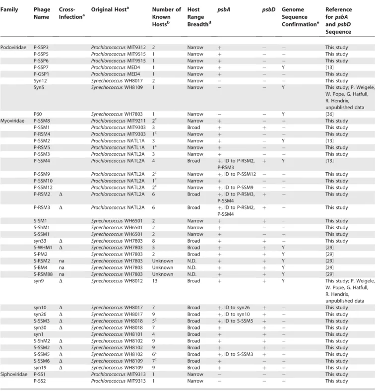
Prevalence of thepsbAandpsbDGenes in Cyanophages The psbA gene was amplified from 28 out of the 33 cyanophage isolates examined (Table 1). Combining these findings with published results (Table 1), we find that thepsbA gene is present in 88% of cyanophage isolates examined, including all myoviruses (n¼32) and all five Prochlorococcus podoviruses included in this study. However, this gene was not detected in Prochlorococcus siphoviruses (n ¼ 2) and Synechococcus podoviruses (n¼ 3), suggesting that there are some combinations of phage family and host genus that do not lead to incorporation of the psbA gene into the phage
genome. Six additional phages yielded ambiguous results and were excluded from these analyses (see Materials and Methods for details).
When present, the psbA gene is likely to be functional, as there is evidence for the conservation of amino acid sequences through purifying selection [13,37], and the gene is expressed during infection [31,32], implying that this gene confers a fitness advantage to the phages that carry it [13,22,29,31]. Sustained photosynthesis is necessary for maximal phage production [31,33,38], and the long latent period of many freshwater and marine cyanophages (8 h or more; [9,31,33,38]) presumably results in energy- and/or carbon-limitation for phage replication. Thus, cyanophage-encoded psbA likely serves to boost the photosynthetic performance of the host during infection, thereby increasing phage production. It is perhaps not coincidental that one of the phages that lackspsbA,Synechococcuspodovirus P60 (Table 1), has a latent period of only 1 h (K. Wang and F. Chen, personal communication), which may be too short for psbA expression to be beneficial. Latent period information for marine cyanophages, however, is sparse. It is not known for theProchlorococcussiphoviruses that lackpsbA, and it has only been shown to be.8 h for a single phage strain from each of the Synechococcus myoviruses [39] and Prochlorococcus podovi-ruses [31]. Further, theory [40–43] and experiments [44] suggest that latent period length may be a transient property that rapidly evolves in response to changes in host cell densities. Thus, further exploration of this hypothesis requires analysis of the latent period of many more phage isolates under variable host cell concentrations.
The psbD gene was amplified from 15 out of the 33 cyanophage isolates examined (Table 1). Again, combining our data with published findings, we observe that psbD is found only in isolates that contain psbA and only in myoviruses, but not in allpsbA-containing myoviruses. Only four of 12 Prochlorococcus myoviruses (as defined by original host strain of isolation; Table 1) containedpsbD,whereas this was the case for 17 of 20Synechococcusmyoviruses. Although it is possible that differences in the photosystem II reaction center betweenProchlorococcusandSynechococcusexist (such as differences in the rate of PsbD degradation) and could explain the biased distribution of thepsbD gene among the myoviruses, there is no evidence that this is the case. The breadth of phage host ranges (as operationally defined in Table 1), however, appears to be a reasonably good predictor of whether a phage will contain psbD: 17 of 18 broad-host-range phages encode it,whereas only one out of 21 narrow-host-range phages do so (Table 1). Perhaps broad-narrow-host-range phages have co-opted bothpsbAandpsbDto better ensure the formation of a functional PsbA–PsbD protein complex in the host during infection.
Origins and Evolutionary History ofpsbAandpsbD in Cyanophages
well-Table 1.Presence or Absence ofpsbAandpsbDamongProchlorococcusandSynechococcusCyanophages
Family Phage
Name
Cross-Infectiona
Original Hosta Number of
Known Hostsb
Host Range Breadthd
psbA psbD Genome
Sequence Confirmatione
Reference forpsbA andpsbD Sequence
Podoviridae P-SSP3 ProchlorococcusMIT9312 2 Narrow þ This study
P-SSP5 ProchlorococcusMIT9515 1 Narrow þ This study
P-SSP6 ProchlorococcusMIT9515 1 Narrow þ This study
P-SSP7 ProchlorococcusMED4 1 Narrow þ Y [13]
P-GSP1 ProchlorococcusMED4 1 Narrow þ This study
Syn12 SynechococcusWH8017 2 Narrow This study
Syn5 SynechococcusWH8109 1 Narrow Y This study; P. Weigele,
W. Pope, G. Hatfull, R. Hendrix, unpublished data
P60 SynechococcusWH7803 1 Narrow Y [36]
Myoviridae P-SSM8 ProchlorococcusMIT9211 2c Narrow þ This study
P-SSM1 ProchlorococcusMIT9303 3 Broad þ þ This study
P-RSM4 ProchlorococcusMIT9303 1c Narrow þ This study
P-SSM2 ProchlorococcusNATL1A 3 Narrow þ Y [13]
P-RSM5 ProchlorococcusNATL1A 1c Narrow þ This study
P-SSM3 ProchlorococcusNATL2A 3 Narrow þ This study
P-SSM4 ProchlorococcusNATL2A 4 Broad þ, ID to P-RSM2,
P-RSM3
þ Y [13]
P-SSM9 ProchlorococcusNATL2A 2c Narrow þ, ID to P-SSM12 This study
P-SSM10 ProchlorococcusNATL2A 1c Narrow þ This study
P-SSM12 ProchlorococcusNATL2A 2c Narrow þ, ID to P-SSM9 This study
P-RSM2 D ProchlorococcusNATL2A 6 Broad þ, ID to P-RSM3,
P-SSM4
þ This study
P-RSM3 D ProchlorococcusNATL2A 6 Broad þ, ID to P-RSM2,
P-SSM4
þ This study
S-SM1 SynechococcusWH6501 2 Narrow þ þ This study
S-ShM1 SynechococcusWH6501 2 Narrow þ This study
S-SSM1 SynechococcusWH6501 2 Narrow þ This study
syn33 D SynechococcusWH7803 8 Broad þ þ This study
S-WHM1 D SynechococcusWH7803 5 Broad þ þ Y [29]
S-PM2 SynechococcusWH7803 2 Broad þ þ Y [29]
S-RSM2 na SynechococcusWH7803 Unknown N.D. þ þ Y [29]
S-BM4 na SynechococcusWH7803 Unknown N.D. þ þ Y [29]
S-RSM88 na SynechococcusWH7803 Unknown N.D. þ þ Y [29]
syn9 D SynechococcusWH8012 13 Broad þ þ Y This study; P. Weigele,
W. Pope, G. Hatfull, R. Hendrix, unpublished data
syn10 D SynechococcusWH8017 7 Broad þ, ID to syn26 þ This study
syn26 D SynechococcusWH8017 9 Broad þ, ID to syn10 þ This study
S-SSM3 D SynechococcusWH8018 5c Broad þ, ID to S-SSM5 þ This study
syn30 D SynechococcusWH8018 7 Broad þ þ This study
syn1 SynechococcusWH8101 4 Broad þ þ This study
S-ShM2 D SynechococcusWH8102 9 Broad þ þ This study
S-SSM2 D SynechococcusWH8102 9 Broad þ þ This study
S-SSM5 D SynechococcusWH8102 6c Broad þ, ID to S-SSM3 þ This study
S-SSM6 D SynechococcusWH8109 7c Broad þ This study
syn19 D SynechococcusWH8109 9 Broad þ þ This study
Siphoviridae P-SS1 ProchlorococcusMIT9313 1 Narrow This study
P-SS2 ProchlorococcusMIT9313 1 Narrow This study
Presence is indicated byþ, and absence by.
Phages that contained identical sequences to other phages are noted as‘‘ID to X.’’
a
Cultured strain used for isolation of phage from natural seawater samples. Phages are defined as eitherProchlorococcusorSynechococcusphages based on original host of isolation, but many of the myoviruses cross-infect both genera. Those phages that cross-infect both genera are marked‘‘D’’, those that do not are left blank, and those that were not available for testing are marked‘‘na’’.
b
The number of host strains infected by each phage out of 21 strains tested [5]. c
Phages whose host ranges are first reported here: P-SSM8,ProchlorococcusMIT9211 and MIT9303; P-RSM4,ProchlorococcusMIT9313; P-RSM5,ProchlorococcusNATL1A; P-SSM9,
ProchlorococcusNATL1A and NATL2A; P-SSM10,ProchlorococcusNATL2A; P-SSM12,ProchlorococcusNATL2A and NATL1A; S-SSM3,ProchlorococcusNATL1A and NATL2A,Synechococcus
WH7803, WH8102 and WH8109; S-SSM5,ProchlorococcusMIT9303 and MIT9313,SynechococcusWH7803, WH8102, WH8103, and WH8109; S-SSM6,Prochlorococcusstrains NATL1A, MIT9215, and MIT9211,Synechococcusstrains WH6501, WH8017, WH8018, and WH8109.
dOperationally defined as narrow if a phage infects less than four hosts within a single cluster determined from 16S-23S rRNA internal transcribed spacer clustering [48] and broad if it infects more than four hosts within a cluster or at least two hosts that span more than one cluster. Small variations in this definition did not significantly affect the conclusions made. e
supported sequence clusters contain only one organism type (Figures 1 and 2), with sequences from high-light adapted (HL) and low-light adapted (LL)Prochlorococcus[45] forming discrete clusters. These well-supported Prochlorococcus clusters are similar to those observed using other host genes such as rRNA, rpoC1,andntcA[46–49], indicating thatpsbAandpsbD have not been transferred betweenProchlorococcuslineages. In contrast, theSynechococcusclusters for bothpsbAandpsbDare poorly supported, a finding different to that obtained using other highly conserved genes [46–49] and thus may have resulted from genetic exchange betweenSynechococcuslineages. ThepsbAsequences fromSynechococcusmyoviruses, Prochlor-ococcus myoviruses, andProchlorococcus podoviruses generally
formed discrete clusters consistent with their host ranges (Figure 1), suggesting that the transfer of photosynthesis genes from host to phage has been largely limited by host range (but see exceptions discussed below). Although many of these phages are capable of infecting both host genera (denoted as ‘‘D’’ in all figures), we designated each cyano-phage isolate as aProchlorococcus orSynechococcuscyanophage based upon its original host strain of isolation (as mentioned above and in Table 1). Given this designation scheme, it appears that transfers were predominantly from Prochlorococ-custo their phages and fromSynechococcusto their phages. This suggests host-range-limited host-to-phage transfer events, Figure 1.Phylogenetic Tree ofpsbAGene Sequences from Cultured Cyanobacteria and Cyanophages
Phages are listed by their name, followed by their original host. Phages that are known to infect bothProchlorococcusandSynechococcushosts are indicated with a‘‘D’’; those that infect only one genus are labeled either P (infect onlyProchlorococcushosts) or S (infect onlySynechococcushosts), while those that are unknown are designated with a‘‘?’’. Phages shown in italics and bracketed with‘‘**’’were isolated on hosts that do not belong to the same cluster and are thus exceptions to the general clustering pattern (see text). Taxa are color coded according to the following biological groupings: myoviruses (red), podoviruses (black), marineSynechococcushosts (light blue), marineProchlorococcushosts (dark green, LL; light green, HL), freshwater cyanobacteria (dark blue). The tree topology was estimated by LogDet analysis of 1st and 2nd codon positions. Sequences where intragenic recombination was detected using other methods (see Materials and Methods) were not included in these phylogenetic analyses. Branch lengths were estimated by maximum likelihood under a model with nonstationary nucleotide frequencies. Numbers at the nodes represent neighbor-joining bootstrapping and maximum likelihood puzzling support. Anab,Anabaena;Gloe, Gleobacter; HL, high-light adapted; LL, low-light adapted; Syncy, Synechocystis;Thermo,Thermosynechococcus.
with subsequent horizontal and vertical transfers occurring among viral lineages.
Two isoforms of the PsbA protein are often found in cyanobacteria [50]. The PsbA.1 (D1.1) isoform is constitutively expressed, whereas the PsbA.2 (D1.2) isoform is upregulated in response to high light and UV stress [51,52]. Many of the differences between the isoforms are found in ten amino acids between position 121 and 312 [50]. Based on which isoform the majority of these ten amino acids were identical to (including glutamine/glutamate at position 130), we determined that PsbA from bothProchlorocococusmyoviruses and podoviruses are more similar to PsbA.1, the only isoform found inProchlorocococushosts so far [53] (unpublished data). Although Synechococcus hosts encode both isoforms (unpub-lished data), Synechococcus myoviruses encode the stress-responsive PsbA.2 isoform exclusively (unpublished data), which may be particularly beneficial during the stress of infection. These findings are consistent with the hypothesis of host-range-limited transfers of the psbA gene (but see exceptions below).
Host-to-phage transfers appear to have occurred at least four times for psbA and twice for psbD, as seen from the
number of discrete clades containing phage-encoded genes in each case (Figures 1 and 2). The fourpsbAgene acquisitions by phage appear to include two transfer events for the Prochlorococcus myoviruses (Prochlorococcus myovirus group 1 and 2 in Figure 1) and a single event for Prochlorococcus podoviruses all from their Prochlorococcus hosts, as well as a single event for Synechococcus myoviruses from their hosts (Figure 1). ThepsbDgene appears to have been acquired once by bothSynechococcusandProchlorococcusmyoviruses from their respective hosts (Figure 2). Interestingly, the three Prochlor-ococcusmyoviruses that containpsbDall encodeProchlorococcus myovirus group 1 psbA sequences, suggesting that this gene was acquired only once by a subset of these myoviruses. Although the specific source is difficult to determine from phylogeny alone, the placement of theProchlorococcus myovi-rus sequence clusters suggests that psbA was derived from either HL Prochlorococcus hosts or LL NATL2A-type hosts, while thepsbDgenes could have been acquired from any of the Prochlorococcus hosts other than MIT9313/9303. The placement of the Prochlorococcus podovirus (psbA only) and Synechococcus myovirus sequence clusters at the base of the Figure 2.Phylogenetic Tree ofpsbDGene Sequences from Cultured Cyanobacteria and Cyanophages
Details as in Figure 1. Sequences where intragenic recombination was detected using other methods (e.g., P-SSM1) were not included in these phylogenetic analyses.
host and virus clades provides little further information about the source of these phage genes.
We found three exceptions to the above host-constrained evolutionary scenario—i.e., cases where phagepsbAandpsbD genes did not cluster with those of their hosts (Figure 1 and Figures S1 and S2) and did not have PsbA isoforms consistent with that of their hosts (unpublished data). These include two narrow host-range Synechococcus myoviruses (ShM1, S-SSM1), which encode psbA sequences most similar to Prochlorococcusmyoviruses (Figure 1) even to the extent that they encode the PsbA.1 isoform, as well as a Prochlorococcus myovirus (P-SSM1) with apsbA sequence that is most similar to those fromSynechococcusmyoviruses (Figure 1) and encodes the PsbA.2 isoform as expected for aSynechococcusmyovirus. Although the latter can cross-infect across Prochlorococcus ecotypes, it has not been shown to infectSynechococcus[5]. The P-SSM1 phage also encodespsbD,which, like itspsbAgene, is more similar toSynechococcus psbDsequences than those of the Prochlorococcushost upon which it was isolated (Figure S2; note that this sequence does not appear in Figure 2 because it was a candidate for intragenic recombination; see Materials and Methods). It is likely that these exceptions to the rather consistent host-phage sequence clustering resulted from horizontal transfer events between a broad-host-range donor phage and a limited-host-range recipient phage during coinfection of a single host, i.e., swapping of genes within the phage gene pool [24]. Whole gene transfers within the phage gene pool are likely to be more common than this, but undetectable when occurring within phages that form a discrete phylogenetic cluster. These observations call for caution when using clustering patterns of psbA and psbD sequences from uncultured phage (obtained from environ-mental genome data) to identify potential hosts.
Intragenic Recombination within Core Reaction Center Proteins
The lack of well-supported clade structure in phylogenetic reconstructions forSynechococcushost strains when using both psbA and psbD differs from those constructed using other genes [46–49], which led us to wonder about underlying mechanisms that could be responsible for such a blurred phylogenetic signal. In a recent study, Zeidner et al. [37] showed thatSynechococcus-phage-likepsbAsequences from the environment had a patchy %GþC distribution, which they suggest is due to intragenic recombination [37]. Their analyses demonstrated that such recombination had occurred within the inferred-phage clusters and within clusters spanning both phage and host psbA sequences. They could not discern, however, whether the signal was caused only by phage-to-phage exchanges, or included phage-to-host ex-changes, because the majority of their sequences were of unknown origin (i.e., they were derived from environment clone libraries), and the test employed does not assess the directionality of intragenic recombination events. Our cultured hosts and phages provide an opportunity to assess recombination partners without ambiguity regarding the source of the genes. In addition, the known host ranges of these phages [5] (Table 1), together with the types of recombination tests we have used (see Materials and Methods), allow us to assess the directionality and the pathways through phages and hosts that these recombination events are likely to have taken.
As a first assessment for potential intragenic recombina-tion, we analyzed the %GþC patterns in all of thepsbAand psbD genes (Figures 3 and 4, respectively). Prochlorococcus phage genes had similar average %GþC contents to those from theirProchlorococcushosts (39%–46%), whereas those of Synechococcusphages had %GþC contents that were lower than those from their Synechococcushosts (46%–51% versus 56%– 62%), but not as low as those fromProchlorococcus hosts and phages. This intermediate %GþC could be the result of intragenic recombination between variants of the two host lineages. Alternatively, it may reflect the current state of mutational amelioration of the acquired gene from a high %GþC source towards the low genome-wide %GþC of the virus (Synechococcusmyoviruses S-PM2 and Syn9 both have low genome-wide %GþC; [28]; P. Weigele, W. Pope, G. Hatfull, R. Hendrix, personal communication). If the latter is the case, we might expect such amelioration to be constant across the gene, resulting in an even %GþC distribution pattern.
To help differentiate between these hypotheses, we mapped the %GþC variation across thepsbAandpsbDgenes using the methodology developed by Zeidner et al. [37]. We detected patchiness of %GþC in SynechococcusmyoviruspsbA sequences dispersed along the length of the gene (Figure 3), confirming the findings reported by Zeidner et al. [37]. We also detected %GþC patchiness among psbA from Prochlor-ococcus podoviruses, but not fromProchlorococcus myoviruses, despite overall similarity of their %GþC content with their Prochlorococcus hosts. This suggests that intragenic recombi-nation has occurred among the podoviruses. In addition, patterns of %GþC were not uniform and even markedly clumped across thepsbD gene fromSynechococcus myoviruses (Figure 4), with the first segment resembling Synechococcus hosts and the last segment resembling Prochlorococcus hosts and their phages. Thus, intragenic recombination is likely to be at least partly responsible for the intermediate %GþC content inSynechococcusmyoviruspsbAandpsbDsequences.
Statistical methods for detecting intragenic recombination (see Materials and Methods) revealed strong evidence for its presence in both thepsbAandpsbDsequence sets (Tables S1 and S2), but the relative frequency of recombination events was not equal for different groups of hosts and phages. Recombination appears most common among the cyanoph-ages, and more so forSynechococcusthanProchlorococcusphages. Exchanges were detected between phages that infect both Synechococcus andProchlorococcus as well as within myoviruses that infect a single genus(Synechococcus).Note that exchanges within a single phylogenetic phage cluster, such as within the Synechococcusmyoviruses, were undetectable by our previous phylogenetic analyses. Interestingly, our analyses also revealed exchanges between Prochlorococcus-specific podoviruses and broad-host-range Synechococcusmyoviruses, with the Prochlor-ococcuspodoviruses serving as the donors (Table S1). Marine cyanobacterial podoviruses contain integrase genes and are thought to have the ability to integrate into the genomes of their hosts as prophages [30] (P. Weigele, W. Pope, G. Hatfull, R. Hendrix, personal communication). If true, genetic exchange could occur between the Prochlorococcus prophage and a Synechococcus lytic phage—a scenario well accepted in other phage-host systems for genetic exchange [14,15].
are evident, however, and appear to have occurred both from host to phage and phage to host for both psbA and psbD. Although such events were not detected between Prochlor-ococcusand their phages, there were cases whereProchlorococcus myoviruses were the recipients of external DNA from an unknown source (i.e., recombination events possibly involv-ing donors outside of our dataset). Thus, phages may be contributing to the intragenic recombination of portions of these genes inSynechococcus, perhaps explaining the lack of phylogenetic structure observed inpsbA and psbD trees for Synechococcus clusters (but not for Prochlorococcus clusters) relative to those obtained when using other phylogenetic
markers [46–49]. Presumably, phage-host intragenic ex-changes occur via homologous recombination during infec-tion. Clearly, the transfer of DNA will be retained in host lineages only if infection fails to lyse the host (e.g., abortive infection [54]).
Finally, intragenic exchanges among hosts were also occa-sionally detected, particularly amongSynechococcus(Tables S1 and S2). This may also play a role in the lack of clade structure among Synechococcus strains in the psbA and psbD trees. Although two possible intragenic recombination events betweenSynechococcusandProchlorocococuswere identified, they were resolved as small regions (15–16 bases) and may be false Figure 3.Visualization of %GþC Content across thepsbAGene
Colors represent the averaged %GþC in sliding windows along the length of the gene (20%–80%); white regions represent windows that included ambiguous bases in which %GþC could not be calculated for that region. The average %GþC content of the amplified sequence is tabulated on the right side of the figure. Phages are listed by phage name followed by their original host. Phages that are known to infect bothProchlorococcusand Synechococcushosts are indicated with a‘‘D’’; those that infect only one genus or the other have no marker, while those that are unknown are designated with a‘‘?’’. Host names are prefaced with Syn or Pro forSynechococcusandProchlorococcushosts, respectively. Scale indicates nucleotide positions relative to thepsbAgene sequence inThermosynechococcus.
positives. Host-to-host transfers may have occurred through the uptake of DNA directly from the environment (e.g., via transformation) or through viral intermediates [37]. Such host-to-host intragenic exchanges via viral intermediates presumably occur through generalized transduction [55].
In summary, our findings suggest that the shuffling of segments ofpsbAandpsbDwithin the cyanophage gene pool has generated significant photosynthesis gene diversity and serves as an extended reservoir of genetic diversity for their hosts, influencing photosystem evolution.
psbAandpsbDGene Diversity in Cultured Isolates Captures Most of the Field Diversity
We next sought to determine how well psbA and psbD sequence diversity observed in culture collections represents that observed in wild phage populations, and whether additional whole-gene host-to-phage transfer events could be identified from these wild sequences from the phage gene pool. Zeidner et al. [37] had previously examined field diversity of the psbA gene sequence from environmental samples where Synechococcus strains were the dominant
phototroph [37]. Thus, we sought to examine genetic diversity of this gene, as well as that of psbD, from an environment whereProchlorococcuscells commonly outnumberSynechococcus cells by orders of magnitude [56]. To this end, we amplified, cloned, and sequenced psbA and psbD gene sequences obtained from the viral-sized fraction (0.02–0.2 lm) of two seawater samples within (25 m) and below (75 m) the mixed layer in the Pacific Ocean off the coast of Hawaii (Figures 5 and 6, respectively). ThepsbAandpsbDsequences from these viral-fraction samples clustered with cultured Prochlorococcus cyanophage isolates (with varying levels of support; Figures 5 and 6), but not withSynechococcuscyanophages. There was not a notable difference in the phylogenetic placement of the psbAorpsbDclones obtained from within or below the mixed layer. Although this suggests a lack of vertical structure in diversity among the sequence types, we did not sequence these samples to saturation; thus, such conclusions are preliminary.
More than half of the wildpsbAsequences (42 of 81) form a large cluster with culturedProchlorococcuspodoviruses (Figure 5). Within this group, all but one cluster of wild sequences Figure 4.Visualization of %GþC Content across thepsbDGene
Details as in Figure 3. Note that the 21-nucleotide indel inProchlorococcushosts and their phages [13] (unpublished data) was excluded from the analysis at the position indicated by the‘‘//’’symbol to maximize the data that could be displayed using the sliding window approach.
contain cultured podovirus sequences (Figure 5). The extensive microdiversity in this cluster (labeled ‘‘ unrepre-sented 1’’) was probably derived from within the podovirus gene pool, as evidenced by the presence of podovirus phage isolates in the more basal branches of the cluster. OtherpsbA sequences from the field samples form subclusters that contain culturedProchlorococcusmyoviruses and form a large group that also containsProchlorococcushosts (Figure 5). One cluster (‘‘unrepresented 2’’in Figure 5) within this group also lacks sequences from cultured hosts or phages. The basal position of this cluster suggests that these sequences may belong to phages that infect as-yet unculturedProchlorococcus hosts [57] and may represent an additional host-to-phage
transfer event. Thus, our work here, together with that of Zeidner et al. [37], suggests that cyanophage culture collec-tions represent much of the naturally occurring Prochlorococ-cusandSynechococcuscyanophagepsbAgene sequence diversity [37].
AllpsbDsequences from wild phages fall into a single well-supported cluster that includes a representative cultured Prochlorococcus cyanophage P-SSM4 (Figure 6). This cluster reveals significant microdiversity within the psbD Prochlor-ococcusphage gene pool in the viral-fraction from this Pacific Ocean site and suggests that phages that encode Prochlor-ococcus-phage-like psbD genes are perhaps not rare in this environment. The four Prochlorococcus cyanophages that Figure 5.Phylogenetic Tree ofpsbAGene Sequences from Representative Cultured Cyanobacterial and Cyanophage Isolates and Cloned Environmental Sequences from the Hawaii Ocean Time Series Site in the Pacific Ocean
Phylogenetic tree ofpsbAgene sequences and cloned environmental sequences were collected from above (25 m, black) and below (75 m, red) the surface mixed layer at the Hawaii Ocean Time Series site in the Pacific Ocean, a region whereProchlorococcusare the dominant phototrophs. Details for naming conventions are as in Figure 1.Synechococcusenvironmental‘‘viral’’sequences from [37]. The tree topology was estimated by LogDet analysis of 1st and 2nd codon positions, with branch lengths estimated using stationary nucleotide frequencies.
contain the psbD gene in our culture collection originated from either the Sargasso Sea or the Red Sea; thus, it is perhaps not surprising that the viral-fraction microdiversity from the Pacific Ocean is largely unrepresented in this collection.
Conclusions
The phage genomic repertoire evolves through the exchange of genetic material from other phages [24] and by co-opting metabolic genes from their hosts [13,20,22]. The prevalence of photosynthesis genes in cyanophages strongly suggests that the capture of these genes provides a significant
fitness advantage among certain cyanophage types. Previ-ously, we have shown that the horizontal transfer ofhligenes from cyanophages to their hosts has likely played a role in driving host niche differentiation [13]. More recently, cyanophages were hypothesized to be involved in partial gene exchanges even for the core photosystem genepsbAof their hosts [37]. Here, we show that genetic exchanges involving cyanophages may have influenced the make-up of both of the core photosystem II genes (psbA and psbD) in Synechococcus,whereas this was less apparent forProchlorococcus. Therefore, mounting evidence indicates that host-like genes acquired by phages undergo a period of diversification in Figure 6.Phylogenetic Tree ofpsbDGene Sequences from Cultured Cyanobacterial and Cyanophage Representatives and Cloned Environmental Sequences from the Pacific Ocean
phage genomes and serve as a genetic reservoir for their hosts. Thus, a complex picture of overlapping phage and host gene pools emerges, where genetic exchange across these pools leads to evolutionary change for host and phage. Fully understanding the mechanisms of microbial and phage coevolution clearly requires an improvement in our ability to quantify horizontal gene transfer at the whole and partial gene level and in our ability to accurately estimate the relative fluxes into and out of these pools.
Materials and Methods
DNA isolation from cultured hosts and phages and environmental samples.Eleven strains ofProchlorococcus,ten strains ofSynechococcus,
and 38 phages ofProchlorococcusandSynechococcus(seven podoviruses, 29 myoviruses, and two siphoviruses) were screened forpsbAandpsbD
sequences for this study. We report here on newpsbAsequences from nineSynechococcus hosts and new psbD sequences from 19 Prochlor-ococcus and Synechococcus hosts (including two from unpublished
Synechococcusgenomes for strains CC9605 and CC9902; available from http://genome.jgi-psf.org/mic_home.html. The 38 phages screened included seven phage templates for which genome sequences are now available (P-SSM2, P-SSM4, P-SSP7, S-PM2, S-WHM1, Syn5, Syn9), enabling us to validate our PCR amplification findings. Host genomic DNA was extracted using a DNeasy Tissue Kit (Qiagen, Valencia, California, United States). Filtered (0.2 lm, Acrodisc supor mem-brane syringe filter) phage lysates in Pro99 medium were used as DNA templates for subsequent PCR amplification experiments.
Environmental samples were collected from the Hawaii Ocean Time Series (HOT) on 15 October 2003 at 458N 1588W from depths of 25 m and 75 m. These samples were filtered through a 0.2-lm filter (Osmonics, Minnetonka, Minnesota, United States, Poretics polycar-bonate 25-mm filter) to remove cellular material and substantially enrich for environmental phages. A 100-ml volume of 0.2-lm filtrate was then filtered onto a 0.02-lm filter (Whatman Anotop 25) to collect phage particles and resuspended in 7 ml of a modified SM storage buffer (600 mM NaCl, 8 mM MgSO4-7H2O, 50mM Tris [pH
7.5], 0.04% gelatin).
Overview ofpsbAandpsbDscreening strategy.PCR screening for
psbA and psbD across a diverse set of samples presented several challenges. These included variable amplification efficiencies, un-certainty about whether amplicons derived from phage or host, and multiple gene copies in hosts. The amplification strategy was as follows: for each virus and host strain, four PCR reactions were carried out, pooled, and analyzed by gel electrophoresis; if the amplification product was not visible, it was diluted 10-fold and used as template for nested or semi-nested PCR and the resulting products analyzed; if still no product was visible, multiple phage stocks were rescreened. Multiple copies of psbA in Synechococcus strains were identified by sequencing many clones and were distinguished from sequencing errors as described below. We did not screen for multiple copies ofpsbAfromProchlorococcusor multiple copies of psbDfrom either Synechococcus or Prochlorococcus, as when present, they are generally indistinguishable from each other [58–60].
Amplification of psbA andpsbD.PCR reactions were performed withTaq DNA polymerase and deoxyribonucleotide triphosphates from New England Biolabs (Beverly, Massachusetts, United States) or Invitrogen (Carlsbad, California, United States) and carried out with a PTC-100 or PTC-200 DNA Engine (MJ Research, Waltham, Massachusetts, United States) or a Robocycler Gradient 96 (Strata-gene, La Jolla, California, United States). Template amounts were 10 ng of genomic DNA forProchlorococcusandSynechococcus,1ll of lysate for cyanophages, and 2ll of filtrate for environmental samples. PCR primers and amplification reaction conditions are shown in Tables S3 and S4.
ThepsbAgene from all sources was amplified using primer pair
psbA-F/R [61] and PCR protocol A (Tables S3 and S4). Four reactions were conducted with each template, and the products were pooled and analyzed by agarose gel electrophoresis. PrimerpsbA-R falls on the intron region in S-PM2 [29]. Therefore, for efficient amplification of phage psbA genes that may contain introns, and for increased sensitivity, we used the Pro-psbA-F/R primer set and protocol B in nested PCR reactions when no PCR product was visible from cyanophage lysates and environmental filtrates. To reduce the incidence of heteroduplex formation, amplification products from environmental samples were subjected to reconditioning PCR [62]:
initial PCR products were diluted 1:10, then amplified using protocol A but for only three cycles.
ThepsbDgene fromProchlorococcus, Synechococcus,and cyanophages was amplified using primer pairpsbD-54F/psbD-308R and protocol D. However, when product yield was low or absent, semi-nested PCR was carried out as follows. Amplification was first conducted using primer pair psbD-26F/psbD-308R and protocol C. Four reactions were conducted with each template, the products were pooled, diluted 1:10, and used as templates for a second round of amplification using primer pairpsbD-54F/psbD-308R and protocol D.psbDfrom environ-mental samples was amplified using primer pairpsbD-26F/psbD-308R and protocol C and subjected to reconditioning PCR as forpsbA(see above).
In preparation for sequencing, PCR products were either purified directly using the QIAquick PCR Purification Kit (Qiagen) or separated on an agarose gel and then purified using the QIAquick Gel Extraction Kit (Qiagen).
To confirm that the absence ofpsbAorpsbDPCR products from phage was not simply due to a lack of amplifiable phage DNA, we screened phage lysates for known phage genes:g20(for myoviruses) andDNApol(for podoviruses).g20was amplified using primer pair g20-F/R and protocol E, andDNApolusing primer pair DNApol-F/R and protocol F, both with 1ll of lysate. In all cases, a product was obtained, suggesting the phage template DNA was present and amplifiable by PCR (unpublished data).
Six phage lysates yielded PCR products with sequences identical to those of a known host. These six phage lysates include five cyanophages previously described (RSP1, SSP1, SSP2, P-ShM1, P-ShM2; [5]), as well as one cyanophage not previously reported in the literature (P-SSP9; M.B.S. and S.W.C., unpublished data). In these cases we could not eliminate the possibility that the amplicon resulted from host DNA, the amplification of which may be more likely to occur when there is no phage template for this gene. Thus, we excluded these phages from further analyses. In contrast, phages with amplicon sequences identical to those of other phages (indicated as‘‘ID to X’’in Table 1) were passed through multiple lysates, and a‘‘fingerprint’’phage gene(g20)was used to confirm that there was a single phage in the lysate. ThepsbAsequence was then re-assayed, increasing our confidence in these results. Even with this precaution, we cannot rule out the possibility of PCR contamination for those few cases where identical sequences were amplified from different phage lysates.
Cloning and sequencing of PCR products.ThepsbAgene is often found in multiple distinct copies in marineSynechococcus[59], whereas inProchlorococcusthepsbAgene is either single copy per genome or encodes multiple copies that are nearly identical to each other [60,63,64]. Among cyanophages, thepsbAgene has only been found in a single copy per genome [28,30]. To allow for the identification of multiplepsbAgene copies inSynechococcusstrains, PCR products from
Synechococcustemplates were cloned prior to sequencing. Cloning was performed using the TOPO TA Cloning Kit for Sequencing (Invitrogen) with the pCR4-TOPO vector. Ligation products were transformed into TOP10 competent cells. Plasmid purification and sequencing were conducted by Genaissance Pharmaceuticals (New Haven, Connecticut, United States). Inserts were sequenced from both forward and reverse directions, using the M13F and M13R primer binding sites in the pCR4-TOPO vector.
Approximately ten psbAclones were sequenced for each Synecho-coccusstrain. The published genome ofSynechococcusWH8102 provides an example of naturalpsbAdiversity in a given strain, as it contains four copies ofpsbA:two copies that are 99.8% identical and a third and fourth copy that are 99.4% and 88% identical, respectively, to the above twopsbAcopies [59]. Considering aTaqpolymerase error rate of 33105
per nucleotide per duplication [65], at most one error could be expected in each psbA gene sequenced. Thus, sequences were considered identical, and removed from the analysis pool, if they were more than 99.8% identical, to avoid data issues stemming from possible PCR error (sequencing error should be nonexistent because consensus sequences were obtained from forward and reverse sequencing of the clones). Sequence identity levels for nonidentical clones from the remaining dataset ranged from about 60% to 99.0%. PCR products from genes presumed not to have multiple distinct copies per genome (psbAfromProchlorococcusand cyanophage;psbD
both forward and reverse directions, using the same primers used for PCR amplification.
Sequence analyses. Previous analyses have raised important concerns about usingpsbAgene sequence datasets that may suffer from large %GþC variability and conflicting phylogenetic signals in phylogenetic reconstructions [37]. To minimize such errors, we followed these steps.
We first performed phylogenetic analyses using sequences from all taxa (80 forpsbAand 50 forpsbD) and all codon positions (Figures S1 and S2). Phylogenetic trees were constructed by using distance and maximum likelihood. Neighbor-joining [66] was used to reconstruct a distance tree under the HKY85 model [67]. Maximum likelihood analysis was performed under HKY85 combined with a gamma model for among sites rate variation, assuming eight rate categories with model parameters estimated from the data [68]. Maximum likelihood trees were obtained by quartet puzzling, as implemented in the program TREE-PUZZLE 5.0 [69]. Bootstrap resampling (1,000 pseudoreplicates) was used to measure the relative support for internal branches of the neighbor-joining trees. For quartet puzzling, support was estimated from 25,000 (psbDtrees) or 50,000 (psbAtrees) pseudoreplicates.
These analyses resulted in trees with high bootstrap support at many critical nodes (Figures S1 and S2). However, fitting a single tree to large datasets containing conflicting phylogenetic signals can lead to reconstruction artifacts (i.e., systematic errors) that result in high bootstrap support [70,71]. We found, using neighbor-nets [72] constructed by using the SplitsTree2 program [73], within-gene conflicting phylogenetic signals in both thepsbAandpsbDdatasets as indicated by the box-like structures in neighbor-nets graphs (Figures S3 and S4). Specifically, networks for both genes revealed substantial conflict involving splits betweenSynechococcus strains, their myovi-ruses, and a complex of sequences comprised ofProchlorococcusand their viruses.
We further investigated whether these large datasets could suffer from systematic errors related to: (i) substitution rate variation among lineages [74], (ii) heterogeneous compositional bias among lineages (e.g., %GþC; [75]), and (iii) within-gene heterogeneity in phylogenetic signals [76]. We found significant substitution rate variation among lineages (Table S5) using likelihood ratio tests. In addition, nucleotide frequencies were nonstationary across these data, with significant differences in equilibrium frequencies for clades defined according to organism types (Table S6; [77]). Not surprisingly, the largest divergence in %GþC across taxa was at the
3rd codon positions of bothpsbAandpsbD.
Zeidner et al. [37] hypothesized intragenic recombination inpsbA
[37]. We attempted to identify this qualitatively through graphical analysis of %GþC and quantitatively using four different tests for intragenic recombination. The %GþC distribution was examined within overlapping sequence windows (a sliding window of 30 nucleotides with a five-nucleotide step) using the GCViz script [37] (available upon request from Dr. Shmoish of Technion–IIT; [email protected]) written in the R-language (http://www. r-project.org). Three of the four different tests for within-gene recombination are based on the distribution of substitutions (GENECONV: [78]; MAXCHI: [79]; CHIMAERA: [80]), while the fourth used
a phylogenetic approach (‘‘RDP,’’ as implemented in [81]). We considered only those recombination events that satisfied all of the following criteria: (i) results were significant after application of Bonferroni correction for multiple tests, (ii) regions were detected by two or more different methods, and (iii) consensus breakpoints could be estimated for a given region identified using different methods. Once a putative recombination event was detected, we inferred the best candidate donor sequence (that most similar to the recombinant segment) using RDP [81].
In summary, to minimize systematic errors in the ultimate phylogenetic analyses, we first processed the dataset as follows: (i) excluded those sequences having a strong signal for intragenic recombination, (ii) excluded 3rd codon positions, which display the largest differences in %GþC and substitution rates among lineages, and (iii) employed LogDet distances [75] to accommodate composi-tional heterogeneity (variable %GþC) in the remaining data. These measures proved to be important. The uncorrected dataset grouped lineages according to evolutionary rates and %GþC bias (Figures S1 and S2), whereas the ultimate analysis did not (see Figures 1 and 2). Statistical analysis of the processed dataset under nonhomogenous evolutionary models [77] revealed that the ultimate phylogenetic hypotheses (see Figures 1 and 2) provided a significantly better fit to the data (Table S7). Prior to processing the data, the alternative phylogenies were indistinguishable (Table S7).
Supporting Information
Figure S1.Phylogenetic Analyses Including AllpsbAGene Sequences from Cultured Cyanobacteria and Cyanophages
Phages are listed by phage name, followed by their original host. Host range information is designated in parentheses. Phages known to infect bothProchlorococcusandSynechococcushosts are indicated with a
‘‘D’’; phages that infect only Prochlorococcus or Synechcococcus are designated by a P or S, respectively; and those host ranges that are unknown have a‘‘?’’. Phages shown in italics and bracketed with‘‘**’’
were isolated on hosts that do not belong to the same cluster and are thus exceptions to the general clustering pattern (see text). Taxa are color coded according to the following biological groupings: myoviruses (red), podoviruses (black), marineSynechococcushosts (light blue), marineProchlorococcushosts (dark green, HL; light green, LL), freshwater cyanobacteria (dark blue). Neighbor-joining tree was inferred under HKY85 mode and using sequences from all taxa and all codon positions. Nucleotide frequencies were assumed to be homogenous across lineages. Numbers at the nodes represent neighbor-joining bootstrapping and maximum likelihood puzzling support. Anab,Anabaena;Gloe, Gleobacter; HL, high-light adapted; LL, low-light adapted; Syncy,Synechocystis;Thermo, Thermosynechococ-cus.
Found at DOI: 10.1371/journal.pbio.0040234.sg001 (79 KB PPT).
Figure S2.Phylogenetic Analyses Including AllpsbDGene Sequences from Cultured Cyanobacteria and Cyanophages
Details are as in Figure S1.
Found at DOI: 10.1371/journal.pbio.0040234.sg002 (59 KB PPT).
Figure S3. Neighbor-Nets Analysis of 80 psbA Gene Sequences (including All Cyanophage and Marine Cyanobacterial Sequences Available)
The analysis was conducted under the HKY85 model of substitution using all codon positions. Taxa color coding and abbreviations are as in Figure S1. The box-like appearance in the basal branches of this phylogeny suggests regions of conflicting phylogenetic signals (see Materials and Methods).
Found at DOI: 10.1371/journal.pbio.0040234.sg003 (272 KB PDF).
Figure S4. Neighbor-Nets Analysis of 50 psbD Gene Sequences (including All Cyanophage and Marine Cyanobacterial Sequences Available)
Taxa color coding and abbreviations are as in Figure S1. Details of the analysis are as in Figure S3.
Found at DOI: 10.1371/journal.pbio.0040234.sg004 (249 KB PDF).
Table S1. Consensus Results from Four Tests for Intragenic Recombination within Gene Sequences in OurpsbADataset The four tests included (1) RDP, (2) GeneConv, (3) MaxChi, and (4) Chimaera (as described in Materials and Methods), and recombina-tion was considered‘‘detected’’only when the following criteria were satisfied: (i) similar regions were detected by two or more methods, (ii) all such regions were significant atp,0.05 after a Bonferroni correction for multiple tests, and (iii) consensus breakpoints could be inferred from the results. Thus,‘‘No recombination detected’’does not preclude that intragenic recombination could be occurring within the sequence, but rather indicates that our stringent criteria have not identified such an event. While we define phages as either
ProchlorococcusorSynechococcusphages depending on the original host of isolation, we note that many of the myoviruses cross-infect both genera (represented with a‘‘D’’where known, a‘‘?’’where unknown, and no symbol for isolates that do not cross-infect across genera). Consensus breakpoints are relative to nucleotide positions in
Thermosynechococcus psbA.
Found at DOI: 10.1371/journal.pbio.0040234.st001 (29 KB XLS).
Table S2. Consensus Results from Four Tests for Intragenic Recombination within Gene Sequences in OurpsbDDataset Details are as in Table S1.
Found at DOI: 10.1371/journal.pbio.0040234.st002 (28 KB XLS).
Table S3.PCR Conditions
Found at DOI: 10.1371/journal.pbio.0040234.st003 (38 KB DOC).
Table S4.PCR Primers
Table S5.Likelihood Ratio Tests for Variable Evolutionary Rates among Branches
For bothpsbAandpsbD,individual sequences exhibiting a signature for intragenic recombination (Tables S1 and S2) were excluded from analysis. Likelihood scores were obtained under a stationary HKY85 model combined with a gamma correction for among-sites rate variation. All model parameters, including nucleotide frequencies, were estimated by using maximum likelihood. Data analysis included all three codon positions. Models were employed as implemented in the baseml program of the PAML package [82]. Tree 1 was obtained by neighbor-joining analysis of LogDet distances estimated from all three codon positions. Tree 2 was obtained by neighbor-joining analysis of LogDet distances estimated from 1st and 2nd codon positions. For both genes, Tree 1 grouped lineages along lines of similarity in evolutionary rates and compositional biases, and Tree 2 did not. Found at DOI: 10.1371/journal.pbio.0040234.st005 (36 KB DOC).
Table S6. Likelihood Ratio Tests for Nonstationary Frequencies among Lineages
H0denotes the null hypothesis of stationary nucleotide frequencies;
this was modeled by specifying one set of nucleotide frequencies for all branches of the tree. H1denotes the alternative hypothesis of
nonstationary nucleotide frequencies; this was modeled by assigning all branches of the tree topology to one of several independent sets of frequency parameters (six sets forpsbAand five sets forpsbD). Apart from nucleotide frequencies, H0 and H1 assumed a substitution
process equivalent to an HKY85 model combined with a gamma model for among-sites rate variation. The transition/transversion ratio was assumed to be homogenous among branches. H1represents
a user-defined version of the nonhomogenous models of Yang and Roberts [77]. All model parameters, including nucleotide frequencies, were estimated by using maximum likelihood. Data analysis included all three codon positions. Models were employed as implemented in the baseml program of the PAML package [82].
Tree 1 was obtained by neighbor-joining analysis of LogDet distances estimated from all three codon positions. Tree 2 was obtained by neighbor-joining analysis of LogDet distances estimated from 1st and 2nd codon positions. For both genes, Tree 1 grouped lineages along lines of similarity in evolutionary rates and compositional biases, and Tree 2 did not. User-defined sets of frequency parameters for H1
were specified in the tree file (shown below) by using the‘‘branch label’’format described in the PAML manual. For bothpsbAandpsbD,
individual sequences exhibiting a signature for intragenic recombi-nation (Tables S1 and S2) were excluded from analysis.
Found at DOI: 10.1371/journal.pbio.0040234.st006 (44 KB DOC).
Table S7. Likelihood-Based Statistical Comparison of Competing Evolutionary Hypotheses under a Model of Nonstationary Nucleotide Frequencies
PKHdenotes thep-value for the KH normal test of [83].PSHdenotes
thep-value for the SH test [84].PRELLdenotes the RELL bootstrap
proportion [83]. Note that although Tree 1 and Tree 2 were not selected independently of the data, neither was selected according to its likelihood score. For both genes, Tree 1 grouped lineages along lines of similarity in evolutionary rates and compositional biases, and Tree 2 did not. For both psbA and psbD, individual sequences exhibiting a signature for intragenic recombination (Tables S1 and S2) were excluded from analysis. Tree 1 was estimated by a neighbor-joining analysis of LogDet distances from all sites, and Tree 2 was estimated by a neighbor-joining analysis of LogDet distances based on only 1st and 2nd codon positions. Likelihood scores were obtained under nonstationary models of nucleotide frequencies (see Table S5 for additional model details).
Found at DOI: 10.1371/journal.pbio.0040234.st007 (46 KB DOC).
Accession Numbers
New sequences from cultured cyanobacteria and cyanophages are deposited in GenBank (http://www.ncbi.nlm.nih.gov/Genbank) under accession numbers DQ473647–DQ473719, whereas new environ-mental sequences are deposited under accession numbers DQ473720–DQ473847.
Acknowledgments
We thank M. Shmoish, R. Fu, and V. Quinlivan for technical assistance; M. Coleman, M. Osburne, J. Waldbauer, and V. Rich for valuable comments on the manuscript; A. Thompson for collecting field samples; and Z. Johnson, K. Armstrong, and B. Tidor for analysis and discussion of possible PsbA/PsbD interactions. We thank P. Weigele, W. Pope, G. Hatfull, and R. Hendrix for providing unpublished genome sequences (Syn5 and Syn9); and F. Chen for sharing his unpublished phage lytic cycle information (P60) with us.
Author contributions.MBS, DL, and SWC conceived and designed the experiments. MBS, DL, JAL, and LRT performed the experiments. MBS, DL, JAL, LRT, and JPB analyzed the data. MBS and DL wrote the paper, with significant contributions from all authors.
Funding.This research was supported by grants from the United States Department of Energy (99ER62814 and DE-FG02-02ER63445), the National Science Foundation and the Gordon and Betty Moore Foundation to SWC, Massachusetts Institute of Technology’s Undergraduate Research Opportunities Program fund-ing to JAL, a National Institutes of Health predoctoral trainfund-ing grant in the biological sciences (GM07287–31) to LRT, and a National Sciences and Engineering Research Council (Canada) Discovery Grant (DG 298394) to JPB.
Competing interests.The authors have declared that no competing interests exist.
References
1. Waterbury JB, Watson SW, Valois FW, Franks DG (1986) Biological and ecological characterization of the marine unicellular cyanobacterium Synechococcus. Can Bull Fish Aquat Sci 214: 71–120.
2. Partensky F, Hess WR, Vaulot D (1999)Prochlorococcus, a marine photosynthetic prokaryote of global significance. Microbiol Mol Biol Rev 63: 106–127. 3. Suttle CA, Chan AM (1994) Dynamics and distribution of cyanophages and
their effects on marineSynechococcusspp. Appl Environ Microbiol 60: 3167– 3174.
4. Waterbury JB, Valois FW (1993) Resistance to co-occurring phages enables marineSynechococcuscommunities to coexist with cyanophage abundant in seawater. Appl Environ Microbiol 59: 3393–3399.
5. Sullivan MB, Waterbury JB, Chisholm SW (2003) Cyanophages infecting the oceanic cyanobacteriumProchlorococcus. Nature 424: 1047–1051. 6. Lu J, Chen F, Hodson RE (2001) Distribution, isolation, host specificity, and
diversity of cyanophages infecting marine Synechococcus spp. in river estuaries. Appl Environ Microbiol 67: 3285–3290.
7. Marston MF, Sallee JL (2003) Genetic diversity and temporal variation in the cyanophage community infecting marineSynechococcusspecies in Rhode Island’s coastal waters. Appl Environ Microbiol 69: 4639–4647.
8. Muhling M, Fuller NJ, Millard A, Somerfield PJ, Marie D, et al. (2005) Genetic diversity of marine Synechococcus and co-occurring cyanophage communities: Evidence for viral control of phytoplankton. Environ Microbiol 7: 499–508.
9. Wilson WH, Joint IR, Carr NG, Mann NH (1993) Isolation and molecular characterization of five marine cyanophages propogated onSynechococcus sp. strain WH 7803. Appl Environ Microbiol 59: 3736–3743.
10. Suttle CA, Chan AM (1993) Marine cyanophages infecting oceanic and
coastal strains ofSynechococcus: Abundance, morphology, cross-infectivity and growth characteristics. Mar Ecol Prog Ser 92: 99–109.
11. Brussow H, Canchaya C, Hardt WD (2004) Phages and the evolution of bacterial pathogens: From genomic rearrangements to lysogenic conver-sion. Microbiol Mol Biol Rev 68: 560–602.
12. Faruque SM, Mekalanos JJ (2003) Pathogenicity islands and phages inVibrio choleraeevolution. Trends Microbiol 11: 505–510.
13. Lindell D, Sullivan MB, Johnson ZI, Tolonen AC, Rohwer F, et al. (2004) Transfer of photosynthesis genes to and fromProchlorococcusviruses. Proc Natl Acad Sci U S A 101: 11013–11018.
14. Canchaya C, Proux C, Fournous G, Bruttin A, Brussow H (2003) Prophage genomics. Microbiol Mol Biol Rev 67: 238–276.
15. Casjens S (2003) Prophages and bacterial genomics: What have we learned so far? Mol Microbiol 49: 277–300.
16. Forterre P (1999) Displacement of cellular proteins by functional analogues from plasmids or viruses could explain puzzling phylogenies of many DNA informational proteins. Mol Microbiol 33: 457–465.
17. Filee J, Forterre P, Laurent J (2003) The role played by viruses in the evolution of their hosts: A view based on informational protein phylogenies. Res Microbiol 154: 237–243.
18. Filee J, Forterre P, Sen-Lin T, Laurent J (2002) Evolution of DNA polymerase families: Evidences for multiple gene exchange between cellular and viral proteins. J Mol Evol 54: 763–773.
19. Hendrix RW (1999) Evolution: The long evolutionary reach of viruses. Curr Biol 9: R914–917.
20. Hendrix RW, Lawrence JG, Hatfull GF, Casjens S (2000) The origins and ongoing evolution of viruses. Trends Microbiol 8: 504–508.