Prevalence of Human Papillomavirus Infection
and Cervical Cancer Screening among Riverside
Women of the Brazilian Amazon
Prevalência da infecção pelo papilomavírus humano e
rastreamento do câncer em mulheres ribeirinhas da
Amazônia brasileira
Daniel Valim Duarte
1Rodrigo Covre Vieira
1Elza Baía de Brito
2Maria da Conceição Nascimento Pinheiro
3Jeniffer do Socorro Valente Monteiro
1Mário Diego Rocha Valente
4Edna Aoba Yassui Ishikawa
1Hellen Thais Fuzii
5Maísa Silva de Sousa
11Molecular and Celular Biology Laboratory, Núcleo de Medicina Tropical, Universidade Federal do Pará, Belém, Pará, Brazil 2Citopathology Laboratory, Núcleo de Medicina Tropical,
Universidade Federal do Pará, Belém, Pará, Brazil
3Human and Environmental Toxicology Laboratory, Núcleo de Medicina Tropical, Universidade Federal do Pará, Belém, Pará, Brazil 4Departament of Education, Departamento de Trânsito do Pará,
Belém, Pará, Brasil
5Immunopathology Laboratory, Núcleo de Medicina Tropical, Universidade Federal do Pará, Belém, Pará, Brazil
Rev Bras Ginecol Obstet 2017;39:350–357.
Address for correspondence Maísa Silva de Sousa, PhD, Laboratório de Biologia Molecular e Celular, Núcleo de Medicina Tropical, Universidade Federal do Pará, Avenida Generalíssimo Deodoro 92, 66055-240–Belém, Pará, Brasil (e-mail: [email protected]; [email protected]).
Keywords
►
human
papillomavirus 16
►
human
papillomavirus 18
►
epidemiology
►
cervical cancer
screening
►
sexually transmitted
diseases
Abstract
Purpose
The aim of this study was to evaluate the overall and type-speci
fi
c prevalence
of human papillomavirus (HPV) infection among females living in riverside
communi-ties in the state of Pará, in the Eastern Brazilian Amazon. These communicommuni-ties are
inhabited by low-income people, and are accessible only by small boats. Cervical
cytology and risk factors for HPV infection were also assessed.
Methods
Cervical samples from 353 women of selected communities were collected
both for Papanicolau (Pap) test and HPV detection. Conventional polymerase chain
reaction (PCR) and real-time PCR were used to assess the overall and type-speci
fi
c
prevalence of HPV-16 and HPV-18, the main oncogenic types worldwide.
Epidemiologi-cal questionnaires were used for the assessment of the risk factors for HPV infection.
Results
The mean age of the participants was 37 years (standard deviation [SD]
13.7). Most were married or with a
fi
xed sexual partner (79%), and had a low
educational level (80%) and family monthly income (
<
U$ 250; 53%). Overall, HPV
prevalence was 16.4% (
n
¼
58), with 8 cases of HPV-16 (2.3%) and 5 of HPV-18 (1.4%).
Almost 70% of the women surveyed had never undergone the Pap test. Abnormal
cytology results were found in 27.5% (
n
¼
97) of the samples, with higher rates of HPV
infection according to the severity of the lesions (
p
¼
0.026).
received
November 14, 2016 accepted
April 19, 2017 published online June 28, 2017
DOI https://doi.org/ 10.1055/s-0037-1604027. ISSN 0100-7203.
Introduction
The human papillomavirus (HPV) is capable of inducing skin or mucosa lesions in various regions of the body, both malignant and benign.1Human papillomavirus infection is the most common sexually transmitted infection (STI) in the world, since most of the sexually active population is ex-posed to the virus at some moment in their lives.2
The infection caused by HPV is usually asymptomatic, having a limited evolution and often regressing spontane-ously in up to 18 months in immunocompetent women.3 However, persistent infections by high-risk oncogenic types, mainly HPV-16 and HPV-18, which cause70% of all cervical cancers (CC) in the world, can progress with the integration of viral DNA into the genome of the target cell, this process being the main carcinogenic factor for the
development of premalignant lesions and, consequently, the evolution to a CC.4
The Brazilian Amazon is home to countless riverside communities, which are, for the most part, lacking in prima-ry care and public health, sanitation and education. This situation occurs basically due to the difficult to access to these communities, usually made only by small boats, and for cultural reasons, like their fear of exams and lack of knowl-edge on the importance of screening. In the state of Pará, which is located in the Eastern Brazilian Amazon, there are several of these communities; however, little is known about HPV infection and CC among this population.
The official estimates indicate that the crude incidence rate of CC in Pará is of 20.52 per 100,000 women, the second most frequent cancer among women in the state, excluding nonmelanoma skin cancer.5This high rate can be linked to
Conclusions
The infections by HPV-16 and HPV-18 were not predominant in our
study, despite the high prevalence of overall HPV infection. Nevertheless, the
oncogenic potential of these types and the low coverage of the Pap test among
women from riverside communities demonstrate a potential risk for the development
of cervical lesions and their progression to cervical cancer, since the access to these
communities is dif
fi
cult and, in most cases, these women do not have access to primary
care and public health services.
Resumo
Objetivo
O objetivo deste estudo foi avaliar a prevalência global e tipo-especí
fi
ca da
infecção pelo papilomavírus humano (HPV) entre mulheres que vivem em
comunid-ades ribeirinhas do estado do Pará, Amazônia oriental, Brasil. Estas comunidcomunid-ades são
habitadas por pessoas de baixa renda, e são acessíveis somente por meio de pequenos
barcos. A citologia cervical e os fatores de risco para a infecção por HPV também foram
avaliados.
Métodos
Amostras cervicais de 353 mulheres de comunidades selecionadas foram
coletadas para a análise citológica e para a detecção do HPV. A prevalência global e
tipo-especí
fi
ca dos HPV-16 e HPV-18, principais tipos oncogênicos no mundo, foram
avaliadas por meio de reação em cadeia de polimerase (PCR) convencional e PCR
em tempo real. Os fatores de risco para a infecção por HPV foram avaliados a partir de
questionários epidemiológicos.
Resultados
A idade média das participantes foi de 37 anos (desvio padrão [DP]
13,7). A maioria era casada ou tinha um parceiro sexual
fi
xo (79%) e baixo nível de
escolaridade (80%) e de renda familiar mensal (
<
U$ 250; 53%). A prevalência global do
HPV foi de 16,4% (
n
¼
58), com 8 casos de HPV-16 (2,3%) e 5 casos de HPV-18 (1,4%).
Aproximadamente 70% das mulheres entrevistadas nunca tinha realizado o exame
preventivo de Papanicolau. Os resultados citológicos anormais foram encontrados em
27,5% (
n
¼
97) das amostras, com taxas mais altas da infecção por HPV de acordo com
a severidade das lesões (
p
¼
0,026).
Conclusões
As infecções por HPV-16 e HPV-18 não foram predominantes em nosso
estudo, apesar da alta prevalência global da infecção por HPV. No entanto, o potencial
oncogênico desses tipos e a baixa cobertura do exame de Papanicolau entre mulheres
de comunidades ribeirinhas demonstram um risco potencial para o desenvolvimento
de lesões cervicais e sua progressão para o câncer de colo do útero, uma vez que o
acesso a essas comunidades é difícil e, na maioria dos casos, estas mulheres não têm
acesso aos serviços de atenção primária e de saúde pública.
Palavras-chave
►
papilomavírus
humano 16
►
papilomavírus
humano 18
►
epidemiologia
►
rastreamento de
câncer do colo do
útero
the low coverage of the Papanicolau (Pap) test offered by the public health system, as well as to the high prevalence of HPV infection in the region.6,7 Therefore, the identification of indicators that can contribute to the development of health-care actions for these communities is essential for the prevention of HPV infection and to reduce CC mortality rates. The objective of this study was to estimate the overall and type-specific (HPV-16 and HPV-18) prevalence of HPV infec-tion and correlate it with cervical intraepithelial lesions and other risk factors for CC among women from riverside communities of several municipalities located in the state of Pará, Brazil.
Methods
Study Site and Participants
In this cross-sectional study, we conducted an investigation of HPV infection and cervical screening in a spontaneous demand of women who have permanent residence in river-side communities in the state of Pará. Sixteen communities in 7 municipalities were visited between February and December 2008. Including all villages, 353 women were enrolled. All of them provided viable samples for the Pap and HPV tests.
Despite the recommendation of the Brazilian National Cancer Institute for the screening of women aged 25–648, we included women of all ages in the present study due to the difficult access to these communities and to the unusually high mortality rates of CC in our region. Thus, the inclusion criteria were sexually active women of all ages who signed the informed consent form (ICF) and who were not pregnant or menstruating.
The participants were instructed about the importance of the study, and were invited to participate in the research. Informed consent was obtained from all participants. The research protocol was approved by the Human Research Ethics Committee of the Center for Tropical Medicine (NMT from Portuguese) – Universidade Federal do Pará (UFPA) (protocol no. 050/2007). All samples were coded to ensure the privacy of the participants.
Socioeconomic, demographic, behavioral, sexual and re-productive history were collected through interviews and by filling out standardized epidemiological questionnaires. The people who performed the interviews were oriented about the study protocols and trained by the physician responsible for the collection of the cervical specimens.
Specimen Collection
Conventional cytological smears were obtained with an Ayres spatula (ectocervical sample) and endocervical brush (endocervical sample), extended in a glass slide,fixed with polyethylene glycol, and stained using the Pap technique. The samples were examined in the Laboratory of Pathology and Cytopathology, and the results were classified according to the 3rd edition of the Brazilian nomenclature for cervical cytological reports.8 A second endocervical brush was washed into microtubes containing 0.5 mL of saline that were stored at -20°C until the molecular analysis. The viral
DNA was extracted using the standard phenol-chloroform protocol, and purified by ethanol precipitation.
General HPV Detection
The HPV polymerase chain reaction (PCR) was conducted using degenerate MY09/11 primers (Invitrogen, Carlsbad, California, US), which amplify a fragment of 449–458 nu-cleotides of a highly conserved region of the L1 viral gene.9 Each assay included positive and negative controls, and the suitability of the samples was assessed through amplifi ca-tion of a 268-bp fragment of theβ-globin gene.10
Detection of HPV-16 and HPV-18
Human papillomavirus typing was performed using reagents from the Platinum qPCR SuperMix-UDG (Invitrogen, Carls-bad, California, US). Each reaction was prepared with a total volume of 15μL (1μL of DNA, 7.30μL of mix, 0.375μL of each probe, 0.15μL of ROX Dye, 0.15μL of magnesium, and 6.07μL of ultrapure water). The amplification reaction consisted of 40 cycles of denaturation at 95°C for 30 seconds, hybridiza-tion at 60°C for 30 seconds, and extension at 72°C for 30 seconds. The results were analyzed using the StepOne software, version 2.0 (Applied Biosystems, Foster City, California, US). Each assay included positive and negative controls. The PCR real-time analyses for the detection of HPV-16 and -18 were conducted in the Immunopathology Laboratory of our institution.
Statistical Analysis
For the descriptive statistics, data were transferred from the questionnaires to Microsoft Excel (Microsoft Corporation, Redmond, WA, US) spreadsheets, in which graphs and tables were generated, as well as measures of central tendency and dispersion. The Chi-squared test was used to assess the association between the variables investigated in the project and the overall prevalence of HPV, and the G test was used to ascertain the association between the same variables and the prevalence of specific virus types (p<0.05 were considered statistically significant for all data analyses conducted), both at a 95% confidence interval (95%CI). Women who did not respond on a given variable were excluded from the analyses. The analyses were per-formed using the BioEstat software, version 5.3 (Instituto Mamirauá, Tefé, AM, Brazil).11
Results
Total 353 women living in riverside communities from 7 municipalities in the state of Pará participated voluntarily in the present study. Among this sample, 16 (4.5%) women were from the city Belém (the capital of the state), 82 (23.3%) were from the region of Marajó Island (from the municipalities of Soure, Salvaterra and Cachoeira do Arari), 177 (50.1%) were from Northeastern Pará (from the towns of Cametá and Abaetetuba) and 78 (22.1%) were from Midwest Pará (from the town of Itaituba) (►Fig. 1).
(68.8% only studied until elementary school or just know how to read and write, and 11.6% were illiterate), and their monthly incomes were lower than the Brazilian minimum wage (approximately U$ 250) (53%). The majority (79%) was married or with afixed sexual partner, with thefirst inter-course after 15 years of age (58.9%), and more than half (56.1%) reported having had only 1 sexual partner their whole lives. Additionally, 91.8% reported 1 or more pregnan-cies, and 22.1% described at least 1 spontaneous or induced abortion. Most also reported they did not smoke (88.4%) drink alcohol (69.7%), or use contraceptive methods (73.7%) (►Table 1). Almost 70% of the women surveyed had never undergone the Pap test.
There was no evidence of invasive carcinoma in any of the samples. Abnormal cytology results were found in a total of 97 samples (27.5%). High-grade squamous intra-epithelial lesions (HSIL) were found in 6 (1.7%) participants, low-grade intraepithelial lesions (LSIL) in 48 (13.6%), atypi-cal squamous cells of undetermined significance (ASCUS) in 28 (7.9%), atypical glandular cells of undetermined signifi -cance (AGUS) in 12 (3.4%), and atypical squamous cells, cannot exclude HSIL (ASCH) were found in 3 (0.9%) partic-ipants. Furthermore, an elevated number of inflammatory cases without the identification of the agent (70.5%) was observed. Only 7 samples (2%) presented normal cytology.
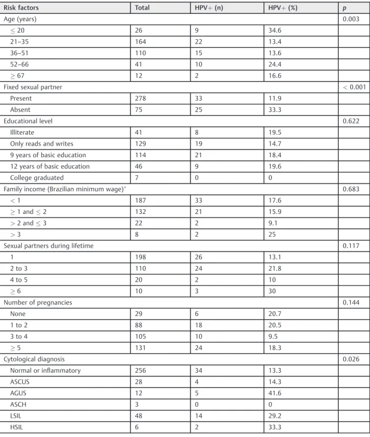
The overall prevalence of HPV infection was of 16.4% (58/353), whereas the prevalence of types 16 and 18 was of 2.3% (8/353) and 1.4% (5/353) respectively (including 1 case of co-infection by HPV-16 and HPV-18) (►Table 2). Human papillomavirus infection was most common among women aged 20 years or younger (34.6%), and among women aged 52 to 66 years (24.4%) (p¼0.003). A statistically significant association between the presence of HPV and the cytologi-cal results, with higher rates of infection in samples with intraepithelial lesions than in normal cytology results (p¼0.026), was evidenced (►Table 1).
The sociodemographic and behavioral variables, as well as the reproductive history surveyed, showed no statistically significant association with HPV infection, except among the women who did not have a steady sexual partner, for whom the infection rates were 3 times higher than among women with afixed sexual partner (33.3% versus 11% respectively) (p<0.01) (►Table 1). No association was found between infection by HPV-16 and HPV-18 and the variables investi-gated in the study (►Table 3).
Discussion
only accessible by river through small boats. Therefore, it is difficult for the inhabitants of these communities to access basic services such as health and education. Almost 70% of the women surveyed had never undergone the screening test
for CC. Although geographically isolated, an elevated HPV prevalence was observed (16.4%), which included HPV-16 and HPV-18, which are associated with 70% of the CC cases worldwide.12
Table 1 Sample characterization
Risk factors Total HPVþ(n) HPVþ(%) p
Age (years) 0.003
20 26 9 34.6
21–35 164 22 13.4
36–51 110 15 13.6
52–66 41 10 24.4
67 12 2 16.6
Fixed sexual partner <0.001
Present 278 33 11.9
Absent 75 25 33.3
Educational level 0.622
Illiterate 41 8 19.5
Only reads and writes 129 19 14.7
9 years of basic education 114 21 18.4
12 years of basic education 46 9 19.6
College graduated 7 0 0
Family income (Brazilian minimum wage)
0.683
<1 187 33 17.6
1 and2 132 21 15.9
>2 and3 22 2 9.1
>3 8 2 25
Sexual partners during lifetime 0.117
1 198 26 13.1
2 to 3 110 24 21.8
4 to 5 20 2 10
6 10 3 30
Number of pregnancies 0.144
None 29 6 20.7
1 to 2 88 18 20.5
3 to 4 105 10 9.5
5 131 24 18.3
Cytological diagnosis 0.026
Normal or inflammatory 256 34 13.3
ASCUS 28 4 14.3
AGUS 12 5 41.6
ASCH 3 0 0
LSIL 48 14 29.2
HSIL 6 2 33.3
Abbreviations: AGUS, atypical glandular cell of undetermined significance; ASCH, atypical squamous cell, cannot exclude HSIL; ASCUS, atypical squamous cell of undetermined significance; HPV, human papillomavirus; HSIL, high-grade squamous intraepithelial lesion; LSIL, low-grade intraepithelial lesion.
Note:
The prevalence of HPV infection demonstrated was simi-lar to the data found in other studies with regional commu-nities with similar characteristics, such as those who live in the towns of Tucuruí (14.2%)6and Abaetetuba (11.4%),13both in the state of Pará. However, the prevalence was lower than that of the indigenous tribes of the Amazon region belonging to Brazil (22.4% to 39.7%)14,15and Paraguay (23.2%).16Little is known about the prevalence of the high-risk HPVs -16 and -18 in these isolated communities in the Amazon, which makes it difficult to compare our results. In fact, to our knowledge, this is thefirst local study to assess the preva-lence of these specific viral types among riverside populations.
It is important to point out that most of the researches reported in the literature identify the rates of infection and
HPV types in a referenced demand of women who have lesions in the cervix, invasive or not, or women who seek healthcare services in order to undergo the Pap test, where there is a tendency to have higher rates of HPV infection and lesions of the uterine cervix associated with viral infection. The overall and specific prevalence of HPV demonstrated in this study was obtained from a spontaneous demand of women who received in their homes healthcare professio-nals and agreed to perform the Pap test. In Spain, a study with sampling features similar to the present one found a preva-lence of HPV lower than 3%.17
The bimodal distribution of HPV infection found in this investigation is in agreement with several studies in the literature.6,18,19Thefirst peak between women aged 20 years or younger may be explained by the greater quantity and Table 2 Status of HPV infection and viral type
Viral type Age (years)
20 21–35 36–51 52–66 67 Total
HPV negative 17 142 95 31 10 295
HPV overall 9 22 15 10 2 58
HPV-16 1 5 1 1 – 8
HPV-18 2 2 1 – – 5
HPV-16 and HPV-18 – 1 – – – 1
Abbreviations: HPV, human papillomavirus; HPV-16, human papillomavirus type 16; HPV-18, human papillomavirus type 18.
Table 3 G test of the association between selected risk factors and specific HPV types (16 and 18)
Risk factors HPVþ HPV-16þ % p HPV-18þ % p
Age (years) 0.59 0.35
20 9 1 11.1 2 22.2
21–35 22 5 22.7 2 9.1
36–51 15 1 6.7 1 6.7
52–66 10 1 10 0 0
67 2 0 0 0 0
Fixed sexual partner 0.73
Present 33 5 15 4 12
Absent 25 3 12 1 4
Cytological Results 0.82 0.77
Normal or inflammatory 33 3 9.1 2 6
Atypical cells 9 3 33.3 1 11.1
LSIL 14 2 14.3 2 14.3
HSIL 2 0 0 0 0
Age atfirst sexual intercourse (years) 0.68 0.07
15 24 3 12.5 4 16.6
16–20 26 4 15.4 1 3.8
21 3 1 33.3 0 0
turnover of sexual partners, the irregularity in the use of a contraceptive barrier, and the fragility of the uterine cervix, which is still in maturation in young women.2The elevated rates of infection in older women (aged between 52 and 66 years) is possibly associated with the gradual loss of immu-nity after menopause, which would facilitate the persistence of viral infection, or with new infections related to new sexual partners.20
The results also demonstrated that women who re-ported a fixed sexual partner had rates of infection 3 times smaller than women without afixed sexual partner (p<0.001). Similar data were found in a study conducted by Foliaki et al21, and they may be explained by limiting the primary route of viral transmission. Moreover, the association between HPV infection and the occurrence of multiple sexual partners is well established, mainly in women who reported having multiple sexual partners over the previous year.22 Additionally, the Pap test re-vealed an elevated number of inflammatory cases that can possibly be associated with concurrent sexually transmit-ted infections, like infections by Chlamydia trachomatis and Neisseria gonorrhoeae. As previously described, the inflammation caused by these pathogens can facilitate the infection by HPV.16,23Unfortunately, it was not possible to perform molecular tests for the detection of these patho-gens in the analyzed samples.
The combination of cytology and HPV screening used in the present study was effective in the identification of women with increased risk of developing CC. The use of primary high-risk HPV screening alone has been evaluated as an alternative to cytology-based CC screening methods in several countries, showing equivalent or superior effective-ness.24However, the use of primary high-risk HPV screening needs to be better evaluated in populations such as riverside communities, and the complementary screening through the Pap test is still essential due to the elevated number of abnormal cytology results and inflammatory cases.
Additionally, the main carcinogenic types -16 and -18 were found only in 24% of the positive cases, and the 2 cytological results of HSIL were not associated with these genotypes, which may reflect in possible limitations to HPV vaccination in this population. Moreover,70% of the participants underwent a Pap screening for thefirst time; the lack of access to these tests contributes to the high CC mortality rates found in Northern Brazil, and demonstrates the necessity of active search in this population.
A limitation of this study is related to the use of questionnaires to collect information on the risk factors of HPV infection. The statistical analyses were composed exclusively of information provided by the participants of the study, which may contain inaccuracies, omissions or lies for various reasons such as shame, fear or forgetful-ness. In addition, the cross-sectional design precluded the use of time as a factor of cause of viral infection, since the risk factors and outcomes were collected at the same time, and the bias of reverse causality could not be extinguished.
Conclusion
Human papillomavirus infections were more frequent among women with multiple sexual partners and those with an abnormal Pap test. A bimodal distribution of the infection according to the age of the participants, with afirst peak of infection among participants aged 20 years or younger, and a second peak among those aged between 52 and 66 years. There was no association between HPV-16 and HPV-18 and the risk factors evaluated in this study.
Despite the isolation of the study population from urban centers, a high prevalence of HPV infection was demonstrat-ed. Infections by HPV-16 and HPV-18 were not predominant. However, due to the oncogenic potential of these types and the elevated percentage of abnormal cytology results, there is an increased risk of CC development among riverside women. These observations highlight the importance of specific actions aimed at preventing transmission, as well as actions to promote the screening of cervical lesions among the riverside communities of the Amazon region.
Conflicts of Interest
The authors have no conflicts of interest to declare.
Authors’Contributions
DD performed the molecular identification of general and specific HPV, being primarily responsible for the analysis, interpretation of data, literature review and production of the manuscript. RV contributed to the draft of the manu-script, data interpretation, and the revision of thefinal version of the manuscript, providing suggestions. EB performed the cytological analysis of the samples and participated in the collection of the questionnaires. MP coordinated the collection of the samples and the applica-tion of the quesapplica-tionnaires. JM performed the molecular analysis of general and specific HPV, and helped in the interpretation of the data. MV was responsible for the statistical analysis of the research, and for the preparation offigures and tables. EI contributed to the analyses, data interpretation and critical review of the manuscript. HF contributed to the analyses, data interpretation and de-tection of HPV-16 and -18. MS was responsible for the study design, interpretation of data, and critical revision of the intellectual content of the manuscript. All authors read and approved thefinal draft of the manuscript.
Acknowledgments
We wish to thank the healthcare professionals and wo-men who participated in the study.
References
2 de Sanjosé S, Diaz M, Castellsagué X, et al. Worldwide prevalence and genotype distribution of cervical human papillomavirus DNA in women with normal cytology: a meta-analysis. Lancet Infect Dis 2007;7(07):453–459
3 Winer RL, Hughes JP, Feng Q, et al. Early natural history of incident, type-specific human papillomavirus infections in newly sexually active young women. Cancer Epidemiol Biomarkers Prev 2011;20(04):699–707
4 Moore MA, Tajima K. Cervical cancer in the asian pacifi c-epide-miology, screening and treatment. Asian Pac J Cancer Prev 2004; 5(04):349–361
5 Brasil. Ministério da Saúde. Instituto Nacional de Câncer José Alencar Gomes da Silva. Estimativa 2016: incidência do câncer no Brasil. Rio de Janeiro: INCA; 2016
6 Pinto DdaS, Fuzii HT, Quaresma JAS. Prevalence of genital HPV infection in urban and rural women in the Eastern Brazilian Amazon. Cad Saude Publica 2011;27(04):769–778
7 Vieira RC, Monteiro JdoS, Manso EP, et al. Prevalence of type-specific HPV among female university students from northern Brazil. Infect Agent Cancer 2015;10:21
8 Brasil. Ministério da Saúde. Secretaria de Atenção à Saúde. Instituto Nacional de Câncer. Nomenclatura brasileira para laudos cervicais e condutas preconizadas. 3a ed. Rio de Janeiro: INCA; 2012
9 Manos MM, Ting Y, Wright DK, Lewis AJ, Broker TR, Wolinsky SM. The use of polymerase chain reaction amplification for the detection of genital human papillomaviruses. Cancer Cells 1989;7:209–214
10 Gage JC, Partridge EE, Rausa A, et al. Comparative performance of human papillomavirus DNA testing using novel sample collection methods. J Clin Microbiol 2011;49(12):4185–4189
11 Ayres M, Ayres Júnior M, Ayres DL, dos Santos ASS. BioEstat 5.3 [Internet]. Tefé: Instituto de Desenvolvimento Sustentável Ma-mirauá; 2011 [cited 2016 Mar 22]. Available at: http://www. mamiraua.org.br/pt-br/downloads/programas/bioestat-versao-53/
12 Muñoz N, Bosch FX, Castellsagué X, et al. Against which human papillomavirus types shall we vaccinate and screen? The inter-national perspective. Int J Cancer 2004;111(02):278–285
13 Duarte DV, Brito EB, Canto ASS, et al. Frequência e genotipagem do Papilomavírus humano em mulheres de comunidades ribeirinhas do Município de Abaetetuba, Pará, Brasil. Rev Pan-Amaz Saúde 2010;1(03):75–82
14 Brito EB, Silva ID, Stávale JN, Taromaru E, Menezess RC, Martins SJ. Amerindian women of the Brazilian Amazon and STD. Eur J Gynaecol Oncol 2006;27(03):279–281
15 Fonseca AJ, Taeko D, Chaves TA, et al. HPV infection and cervical screening in socially isolated indigenous women inhabitants of the Amazonian rainforest. PLoS One 2015;10(07):e0133635 16 Mendoza L, Mongelos P, Paez M, et al. Human papillomavirus and
other genital infections in indigenous women from Paraguay: a cross-sectional analytical study. BMC Infect Dis 2013;13:531 17 de Sanjose S, Almirall R, Lloveras B, et al. Cervical human
papillomavirus infection in the female population in Barcelona, Spain. Sex Transm Dis 2003;30(10):788–793
18 Dursun P, Senger SS, Arslan H, Kuşçu E, Ayhan A. Human
papillo-mavirus (HPV) prevalence and types among Turkish women at a gynecology outpatient unit. BMC Infect Dis 2009;9:191 19 Lazcano-Ponce E, Herrero R, Muñoz N, et al. Epidemiology of HPV
infection among Mexican women with normal cervical cytology. Int J Cancer 2001;91(03):412–420
20 Molano M, Posso H, Weiderpass E, et al; HPV Study Group HPV Study. Prevalence and determinants of HPV infection among Colombian women with normal cytology. Br J Cancer 2002; 87(03):324–333
21 Foliaki S, Brewer N, Pearce N, et al. Prevalence of HPV infection and other risk factors in a Fijian population. Infect Agent Cancer 2014;9:14
22 Dunne EF, Unger ER, Sternberg M, et al. Prevalence of HPV infection among females in the United States. JAMA 2007; 297(08):813–819
23 Shew ML, Fortenberry JD, Tu W, et al. Association of condom use, sexual behaviors, and sexually transmitted infections with the duration of genital human papillomavirus infection among adoles-cent women. Arch Pediatr Adolesc Med 2006;160(02):151–156 24 Huh WK, Ault KA, Chelmow D, et al. Use of primary high-risk