Iezzi LE, Medeiros BA, Feitosa MR, Almeida ALNR, Parra RS, Rocha JJR, Feres O. Association between celiac disease and Crohn’s disease – a challenge to the coloproctologist. J Coloproctol, 2012;32(3): 329-333.
ABSTRACT: Over the past few years, many studies on the association between celiac disease and inlammatory bowel disease have been re -ported. The genetic origin of this association has prompted research that searches for a common link for the concomitant manifestation of these pathologies. Clinical studies aim not only to demonstrate this relation, but also to establish the epidemiological frequencies among affected individuals and their relatives as compared to the general population. The similar clinical symptoms, dificulties, diagnoses, and therapeutics are still a challenge, since this association is unknown to most coloproctologists, thereby culminating in treatments and surgical procedures with no beneits for the patient.
Keywords: celiac disease; Crohn’s disease; proctocolitis; association.
RESUMO: Nos últimos anos, muitos estudos foram relatados sobre a associação entre a doença celíaca e as doenças inlamatórias intestinais. A origem genética dessa associação desperta pesquisas que buscam o elo comum para a manifestação concomitante das patologias. Estudos clínicos visam não apenas demonstrar essa relação mas também estabelecer as frequências epidemiológicas entre os indivíduos acometidos e seus familiares em relação à população geral. À semelhança do quadro clínico, as diiculdades diagnósticas e terapêuticas são ainda desaios, já que tal associação ainda é desconhecida para a maioria dos coloproctologistas, podendo resultar em tratamentos e cirurgias sem benefícios ao paciente.
Palavras-chave: doença celíaca; doença de Crohn; proctocolite; associação.
Association between celiac disease and Crohn’s disease – a challenge
to the coloproctologist
Leonardo Estenio Iezzi
1, Bruno Amaral Medeiros
1, Marley Ribeiro Feitosa
1, Ana Luiza Normanha Ribeiro de Almeida
2,
Rogério Seraim Parra
2, José Joaquim Ribeiro da Rocha
3, Omar Feres
31
Doctor, Sector of Coloproctology, Hospital das Clínicas, Medical School of Ribeirão Preto, Universidade de São
Paulo (USP) – Ribeirão Preto (SP), Brazil;
2Assistant Doctor and Post-Graduate Student, Sector of Coloproctology,
Hospital das Clínicas, Medical School of Ribeirão Preto, USP – Ribeirão Preto (SP), Brazil;
3Doctor Professor, Sector of
Coloproctology, Hospital das Clínicas, Medical School of Ribeirão Preto, USP – Ribeirão Preto (SP), Brazil.
Study carried out at the Hospital das Clínicas, Medical School of Ribeirão Preto, Universidade de São Paulo (USP) – Ribeirão Preto (SP), Brazil. Financing source: none.
Conlict of interest: nothing to declare.
Submitted on: 07/01/2011 Approved on: 07/15/2011
INTRODUCTION
The celiac disease (CD) is characterized as an
autoimmune disease caused by the permanent gluten
intolerance in genetically susceptible individuals
1.
In the past few years, its clinical and
ethiopathogen-ic features have been cleared. Nowadays, it is known
that in order for it to occur the association of three
factors is determinant: genetic changes, exposure to
gluten and altered immune response
3.
Histocompatibility antigens class II,
HLA-DQ2 and HLA-DQ8 present changes in celiac
pa-tients
3-5. When these subjects are exposed to gluten,
their immune response is exaggerated, with increase
in intraepithelial T lymphocytes – in the proximal
intestine mucosa – and inlammatory cytokines,
leading to the villous atrophy and poor absorption
of nutrients
6. Excluding gluten from the diet usually
clinical variants of the disease and its intensity, so
the onset of early symptoms can occur during
child-hood or adultchild-hood
8,9. Thus, CD its the differential
diagnosis of other pathologies that present pain and
abdominal distension, vomit, iron deiciency and
malnutrition
10.
In adult subjects, the diagnosis of CD should
include detailed anamnesis, serological and
histo-pathological studies
11. However, in this age group
the disease can be subclinical, and diagnosis can be
late – sometimes, after surgical approaches, since it
seems like a picture of acute abdomen, or after other
therapies that provided no beneits to the patient
12,13.
The immunological characteristics of CD establish a
relation with other autoimmune conditions, such as
dermatitis herpetiformis, thyroid diseases, Addison’s
disease, autoimmune thrombocytopenia, sarcoidosis,
IgA nephropathy and selective IgA deiciency
14-16.
Approximately 2 to 4% of the insulin dependent
pa-tients with diabetes mellitus present with CD
17.
The relation with other autoimmune diseases
led to studies on the association between CD and
inlammatory bowel diseases (IBD) in the past few
years (proctocolitis and Crohn’s disease)
18-21.
Crohn’s disease is more common in the
termi-nal ileum. It presents segmental lesions in the
diges-tive tract, affecting all the layers of the organ wall
and possibly leading to stenosis or istula. Procto
-colitis is the IBD more commonly associated with
other autoimmune diseases, such sclerosing
cholan-gitis, and other vasculitis. Anemia and malnutrition
are systemic repercussions that are present in most
patients with the disease. The iron-deiciency ane
-mia, which is refractory in patients with IBD, should
lead to the suspicion of associated CD
22.
Many clinical studies and case reports have
been described in the past few years to relate CD
and IBD. The prevalence of IBD in CD has been
described as ive to ten times higher in the general
population
23. Shah et al. described a risk of
procto-colitis ive times higher relative in irst-degree rela
-tives of patients with CD
24. Likewise, Cottone et al.
described the high incidence of proctocolitis in 600
irst-degree relatives of patients with CD
25.
Between January 2002 and December 2004,
the Italian Group of Inlammatory Bowel Diseases
performed a multicentric study aiming to establish
the prevalence of IBD among celiac patients. Out
of the 1,711 patients, 9 (0.5%) serological and
his-tological results that were compatible with the CD
diagnosis – 6 patients presented with proctocolitis
and three with Crohn’s disease, lower prevalence in
comparison to the general population
26.
In 2007, during 1 year period, Masachs et al.
followed-up three groups of celiac patients, their
irst-degree relatives and a control group, in order to
identify the prevalence of IBD in celiac patients and
their relatives. Three cases of Crohn’s disease were
reported in 86 celiac patients; four cases of Crohn’s
disease in irst-degree relatives; and one case in the
control group (809 people); no case of proctocolitis
was reported, which led to the conclusion that celiac
patients and their irst-degree relatives have higher
chances of having Crohn’s disease if compared to
the general population
27.
Lopez-Vasquez et al. identiied mutations in
the A MICA gene (major histocompatibility
com-plex class I chain related gene A) expressed in the
gastrointestinal epithelium of patients with IBD
28.
Changes in the MYOIXB gene, responsible for the
production of myosins and for the intestinal
epithe-lial integrity, are found in celiac patients
29and these
mutations can also be found in 40% of the patients
with IBD
30,31.
The presence of binding regions shared by
these diseases, such as 5q31-33 (IBD5 and
CELI-AC2) and 19p13 (IBD6 and CELIAC4)
32,33has
sup-ported this idea. Recently, the description of
poly-morphisms in the genes IL2, region IL21, in 4q27
and in the gene IL18RAP, in 2q12, reinforces the
evidence of association between CD and IBD.
Be-sides, the well established relation between Crohn’s
disease and the gene IL23R
34has been associated
to CD in the Finn
35and Spanish
36population. Based
on these indings, Dema et al.37 studied the genes
NKX2-3, IRGM and ATG16L1, whose changes are
clearly deined in the IBDs, in patients with CD and
irst-degree relatives; however, no evidence has been
established among celiac patients.
In the service of
Hospital das Clínicas de
Ribeirão Preto
(HCRP) at
Universidade de São Paulo
,
we had a case of association between CD and Crohn’s
disease. The patient was 37 years old at the time, and
pain and abdominal distension, vomit, weight loss
(10 kg in 3 months) and refractory anemia to
treat-ments prescribed in other medical services.
There had been an intestinal subocclusion
(ferring to enterectomy), however, there were no
ports concerning the removed piece. She had the
re-port of an upper digestive endoscopy (UDE) with a
duodenal biopsy compatible with CD. However, up
until then she had not been advised to rule out
glu-ten from her diet. We repeated the UDE with new
biopsies of the second part of the duodenum, and
the CD diagnosis was conirmed. Admitted to our
nursery for clinical and nutritional rehabilitation,
her picture of intestinal occlusion got worse, and
trafic demonstrated areas of stenosis and dilatation
of the small intestine (Figure 1). After being
sub-mitted to exploratory laparotomy, segments of the
jejunum and ileum were shown with stenosis and
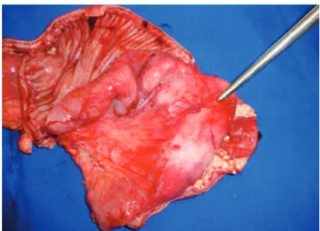
dilatation (Figure 2). The exploration of the
surgi-cal piece demonstrated an inlammatory iniltrate in
the mesentery, intestinal wall thickening with ulcers
and ibrin deposition (Figures 3 and 4). The surgi
-cal piece was analyzed twice at the Department of
Pathological Anatomy at HCRP and the diagnosis
of Crohn’s disease was conirmed. At the postop
-erative, we initiated nutritional guidance and
sus-pended gluten from the diet. Thirty days after the
surgery, we started treating the patient for Crohn’s
disease. An immunomodulator was prescribed and,
nowadays, the patient had signiicant clinical im
-provement, without new intercurrences.
Figure 1. Bowel transit showing areas of stenosis and small intestine dilatation.
Figure 2. Stenoses and dilatations.
Figure 3. Inlammatory iniltrate in the mesentery.
Figure 4. Ulcers and ibrin deposition.
REFERENCES
1. Troncone R, Bhatnagar S, Butzner D, Cameron D, Hill I, Hoffenberg E, Maki M, Mendez V, de Jimenez MZ; European Society for Paediatric Gastroenterology, Hepatology and Nutrition. Celiac disease and other immunologically mediated disorders of the gastrointestinal tract: Working Group report of the second World Congress of Pediatric Gastroenterology, Hepatology, and Nutrition. J Pediatr Gastroenterol Nutr 2004;39 Suppl 2:S601-10.
2. Baptista ML. Doença celíaca: uma visão contemporânea. Pediatria (São Paulo) 2006;28(4):262-71.
3. Ciclitira PJ, King AL, Fraser JS. AGA technical review on Celiac Sprue. American Gastroenterological Association. Gastroenterol 2001;120(6):1526-40. Erratum in: Gastroenterology 2001;121(1):234. Comment in: Gastroenterology. 2002;122(1):246-7.
4. Farré C, Humbert P, Vilar P, Varea V, Aldeguer X, Carnicer J, et al. Serological markers and HLA-DQ2 haplotype among irst-degree relatives of celiac patients. Catalonian Coeliac Disease Study Group. Dig Dis Sci 1999;44(11):2344-9. 5. Sollid LM, Thorsby E. HLA susceptibility genes in
celiac disease: genetic mapping and role in pathogenesis. Gastroenterology 1993;105(3):910-22.
6. Sollid LM. The molecular basis of coeliac disease. Annu Rev Immunol 2000;18:53-81.
7. Stern M, Ciclitira PJ, van Eckert R, Feighery C, Janssen FW, Méndez E, et al. Analysis and clinical effects of gluten in coeliac disease. Eur J Gastroenteol Hepatol 2001;13(6):741-7. 8. Martucci S, Biagi F, Di Sabatino A, Corazza GR. Coeliac
disease. Digest Liver Dis 2002;34 Suppl 2:S150-3.
9. Catassi C, Rätsch IM, Fabiani E, Rossini M, Bordicchia F, Candela F, et al. Coeliac disease in the year 2000: exploring the iceberg. Lancet 1994;343(8891):200-3.
10. Polanco I. Enfermedad celíaca. Pediatria Integral 1995;1(2):124.
11. Fasano A, Catassi C. Current approaches to diagnosis and treatment of celiac disease: An evolving spectrum. Gastroenterol 2001;120(3):636-51.
12. Logan Rf, Rifkind EA, Turner ID, Ferguson A. Mortality in celiac disease. Gastroenterology 1989;97(2):265-71.
13. Corrao G, Corazza GR, Bagnardi V, Brusco G, Ciacci C,
Cottone M, Sategna Guidetti C, Usai P, Cesari P, Pelli MA, Loperido S, Volta U, Calabró A, Certo M; Club del Tenue Study Group. Mortality in patients with celiac disease and their relatives: a cohort study. Lancet. 2001;358(9279):356-61. 14. Holmes GKT. Coeliac disease and type 1 diabetes mellitus –
the case for screening. Diabet Med 2001;18(3):169-77. 15. Arvola T, Mustalahti K, Saha MT, Vehmanen P, Partanen J,
Ashorn M. Celiac disease, thyrotoxicosis and autoimmune hepatitis in a child. J Pediatr Gastroenterol Nutr 2002;35(1):90-2.
16. Kaukinen K, Collin P, Mykkänen AH, Partanen J, Mäki M, Salmi J.. Celiac disease and autoimmune endocrinologic disorders. Dig Dis Sci. 1999;44(7):1428-33..
17. Savilahti E, Simell O, Koskimies S, Rilva A, Akerblom HK. Coeliac disease in insulin-dependent diabetes mellitus. J Pediatr. 1986;108(5 Pt 1):690-3.
18. Cheikh I, Maamouri N, Chouaib S, Chaabouni H, Ouerghi H, Ben Ammar A. [Association of celiac disease and Crohn’s disease. A case report]. Tunis Med 2003;81(12):969-71. Article in French.
19. Chakraborty A, Bremner AR, Moore I, Beattie RM. Coeliac disease and Crohn’s disease: an association not to be forgotten. Hosp Med 2003;64(11):684-5.
20. Schedel J, Rockmann F, Bongartz T, Woenckhaus M, Schölmerich J, Kullmann F. Association of Crohn’s disease and latent celiac disease: a case report and review of the literature. Int J Colorectal Dis 2005;20(4):376-80.
21. Tursi A, Giorgetti GM, Brandimarte G, Elisei W. High prevalence of celiac disease among patients affected by Crohn’s disease. Inlamm Bowel Dis 2005;11(7):662-6. 22. Takei N, Mukai Y, Hasegawa Y, Suzukawa K, Nagata M,
Noguchi M, et al. Refractory iron deiciency anemia as the primary clinical manifestation of celiac disease. Ann Hematol 2003;82:53.
23. Gillberg R, Dotevall G, Ahren C. Chronic inlammatory bowel disease in patients with coeliac disease. Scand J Gastroenterol 1982;17(4):491-6.
24. Shah A, Mayberry JF, Williams G, Holt P, Loft DE, Rhodes J. Epidemiological survey of coeliac disease and inlammatory bowel disease in irst degree relatives of coeliac patients. Q J Med 1990;74(275):283-8.
25. Cottone M, Marrone C, Casà A, Oliva L, Orlando A, Calabrese
CONCLUSION
In the past few years, with the advances in
di-agnostic methods and molecular evaluations, the
analysis between diseases whose ethiopathogens
in-volve genetic changes and autoimmune mechanisms
has improved. Thus, we can cite the studies that
tried to relate CD and IBDs. It is important to
re-member that similarities are not restricted to
E, et al. Familial occurrence of inlammatory bowel disease in celiac disease. Inlamm Bowel Dis 2003;9(5):321-32. 26. Casella G, D’Incà R, Oliva L, Daperno M, Saladino V,
Zoli G, Annese V, Fries W, Cortellezzi C; Italian Group – IBD. Prevalence of celiac disease in inlammatory bowel diseases: an IG-IBD multicentre study. Dig Liver Dis 2010;42(3):175-8.
27. Masachs M, Casellas F, Malagelada JR. Enfermedad inlamatoria intestinal en pacientes celíacos. Rev Esp Enferm Dig. 2007;99(8):446-50.
28. Lopez-Vazquez A, Rodrigo L, Fuentes D, Riestra S, Bousoño C, Garcia-Fernandez S, et al. MHC class I chain related gene A (MICA) modulates the development of celiac disease in patients with the high risk heterodimer DQA1 0501/DQB1 0201. Gut 2002;50(3):336-40.
29. Monsuur AJ, de Bakker PI, Alizadeh BZ, Zhernakova A, Bevova MR, Strengman E, et al. Myosin IXB variant increases the risk of celiac disease and points toward a primary intestinal barrier defect. Nat Genet 2005;37(12):1341-4. 30. van Bodegraven AA, Curley CR, Hunt KA, Monsuur AJ,
Linskens RK, Onnie CM, et al. Genetic variation in myosin IXB is associated with ulcerative colitis. Gastroenterology 2006;131(6):1768-74.
31. Latiano A, Palmieri O, Valvano MR, D’Incà R, Caprilli R, Cucchiara S, et al. The association of MYO9B gene in Italian patients with inlammatory bowel diseases. Aliment Pharmacol Ther 2008;27(3):241-8.
32. Rioux JD, Silverberg MS, Daly MJ, Steinhart AH, McLeod RS, Grifiths AM, et al. Genomewide search in Canadian families with inlammatory bowel disease reveals two novel susceptibility loci. Am J Hum Genet 2000;66(6):1863-70.
33. Van Belzen MJ, Meijer JW, Sandkuijl LA, Bardoel AF, Mulder CJ, Pearson PL, et al. A major non-HLA locus in celiac disease maps to chromosome 19. Gastroenterology 2003;125(4):1032-41.
34. Duerr RH, Taylor KD, Brant SR, Rioux JD, Silverberg MS, Daly MJ, et al. A genome-wide association study identiies IL23R as an inlammatory bowel disease gene. Science. 2006;314(5804):1461-3..
35. Einarsdottir E, Koskinen LL, Dukes E, Kainu K, Suomela S, Lappalainen M, et al. IL23R in the Swedish, Finnish, Hungarian and Italian populations: association with IBD and psoriasis, and linkage to celiac disease. BMC Med Genet 2009;10:8
36. Nunez C, Dema B, Cenit MC, Polanco I, Maluenda C, Arroyo R, et al. IL23R: a susceptibility locus for celiac disease and multiple sclerosis? Genes Immun 2008;9(4):289-93.
37. Dema B, Fernández-Arquero M , Maluenda C, Polanco I, Figueredo M, de la Concha EG, et al. Lack of association of NKX2-3, IRGM, and ATG16L1 inlammatory bowel disease susceptibility variants with celiac disease. Human Immunology 2009;70(11):946-9.
Correspondence to:
Omar Feres
Divisão de Coloproctologia do Departamento de Cirurgia e Anatomia da Faculdade de Medicina de Ribeirão Preto da Universidade de São Paulo