Chloroaluminium phthalocyanine polymeric nanoparticles as photosensitisers:
Photophysical and physicochemical characterisation, release and phototoxicity
in vitro
Carina Silva de Paula
a,b, Antonio Cláudio Tedesco
c, Fernando Lucas Primo
c, José Mário Carneiro Vilela
d,
Margareth Spangler Andrade
d, Vanessa Carla Furtado Mosqueira
a,b,e,⇑aPrograma de Pós-Graduação em Nanotecnologia Farmacêutica, Universidade Federal de Ouro Preto, Campus Universitário Morro do Cruzeiro, Ouro Preto, MG 35400-000, Brazil bPrograma de Pós-Graduação em Ciências Biológicas, Universidade Federal de Ouro Preto, Campus Universitário Morro do Cruzeiro, Ouro Preto, MG 35400-000, Brazil
cDepartamento de Química, Centro de Nanotecnologia e Engenharia de Tecidos, Laboratório de Fotobiologia e Fotomedicina, Faculdade de Filosofia, Ciências e Letras de Ribeirão Preto, Universidade de São Paulo, 14040-901 Ribeirão Preto-SP, Brazil
dFundação Centro Tecnológico de Minas Gerais – CETEC, Avenida José Cândido da Silveira, 2000, Belo Horizonte, MG 31170-000, Brazil
eDepartamento de Farmácia, Escola de Farmácia, Universidade Federal de Ouro Preto, Rua Costa Sena, 171 Centro, Ouro Preto, MG 35400-000, Brazil
a r t i c l e
i n f o
Article history:
Received 28 June 2012
Received in revised form 17 March 2013 Accepted 20 March 2013
Available online 29 March 2013
Keywords:
Photodynamic therapy Polymeric nanoparticles Chloroaluminium phthalocyanine Photophysical characterisation Release kinetic
Phototoxicityin vitro
a b s t r a c t
Nanoparticles of poly(D,L-lactide-co-glycolide), poly(D,L-lactide) and polyethylene glycol-block-poly(D,L-lactide) were developed to encapsulate chloroaluminium phthalocyanine (AlClPc), a new hydrophobic photosensitiser used in photodynamic therapy (PDT). The mean nanoparticle size varied from 115 to 274 nm, and the encapsulation efficiency ranged from 57% to 96% due to drug precipitation induced by different types of polymer. All nanoparticle formulations presented negative zeta potential values (–37 mV to –59 mV), explaining their colloidal stability. The characteristic photophysical param-eters were analysed: the absorption spectrum profile, fluorescence quantum yield and transient absorbance decay, with similar values for free and nanoparticles of AlClPc. The time-resolved spectros-copy measurements for AlClPc triplet excited state lifetimes indicate that encapsulation in nanocapsules increases triplet lifetime, which is advantageous for PDT efficiency. A sustained release profile over 168 h was obtained using external sink method. Anin vitrophototoxic effect higher than 80% was observed in human fibroblasts at low laser light doses (3 J/cm2) with 10
lM of AlClPc. The AlClPc loaded within poly-meric nanocapsules presented suitable physical stability, improved photophysical properties, sustained released profile and suitable activityin vitroto be considered a promising formulation for PDT.
Ó2013 Elsevier B.V. All rights reserved.
1. Introduction
Photodynamic therapy (PDT) is an innovative and attractive modality for the treatment of oncologic disease (Allison and Sibata, 2010). The technique involves the topical or systemic administra-tion of a photosensitiser (PS) that should concentrate more in tumour tissues than in normal tissues, followed by illumination of the tumour with visible light in a wavelength range matching the absorption spectrum of the photosensitiser (Allison and Sibata, 2010; Konan et al., 2002).
The resulting photodynamic reactions give rise to singlet oxy-gen (1O
2) and to other active oxygen species that lead to tumour
destruction. The PDT efficacy could be improved with the use of
photosensitisers that strongly absorb red light above 650 nm, where tissue exhibits optimal transparency. To this end, several new classes of potential sensitisers for PDT have been developed; among these, phthalocyanines have been found to be highly prom-ising because of their high absorbance coefficient in the region of 650–680 nm (Konan et al., 2003; Sibata et al., 2004). Phthalocya-nines can be chelated with a variety of metals, usually aluminium and zinc because these diamagnetic metals enhance their photo-toxicity (Kluson et al., 2009).
Chloroaluminium phthalocyanine (AlClPc) is a photosensitiser
with adequate photophysical properties for PDT (Nunes et al.,
2004) (Fig. 1). Unfortunately, AlClPc is insoluble in water and bio-logically compatible solvents, which renders its systemic adminis-tration problematic and restricts possible medical applications (Nunes et al., 2004). To overcome this limitation, phthalocyanines have been associated to cyclodextrines (Silva et al., 2011) and to colloidal carriers such as liposomes (Nunes et al., 2004; Rocha et al., 2012), nanoemulsions (Rodrigues et al., 2012) and
0928-0987/$ - see front matterÓ2013 Elsevier B.V. All rights reserved. http://dx.doi.org/10.1016/j.ejps.2013.03.011
⇑ Corresponding author. Address: Escola de Farmácia-Universidade Federal de Ouro Preto, R. Costa Sena, 171 Centro CEP 35400-000 Ouro Preto, Minas Gerais, Brazil. Tel.: +55 31 35 59 10 32; fax: +55 31 35 59 16 28.
E-mail addresses: [email protected], [email protected] (V.C.F. Mosqueira).
Contents lists available atSciVerse ScienceDirect
European Journal of Pharmaceutical Sciences
nanocapsules (Moura-Siqueira et al., 2010; Oliveira et al., 2011). Polymeric nanoparticles (NPs) act as a drug vehicle to specifically target tumour tissues or cells in order to reduce the toxicity effect on normal tissue through the enhanced permeation and retention (EPR) effect (Maeda, 2010). Nanocapsules (NCs) are a carrier of choice for the intravenous administration of highly lipophilic drugs because they can dissolve or disperse drugs with high payload (Legrand et al., 1999). In NC, the oily nanodroplets are surrounded by a biodegradable polymeric wall that can be chemically modified to prolong blood circulation time or to improve the targeting properties of the carrier (Mosqueira et al., 2001b). To reduce undesirable uptake of NP by phagocytes, nanospheres (NSs) and NC were surface-modified with hydrophilic polymers such as polyethylene glycol (PEG) (Gref et al., 1994; Mosqueira et al., 2001a). The PEG corona on the NP surface decreased recognition by the MPS, which thereby increased the half-life of NC circulation in the blood (Mosqueira et al., 2001a, 2001b).
Based on these findings, the objective of this work was the development of formulations of AlClPc-loaded NC and NS prepared from different polymers. The AlClPc was associated with poly(D,L
-lactide-co-glycolide) (PLGA) or poly-D,L-lactide (PLA) to prepare
conventional NC or NS and with the copolymer monomethoxy-polyethylene glycol-co-poly-D,L-lactide (PLA-PEG) to prepare NC
with prolonged blood circulation time. The effect of the formula-tion parameters on the photophysical and physicochemical proper-ties was investigated. Furthermore, the influence of these variables on release kinetics and on phototoxicityin vitrowas studied. This is the first report of the development of AlClPc photosensitiser in polymeric nanoparticles with a detailed investigation of its photo-physical and physicochemical properties.
2. Materials and methods
2.1. Drugs and reagents
Chloro(29H,31H-phthalocyaninato)aluminium (AlClPc) was
purchased from Aldrich Chemical Company Inc. (Milwaukee, WI, USA). Poly(D,L-lactide) (PLA, average Mw 42,000 g/mol) was
pro-vided by Phusis (France). Poly(D,L-lactide-co-glycolide) (85:15)
(PLGA) and Poloxamer 188 (a non-ionic surfactant) were provided by Sigma–Aldrich (USA). PLA-PEG diblock copolymer (average Mw of 66,000 g/mol with approximately 7.5% PEG at a Mw of 5000 g/ mol) was provided by Alkermes (USA). Epikuron 170 (70% soy phosphatidylcholine) was purchased from Lucas Meyer (France).
Miglyol 810N was a kind gift from Hulls (Germany). Polyethylene glycol (PEG 300) was provided by Synth (Brazil). Dimethylacet-amide (DMA) was purchased from Vetec (Brazil). All of the solvents used were analytical grade, and other chemicals were commer-cially available reagent grade and were used without further puri-fication. Milli-QÒ
water (Simplicity 185, Millipore, Bedford, USA) was used throughout. All experiments were carried out with stock solutions of AlClPc prepared in UV/HPLC-grade ethanol and stored in the dark at 4°C. The concentrations of phthalocyanine were as-sayed by HPLC (Waters Alliance 2695) with fluorescence detection (Waters 2475), using a 150 mm4.6 mm, 4.6
l
m particle size C18 Gemini Phenomenex column protected by a Phenomenex securityguard AJO 7597 C18 column (2 mm4.6 mm, 3
l
m) as thesta-tionary phase.
2.2. Preparation of AlClPc nanocapsules
Conventional nanocapsules were obtained by interfacial poly-mer deposition following solvent displacement, which was
previ-ously described byFessi et al. (1989)and was modified here to
include AlClPc in the formulation. Conventional NC were prepared using the polymers PLA and PLGA. Briefly, 60 mg of polymer was dissolved in 10 ml of organic solution composed of (0–70%) etha-nol/acetone containing 75 mg of Epikuron 170, 250
l
l of Miglyol 810N and AlClPc (0.1–0.2 mg/ml). This organic solution was poured into 20 ml of external aqueous phase containing 75 mg of Poloxamer 188 under magnetic agitation. The solvents were evap-orated under reduced pressure (Laborota 4000, Heidolph Instru-ments, Germany) until a final volume of 10 ml was attained. Surface-modified NC were prepared using the diblock polymer of PLA-PEG without Poloxamer surfactant. Briefly, the polymer (75 mg) was dissolved in 10 ml of organic solution composed of (0–70%) ethanol/acetone containing 75 mg of Epikuron 170, 250l
l of Miglyol 810 N and AlClPc (0.1–0.2 mg/ml). This organic solution was poured into the external aqueous phase (20 ml). The solvents were evaporated under reduced pressure to a volume of 10 ml (Mosqueira et al., 2001a). After 24 h, the NC suspension was filtered through a 0.8l
m sterile filter (NalgeneÒ) to remove precipitated drug crystals that were not encapsulated. Nano-spheres were prepared using the PLA polymer in the same way as described above but for the absence of Epikuron 170 and Miglyol 810N.
2.3. Nanocapsule characterisation
2.3.1. Zeta potential and size
The mean size and size distribution of the NC were determined by photon correlation spectroscopy using an N5-Submicron Parti-cle Size Analyser (Beckman Coulter, Florida, US). This method al-lows the determination of the mean diameter of the particle (hydrodynamic radius) and the polydispersity index (PI) of the par-ticle population, which is a dimensionless measure of the broad-ness of the particle size distribution. Samples were analysed after appropriate dilution in ultra-pure Milli-Q water. The reported val-ues were expressed as the means ± standard deviations of at least three different batches of each nanocapsule formulation. The zeta potential was determined by Laser Doppler Anemometry (LDA) using the Zetasizer 3000 HS (Malvern Instruments, UK) after 250-fold dilution of the colloidal suspension in 1 mM NaCl. The conductivity of all NC dilutions was maintained close to 120
l
S/ cm. The measurements were performed at a constant pH of 7.1, as determined by the instrument.2.3.2. Atomic force microscopy (AFM)
To examine the NC morphology, a 5
l
l sample droplet wasdeposited on a freshly cleaved mica surface and then it was spread
and dried by argon flow. The measurements were performed at room temperature and in air on a Dimension 3000 and with Mul-timode Equipment; both instruments were monitored by a Nano-scope IIIa controller from Digital Instruments (Santa Barbara, CA).
The images were obtained intapping modeusing commercial
sili-con probes from Nanosensors with cantilevers of 228
l
m length,resonance frequencies of 75–98 kHz, spring constants of 3.0– 7.1 N/m and a nominal tip radius of curvature of 8 nm. The ‘‘scan rate’’ used was 1 Hz. Dimensional analyses were carried out using the ‘‘section of analyses’’ application on the system. The values rep-resent the mean ± standard deviation derived from approximately 40 particle measurements.
2.3.3. Determination of AlClPc encapsulation
A determination of AlClPc content was performed using a meth-odology previously described and validated for the determination
of chloroaluminium phthalocyanine in nanocarriers (Oliveira
et al., 2011). The system consisted of HPLC (Waters Alliance 2695) with fluorescence detection (Waters 2475) using a
150 mm4.6 mm and 4.6
l
m particle size C18 GeminiPhenome-nex column protected by a PhenomePhenome-nex security guard AJO 7597
C18 column (2 mm4.6 mm, 3
l
m) as a stationary phase andmethanol/acetone/dimethylformamide (90:5:15 v/v/v) as the mo-bile phase at a flow rate of 1 ml/min. The samples were injected using the HPLC autosampler at an injection volume of 10
l
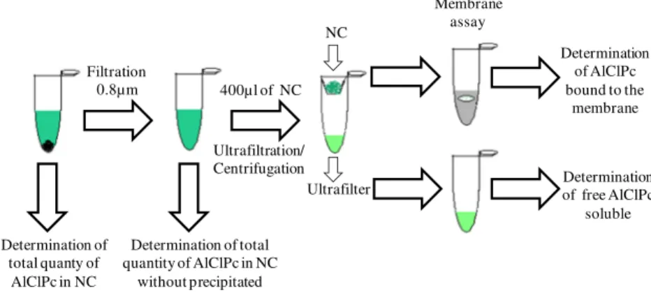
l. The elution was monitored by a fluorescence detector via excitation and emission at 610 nm and 675 nm, respectively, and the quanti-fication limit was 1.12 ng/ml.The encapsulation yield in the nanoparticles was calculated by the difference between the total quantity of drug in the final colloi-dal suspension after filtration in a filter syringe (Filter syringe, 0.8
l
m, NalgeneÒ) minus the free drug in the external aqueous phase divided by the total quantity of drug in the colloidal suspen-sion after filtration in the filter syringe100 (Eq.(1)). The encap-sulation yield, or drug loading, in the nanoparticles was determined from Eq.(1), where A = concentration:
Encapsulation yieldð%Þ ¼A
total
Aultrafiltrate100
Atotal in NP suspension ð1Þ The encapsulation efficiency in the nanoparticles was calculated as the amount of drug truly encapsulated per ml divided by the
to-tal drug weight needed to prepare 1 ml of NP formulation100
(Eq.(2)). In this way, encapsulation efficiency takes into account the drug losses during the encapsulation process:
Encapsulation efficiencyð%Þ ¼A
total
Aultrafiltrate100
Aweight ð2Þ
whereAtotal in NP suspensionis the total quantity of drug in the final
col-loidal suspension after filtration in the filter syringe,Aultrafiltrateis
the free drug in the external aqueous phase andAweightis the total drug weight needed to prepare the NP formulation.
The percentage of precipitated drug was calculated by the dif-ference between the drug content/ml in the total suspension minus the drug content/ml in the suspension after filtration by a 0.8
l
m filter divided by the drug content/ml in the total suspension100 as shown inFig. 2.Total AlClPc in the colloidal suspension was determined by full dissolution of 30
l
l in 970l
l of ethanol/acetonitrile (1:1). The free drug that was soluble in the external aqueous phase was obtained by an ultrafiltration/centrifugation method wherein 400l
l of theNC suspension was spun at 500gfor 30 min in an AMICON device
(MicroconÒ
, molecular weight cut-off = 100,000, MilliporeÒ
). The amount of AlClPc bound to the ultrafiltration membrane was esti-mated by removing the membrane from the device. The membrane was rinsed with Milli-Q water, immersed in 500
l
l acetonitrile,vortex-mixed for 15 min and centrifuged, and the supernatant was assayed for AlClPc (Fig. 2).
2.4. Spectroscopic and photophysical characterisations
The NC samples were analysed for spectral absorption, fluores-cence emission, time-resolved fluoresfluores-cence and fluoresfluores-cence quan-tum yield (/f), and an analysis of the transient absorption spectra and the AlClPc triplet excited-state lifetimes was performed.
2.4.1. Absorption spectroscopy and fluorescence emission
A known amount of AlClPc-loaded NC was extracted with di-methyl sulphoxide. Absorption measurements of the AlClPc ex-tracted from NC and of AlClPc in ethanol were performed on a spectrophotometer (Perkin–Elmer Lambda 20) setup that scanned over the wavelength range from 300 to 800 nm and whose back-ground was corrected using matched quartz cuvettes. Fluorescence emission spectra were performed on a Hitachi F-4500 spectrofluo-rimeter and a Fluorog 3 SPEX (Jobin Yvon, France) working at an excitation of 610 nm, and fluorescence emission spectra were re-corded between 650 and 750 nm.
2.4.2. Time-resolved fluorescence measurement (
s
f)Time-correlated single-photon counting (TCSPC) was used to analyse the fluorescence lifetime (
s
f) of the AlClPc inside nanocap-sules. The system used 2 banks of diode lasers emitting at 809 nm(24 W) to pump a solid-state laser crystal made of Nd:YVO4
(Millenia Xs, Spectra Physics, Mountain View, California) with a fundamental emission wavelength of 1064 nm. The laser beam then passed through a crystal that doubled the frequency, and the resulting beam had an intensity of 10 W with a 532 nm wavelength. This selected wavelength was used to irradiate a titanium-sapphire laser (Tsunami, Spectra Physics, Mountain View, California) that generated laser pulses (with 5 ps width) in the range of 840–1080 nm. The maximum repetition frequency of the pulses was 82 MHz. This laser pulse frequency was divided by up to 8000 times, which gives a more appropriate frequency for the single photon count protocols. After the pulse selector, the beam passed through a third- and second-harmonic generator, which generated an output beam in the 280–330 nm wavelength range. The signal was detected by a setup board capable of measur-ing IRF (instrument response function) with a repetition rate of 60 ps. Software provided by the instruments was used to analyse the decay curves; the fit to an exponential decay was judged by inspection of the plots of the weighted residuals and by statistical parameters such as the reduced chi-square (v2).
2.4.3. Fluorescence quantum yield (/f)
The fluorescence quantum yield (/f) of the AlClPc was measured using the ratio method described by (Eaton, 1988) that utilised ZnPc in ethanol as a standard (/f= 0.28) (Oliveira et al., 2005;
Sibata et al., 2004). Optical densities were set below 0.1 a.u. for NC at the wavelength of absorbance (610 nm), and corrected fluo-rescence emission spectra between 650 and 750 nm were recorded on a Fluorog 3 spectrofluorimeter, following the technique
de-scribed byEaton (1988). Fluorescence quantum yield was
calcu-lated with the following equation:
/u¼ AsFun2 AuFsno
/s ð3Þ
2.4.4. Analysis of transient absorption spectra and AlClPc triplet excited state lifetimes
The triplet state lifetime (
s
T) of AlClPc nanoparticles was calcu-lated from a kinetics analysis of the mono-exponential decay for transient species obtained at the maximum absorption of these species at 480 nm, as previously reported byTedesco et al., 2003. The technique uses a laser flash photolysis spectrometer that al-lows the simultaneous capture of the transient absorptionspec-trum (k= 300–800 nm) and the transient kinetics at a single
wavelength. The system used the third-harmonic (355 nm) of an Nd-YAG laser from Continuum (SURELITE I-10, Continuum, Santa Clara, California). The pulse length was 8 ns, the diameter of the beam incident on the sample was 6 mm and the repetition rate was 10 Hz. The pulse energy was 15 mJ, as measured by a power metre (Field Master, Coherent, Santa Clara, California). A tung-sten-halogen lamp (400 W) was used as a probe. The lamp beam was collimated (1 mm in diameter) through the sample in the cell holder, which was held in a 1 cm cuvette. The growth-decay kinet-ics were measured at a single wavelength using a monochromator M300 and a photomultiplier R928P from Benthon Instruments (Livingston, UK) Transient decays were averaged using a digital oscilloscope (TDS 340A, Tektronix, Wilsonville, Oregon). The stored, digitised kinetic decays were analysed with software sup-plied by Edinburgh Analytical Instruments (Livingston, UK).
2.5. Solubility and in vitro release studies
The solubility of AlClPc inn-octanol was determined at room
temperature. An excess of free AlClPc (5 mg) was equilibrated with the n-octanol (500
l
l) for 24 h under vortex agitation (Vortex Instrument, IKA, Germany). Then, the supernatant was centrifuged at 800g for 30 min and filtered in a filter syringe (Filter syringe, 0.45 m, NalgeneÒ), and aliquots (5
l
l) were taken and analysed by HPLC. The analysis was performed in triplicate.Release kinetic studies were carried out in two different media: phosphate buffered saline (PBS) andn-octanol. A theoretical sink condition (10% of the saturation concentration) was maintained in then-octanol medium. The NP suspensions were first filtrated in a filter syringe (Filter syringe, 0.8
l
m, NalgeneÒ) to remove any precipitated drug.
The direct dialysis method was used for AlClPc release studies in PBS. Aliquots of 1.0 ml of NP suspension containing 0.2 mg/ml of AlClPc were enclosed in dialysis bags (cellulose membrane, Mw cut-off of 12,400, Sigma) and incubated in 30 ml of PBS (pH 7.4) at 37°C under mild agitation in a water bath (n= 3). At prede-termined time intervals, 500
l
l samples were withdrawn from the incubation medium and analysed for AlClPc with HPLC, asdescribed above. After sampling, the incubation medium was re-placed by fresh PBS.
The external sink method was chosen for the AlClPc release studies because AlClPc has extremely low solubility in water and this medium (n-octanol) is capable of providing perfect sink condi-tions (Chorny et al., 2002). Thus, aliquots of 0.5 ml of NS and NC aqueous suspension containing 0.2 mg/ml of AlClPc were put in a
centrifuge tube with 1.5 mln-octanol. The tubes were immersed
in a water bath at 37°C under agitation. At each prescribed time
interval, three tubes were taken out and centrifuged at 8000 rpm for 5 min. The organic layer was withdrawn and transferred into a test tube for analysis. A control experiment to determine the re-lease behaviour of the free drug was also performed. The amount of drug released was determined by HPLC with fluorescence detec-tion as previously cited in Secdetec-tion 2.3.3.
2.6. Assay of in vitro phototoxicity
Human fibroblasts were allowed to grow to confluence in
Dul-becco’s Modified Eagle Medium(DMEM) with glucose (GibcoÒ
) with-out glutamine and supplemented with 10% (v/v) foetal bovine serum (Cultilab) and antibiotics. Cells were detached from plates with 0.05% trypsin (GibcoÒ
). Fibroblasts were seeded into 24-well plates at 5106cells/ml (1 ml per well) and allowed to grow for
24 h in an incubator (5% CO2, 37°C and humidified atmosphere).
On the day of the experiment, the culture medium was removed. A different culture medium containing some formulation of AlClPc nanoparticles at a final concentration of 10
l
M of AlClPc was added to each well. Free AlClPc (1 mg) was dissolved in 1 ml of DMA/PEG 300 (2:3) and diluted 80-fold in cell culture medium to render a 10l
M concentration (Krishna et al., 1993). Cells were incubated for 4 h at 37°C in a 5% CO2atmosphere. In the photocytotoxicityassay, the DMEM was removed and DMEM without Phenol Red was added before irradiation. Irradiation was performed with a diode laser (Eagle Quantum-tech) equipped with an optical fibre (750 mW and 140 mJ/cm2, 3.0 J/cm2 or 10.0 J/cm2) under sterile
conditions. Cell viability was measured immediately after irradia-tion by determining mitochondrial activity using a colorimetric
MTT assay according to the method described in Mosmann
(1983). Optical densities on the microplates were determined at 540 nm using a Safire2-TECAN instrument. The dark toxicity (no light exposure) was also determined.
2.7. Statistics
All experiments were performed in triplicate and were ex-pressed as mean values ± standard deviations. Mean sizes, zeta potentials and drug release data at each time point were compared
NC
Ultrafilter
Membrane assay
Determination of total quanty of AlClPc in NC
Determination of total quantity of AlClPc in NC
without precipitated Ultrafiltration/ Centrifugation
400µl of NC Filtration
0.8µm
Determination of free AlClPc
soluble Determination
of AlClPc bound to the
membrane
by an ANOVA test using the EpiInfo 6.04 program, and a probability of 5% was considered to be significant.
3. Results and discussion
3.1. Nanocapsules characterisation
The nanoparticles were prepared by the modified solvent dis-placement method first reported byFessi et al. (1989). This prepa-ration technique offers considerable advantages in the prepaprepa-ration of nanodevices for intravascular delivery due to the use of excipi-ents of low toxicity and non-chlorinated solvexcipi-ents that can be easily removed from the preparation (Fessi et al., 1989). Furthermore, it produces nanometric carriers with a suitably narrow distribution
of sizes using biodegradable polymers such as PLA (Legrand
et al., 2007). The obtained NC showed high AlClPc concentration in the final colloidal suspension (0.2 mg/ml) when considering the low solubility in water of AlClPc (Oliveira et al., 2011). Typi-cally, a NC has an oily core in which lipophilic drugs can be dis-solved or dispersed and which is surrounded by a polymeric biodegradable wall (Legrand et al., 1999). In the present work, the AlClPc encapsulation yield was close to 100%. The amount of free AlClPc dissolved in the aqueous phase of the colloidal suspen-sion or adsorbed onto the ultrafiltration membrane was not able to be quantified because it was lower than the quantification limit of the method (Oliveira et al., 2011) (Table 1).
The amount of ethanol incorporated in the organic phase during the preparation of the NC alters the encapsulation efficiency of the nanoparticles (Labib et al., 1991andChorny et al., 2002). Because the drug is highly soluble only in absolute ethanol, it is likely that the ethanol concentration significantly altered drug partition be-tween the phases.Table 1shows that the encapsulation efficiency was higher when the formulations of PLGA NC, PLA NC and PLA-PEG NC were prepared with 70% ethanol in organic phase (p< 0.05). These results are in agreement with the data obtained byLabib et al. (1991)for nanocapsules of ZnPc. Moreover, the for-mulations of PLA-PEG NC and PLA NC prepared with 70% ethanol presented a smaller % of precipitate drug than those prepared with 50% of ethanol in the organic phase (p< 0.05).
When PLGA was used as the polymeric wall of the NC, the % of precipitate was increased because the PLGA has lower solubility in ethanol (Table 1). However, the amount of non-precipitated AlClPc increases in encapsulation efficiency because AlClPc has good sol-ubility in ethanol. An increase in ethanol concentration is also responsible for reduction of the size of the PLGA NC (p< 0.05),
which is most likely influenced by the rapid precipitation of poly-meric nanovesicles during the diffusion of both solvents. As the formation of nanoparticles is kinetically controlled in the nanopre-cipitation method, this fast prenanopre-cipitation of polymers prevents par-ticle aggregation and keeps parpar-ticles in a lower size range (Labib et al., 1991andChorny et al., 2002). This effect was not observed
with PLA and PLA-PEG (p< 0.05), which are more soluble in
ethanol.
Increases in drug concentration produced a reduction in size and increased the drug association with the nanocarrier for PLA-PEG and PLGA. A possible explanation for this is an interaction of the AlClPc with the polymeric wall on the NC surface to produce surface active complexes. This interaction can also be observed in the release profile of NC, shown inFig. 7, in which a burst effect was obtained for the PLGA and PLA-PEG formulations but not for PLA nanocapsules. In the case of PLA, these complexes are most likely formed to a lower extent, and the largest amount of AlClPc is associated with the oily core and increases in size as drug con-centration increases (p< 0.05). This is in accordance with the lower encapsulation efficiency obtained for PLA nanospheres because of the absence of an oily nucleus (Table 1). These results indicate that the oily core of an NC can dissolve or disperse hydrophobic molecules with higher payload, as has been previously discussed byLegrand et al. (1999).
Particle sizes must be strictly controlled in nanoparticulate for-mulations intended for intravascular delivery. Because the esti-mated diameter of the smallest blood capillaries in the human body is in the 4–7
l
m range (Kreuter, 1996), the particle size should optimally be kept in the submicron range to prevent occlu-sion of capillaries. The formulations of AlClPc loaded in NP show a mean polydispersity index (P.I.) of 0.128 ± 0.02. PI is used to judge the sample’s quality; a P.I. near 0.1 indicates that samples have excellent quality with a narrow size distribution. The P.I. values for the different NP formulations are shown inTable 1.The NC prepared in this study show negativefpotential values
ranging from59.6 to37.2 mV. The absolute mean values off
potential for the PLA NC and PLGA NC were not significantly mod-ified with increases in drug concentration (Table 1) (p> 0.05) in either type of nanoparticles. This most likely occurs due to the ef-fect of surface masking provided by the Poloxamer surfactant, which is present in the PLA and PLGA NC formulations (Mosqueira et al., 2000). However, an increase of the AlClPc concentration in formulations of PLA-PEG NC from 0.1 to 0.2 mg/ml decreased the absolute mean value offpotential (p< 0.05) because Poloxamer is absent. One possible explanation for this is an interaction of
Table 1
Characterisation of AlClPc nanoparticles obtained from different polymers.
NP formulation
AlClPc (mg/ ml)
Ethanola
(%)
Encapsulation efficiency (%)
AlClPc precipitate (%)
Mean size ± SD (nm)b
PIc fpotential ± SD
(mV)d
NC PLGA 0.1 70 84.3 ± 3.9 9.2 ± 0.6 226.6 ± 2.6 0.120 55.4 ± 3.7 0.1 50 76.0 ± 0.3 2.7 ± 0.3 273.7 ± 3.0 0.095 54.7 ± 3.0 0.2 50 95.6 ± 2.0 4.3 ± 2.0 178.0 ± 0.1 0.116 50.7 ± 1.4 NC PLA 0.1 70 80.9 ± 2.0 2.0 ± 2.0 180.3 ± 0.6 0.143 51.9 ± 4.2 0.1 50 59.5 ± 2.0 8.8 ± 0.6 182.9 ± 2.0 0.116 50.7 ± 1.4 0.2 50 89.8 ± 1.5 5.3 ± 1.7 198.9 ± 2.3 0.141 50.5 ± 0.8 NC PLA-PEG 0.1 70 87.5 ± 1.4 0.13 ± 0.1 207.0 ± 1.9 0.144 59.6 ± 2.0 0.1 50 66.5 ± 0.6 17.3 ± 1.3 189.6 ± 0.1 0.126 47.9 ± 3.8 0.2 50 73.6 ± 2.0 7.1 ± 3.4 138.6 ± 0.7 0.126 37.2 ± 4.1 NS PLA 0.2 50 56.7 ± 0.4 10.0 ± 2.7 115.3 ± 2.5 0.148 nd
nd = Not determined. The percentage of loading was 100 ± 0.1 for all formulations.
aEthanol % in organic phase of the formulations.
bStandard deviation (n = 3) of the population reported by the instrument. c Polydispersity index.
the AlClPc with the polymeric wall in PLA-PEG NC, which decreases the absolute value of the mean zeta potential because PEG chains in PLA-PEG polymer show low ‘‘PEG coating efficiency’’ as dis-cussed previously byVila et al. (2004). Furthermore, the influence of AlClPc on reducing the size of PLA-PEG NC can play an important role in the zeta potential values because the surface potential in-creases as AlClPc interactions with the NC wall increase; this can reduce the absolute value of the mean zeta potential. This hypoth-esis is reinforced by the burst effect in the PLA-PEG release profile observed inFig. 7. Another influencing factor is the amount of eth-anol used in the NC preparation process, which significantly (p< 0.05) altered the zeta potential of PLA-PEG NC. Ethanol most likely interferes in the PEG’s conformational arrangement (solva-tion effect) at the NC surface in PLA-PEG NC, which influences the zeta potential by providing lower surface charge shielding as previously reported byMosqueira et al. (2001a). This effect could explain the increase in surface charge with increasing ethanol content.
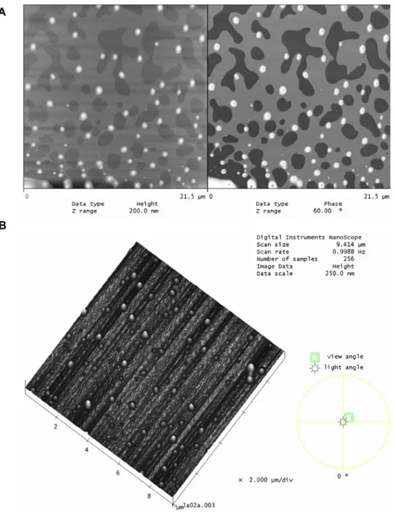
The morphology of the nanoparticles was examined by AFM. The NC suspensions appeared as a homogeneous population of par-ticles in AFM images (Fig. 3A and B). The AFM allows direct mea-surement of the size of the NP in samples deposited on freshly cleaved mica plates, and it permits simultaneous characterisation of particle shape and stiffness. The PLA-PEG NC external morpho-logical analyses (shown inFig. 3A) revealed that all nanostructures were spherical, had a regular surface and presented a halo sur-rounding the internal core, which is evidenced by a different tex-ture in the phase image (right side). The NC presented a homogeneous distribution in height and in three-dimensional images (Fig. 3A and B). An analysis of AFM data indicates that the sizes of PLA NC and PLA-PEG NC decreased with drug loading. These differences were significant (p< 0.05). Analysis of the data indicates that the diameter of the nanocapsules is much larger than the height (Table 2) for all NC formulations (Fig. 4). The PLA nano-spheres’ diameter/height ratio was 1.5, which was calculated from the topographical profile of the AFM images (Fig. 4andTable 2). NS are generally harder than nanocapsules. These results are attrib-uted to the flattening of the NC on a mica surface (Leite et al., 2005) due to the liquid nature of the core. NP stiffness was esti-mated by the diameter/height ratio (Table 2). The results show that association of AlClPc with NP has no significant effect (p> 0.05) on NP stiffness. The same can be observed with the polymeric wall (PLA or PLA-PEG) for those preparations. Only the NP type (NC or NS) affects significantly the flattening of the NP.
3.2. Photophysical characterisation
The absorption spectra of an AlClPc standard is compared to that of AlClPc extracted from NC in dimethyl sulphoxide inFig. 5. In all NC preparations, AlClPc exhibits a strong absorbance in the red region with a maximum wavelength at 676 nm (670 nm in eth-anol) as shown inTable 3. Furthermore, it presents a group of Q bands in the region from 600 to 710 nm and a Soret band at 340 nm. Notably, incorporation of the drug into a NC causes a red-shift in these bands. The incorporation of dyes into micro-hetero-geneous media produces redshifts, which is in agreement with previously reported work (Sibata et al., 2004). As shown inFig. 5
the absorption spectrum profile for the AlClPc in NC was identical to the spectrum observed for AlClPc in ethanol. In all NC formula-tions, the results indicate the absence of dimeric AlClPc or its aggregation states.
The fluorescence emission spectrum shows a maximum emis-sion for AlClPc at 679 nm in ethanol and 680 nm in NC after light excitation at 610 nm (Fig. 6). A redshift in the fluorescence maxi-mum is observed due to the environmental change from homoge-neous (ethanol) to heterogehomoge-neous media, in this case the NC. The
redshift observed for the encapsulated phthalocyanines is a mini-mal value (<10 nm). Generally, this phenomenon is related to the drug-polymer interaction from non-bonded intermolecular forces. The single photon counting technique was used to determined fluorescence lifetime for AlClPc in ethanol and in NC (Table 3). Sin-glet excited-state lifetimes were determined through the decay curve profiles of AlClPc in NC and in organic media. Mono-expo-nential decay was observed for the AlClPc standard in ethanol and for AlClPc in PLGA NC, PLA NC and PLA-PEG NC, which indi-cates that the AlClPc is distributed into one site (or population) with one singlet excited-state lifetime. The AlClPc in NC presented lifetimes of 5.77, 5.80 and 5.85 ns for PLGA NC, PLA NC and PLA-PEG NC, respectively (Table 3).
The relationship between emitted and absorbed photons results in a fluorescence quantum yield (/f) as calculated by Eq.(1). The fluorescence quantum yield for the AlClPc standard and for the NC formulations are similar (Table 3), which indicates that the photosensitiser does not suffer degradation during the encap-sulation. The fluorescence quantum yield of the formulations was evaluated using ZnPc in ethanol as a standard (/f= 0.28) (Oliveira
et al., 2005;Sibata et al., 2004). The fluorescence quantum yields obtained for AlClPc in ethanol were 0.81, 0.72 and 0.80 for PLGA NC, PLA NC and PLA-PEG NC, respectively. The value of the fluores-cence quantum yield is directly related to the photodynamic effi-ciency of the photosensitiser. Values near unity indicate that the fluorophores are more efficient as fluorescence probes for diagnos-tic use than as photosensitisers for PDT. The fluorescence quantum yield of AlClPc (0.8) was higher than that of ZnPc (0.28), which indicates it can be usefully applied to photo-diagnosis procedures. However, the compound also has an appropriate oxygen quantum yield in the homogeneous medium (/D= 0.3) as described by Ido-wu and Nyokong (2007). This characteristic is an important factor and is essential to produce PDT-effectiveness based on the tumour inactivation mechanisms (Type I/II).
The transient absorbance spectra for the drugs were obtained by laser flash photolysis, and triplet lifetimes (
s
T) were calculated from a kinetic analysis of the transient decays (Table 3). The triplet excited-state absorbed in the wavelength range from 380 to 650 nm with a maximum transient absorption at 470 nm for AlClPc in ethanol and AlClPc in NC. Beyond the triplet absorption, ground-state photobleaching of the Q and Soret bands of the AlClPc during the excitation process can be observed (data not shown). The decay curve adjustments were performed according to the chi-square function. The obtained transient absorbance spectra showed the presence of transient species that decayed mono-exponentially with characteristic lifetimes and resulted in significantly higher lifetimes for the triplet state of AlClPc in NC (Table 3). The tripletstate reacts with forms of molecular oxygen (O2) by an energy
transfer process leading to singlet oxygen, which is the key agent in cell damage in PDT. Therefore, the longer the triplet excited state lifetime, the better is the probability that drug energy can be trans-ferred to molecular oxygen and, consequently, to produce singlet oxygen.
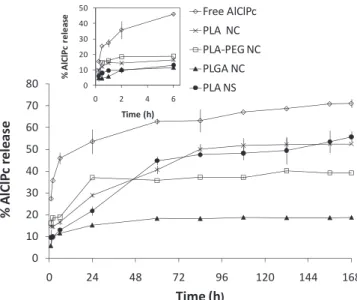
3.3. Solubility and in vitro release studies
avoids contact with the aqueous medium (Dhami and Phillips, 1996). The release study using the direct dialysis method results in no detectable AlClPc in the external medium.
Therefore, the external sink method was used to study the
in vitrorelease of AlClPc usingn-octanol as described byChorny
et al. (2002). The solubility of AlClPc inn-octanol at room temper-ature was 1.2 mg/ml. This method is not sensitive enough to study rapid release formulations, but can be used to release formulations having a long release time (Soppimath et al., 2001).N-octanol was established as the external medium of choice due to the similarity between its lipophilicity and the lipophilicity of biological mem-branes (Chorny et al., 2002).
Fig. 7shows thein vitrodrug release profiles of free AlClPc and AlClPc-loaded PLA NS, PLA NC, PLA-PEG NC and PLGA NC in the first 7 days. Thesein vitrorelease studies demonstrated that these types of drug carriers allow for extended delivery of the drug over more than 1 week (Fig. 7) compared to the solubility profile of free AlClPc. The analysis of the AlClPc release profile from PLGA NC shows approximately 10% released in a burst in the first 6 h fol-lowed by a slow release over 7 days that does not exceed 18%. A
Fig. 3.(A) AFM images of height (left) and phase (right) of PLA-PEG NC showing spherical nanostructures. Scan size: 21.5lm21.5lm. (B) AFM image giving a three-dimensional view of PLA NC containing AlClPc (0.2 mg/ml) spread on mica. Scan size: 10lm10lm.
Table 2
Characterisation of the nanoparticles by Atomic Force Microscopy.
Nanoparticles formulation
AlClPc (mg/ ml)
Mean size ± SD (nm)a
Ratio diameter/ height PLA NC 0.0 265.3 ± 37.5 8 AlClPc-PLA NC 2.0 211.8 ± 31.4 11 PLA-PEG NC 0.0 270.7 ± 68.3 11 AlClPc-PLA-PEG NC 2.0 153.5 ± 49.9 8 PLA NS 2.0 98.0 ± 25.2 1.5
good association of AlClPc with PLGA NC, which was also evi-denced by the encapsulation efficiency, seems to explain this pro-file. The AlClPc releases from PLA NC and PLA-PEG NC displayed an initial burst of 14.7% and 18.5% up to 2 h, respectively. These effects were followed by the slow release of 52.5% and 39.1% after 7 days for PLA NC and PLA-PEG NC, respectively. After the burst effect, the release of all NC formulations follows a first-order release profile. The initial burst may be caused by AlClPc molecules associated to the surface of the NC, particularly with PLA-PEG NC. Zeta poten-tial data (Table 1) corroborate this hypothesis. The release of AlClPc from the PLA NS also involved an initial rapid release phase that was followed by a phase of relatively slow release. Approximately 55% of the AlClPc was released after 7 days from the nanospheres (Fig. 7). On the other hand, when free AlClPc was incubated with
the acceptor media, 35.7% was rapidly dissolved in the first 120 min. Thus, more than 70% of the free AlClPc was dissolved in n-octanol over 7 days (Fig. 7) in contrast to the 55% released from PLA nanospheres over that time. Entrapment of AlClPc in nanopar-ticles, particularly in PLGA and PLA-PEG, significantly prolongs its
in vitrorelease (p< 0.05).
3.4. Assay of in vitro phototoxicity
Photodynamic therapy requires three components to be present simultaneously for cytotoxicity: a sensitiser, light and oxygen. In this study, thein vitrophototoxicity of AlClPc using different for-mulations of NP was compared. The phototoxic effect was ob-served in human fibroblasts in the presence of laser light doses
of 140 mJ/cm2, 3 J/cm2 and 10 J/cm2 at AlClPc concentrations of
10
l
M. Fibroblasts are an excellent biological model to evaluate the biocompatibility of new biomaterials and innovative pharma-ceutical formulations. First, it must be noted that light alone did not result in a significant decrease in cell viability under the exper-imental conditions used for this study (data not shown). The cyto-toxicity of free AlClPc and unloaded and AlClPc-loaded NC and NS were also evaluated in the dark.No significant cytotoxicity in the dark was observed after treat-ment with free AlClPc and unloaded or AlClPc-loaded PLA NC, PLA NS, PLA-PEG NC and PLGA NC formulations (p> 0.05). As shown in
Fig. 8, cellular viability was not significantly affected after treat-ment with unloaded PLA NC, PLA NS, PLA-PEG NC and PLGA NC in the absence of light (p >0.05). This is consistent with the known biocompatibility and safety of PLA and PLGA polymers (Davda and Labhasetwar, 2002; Konan et al., 2003). EMT-6 cell viability was not affected, for example, after treatment with drug-free PLA and PLGA nanoparticle formulations with irradiation at 9 J/cm2(Konan et al., 2003).
Fig. 9 shows the in vitro cytotoxic effect of free AlClPc and loaded-NP at light doses of 140 mJ/cm2, 3 J/cm2and 10 J/cm2and
AlClPc concentrations of 10
l
M. After irradiation at 140 mJ/cm2,all formulations were effective at inducing cell damage irrespective of the delivery vehicle as demonstrated by their similar phototox-icity; there were no significant differences among them (p> 0.05). The photoactivity of AlClPc-loaded NP was compared to the free AlClPc solution after irradiation at a light dose of 140 mJ/cm2
(Fig. 9). The viabilities of treated cells with free AlClPc, PLA NC, PLA NS, PLGA NC and PLA-PEG NC containing AlClPc and a light
dose of 140 mJ/cm2 were reduced to 58.7%, 51.3%, 41.3%, 57.5%
and 46.4%, respectively. There was no significant difference be-tween free AlClPc and AlClPc-loaded NP at 140 mJ/cm2(p> 0.05).
The cells treated with free AlClPc demonstrated a reduction of cell viability proportional to an increase of dose of light (p< 0.001). Increasing the dose to 3 J/cm2with free AlClPc solution
reduced the cell viability to 33.3%; the value was only 15% at 10 J/ cm2 (Fig. 9). At the same time, free AlClPc presented similar
(p> 0.05) phototoxicity to all NP formulations at 140 mJ/cm2.
AlClPc-loaded nanoparticles at 3 J/cm2 were more efficient at
inducing cellular death than free AlClPc (p< 0.001). This may be explained by the interaction of the NP with cells, which would re-sult in a high AlClPc association to cells and improved PDT efficiency.
Concerning cells treated with NP formulations, there were no significant differences (p> 0.05) between the values of cellular viability at laser light doses of 3 J/cm2 or 10 J/cm2 based on the Fig. 5.Absorption spectra of AlClPc in ethanol: (—) AlClPc standard, (–––) AlClPc
in PLGA NC, (––) AlClPc in PLA NC and (—————) AlClPc in PLA-PEG NC.
Table 3
Photophysical characterisation.
Samples UV–visa(nm) k
EXb(nm) kEMc(nm) sfd(ns) ± SD sfe(%) /ff sTe(ls)g± SD
Free AlClPc 670 610 679 7.57 ± 0.06 100 0.81 0.80h
AlClPc loaded PLGA NC 676 610 680 5.77 ± 0.09 100 0.81 1.38 ± 0.05 AlClPc loaded PLA NC 676 610 680 5.80 ± 0.09 100 0.72 1.37 ± 0.03 AlClPc loaded PLA-PEG NC 676 610 680 5.85 ± 0.08 100 0.80 1.41 ± 0.06
a Maximum wavelength UV–vis absorption. b Excitation wavelength.
c Maximum wavelength fluorescence emission. d Fluorescence lifetimes, mean ± SD.
e Population distribution.
f Fluorescence quantum yield using ZnPc (/
f= 0.28) in ethanol as a standard.
g Triplete lifetime, mean ± SD (n= 3). h Nunes et al., 2004.
mitochondrial activity test (MTT) (Fig. 9). The viabilities of treated cells with AlClPc-loaded PLA NC, PLA NS, PLGA NC and PLA-PEG NC that were irradiated at a light dose of 3 J/cm2were 12.9%, 15.5%,
13.7% and 11.3%, respectively. After irradiation at a dose of 10 J/ cm2, the viabilities of incubated cells with PLA NC, PLA NS, PLGA
NC and PLA-PEG NC containing AlClPc were reduced to 13.5%, 15.2%, 18.4% and 13.5%, respectively (Fig. 9). The results analysis indicates that no significant difference occurs in the phototoxic ef-fect observed for AlClPc incorporated into different formulations (p> 0.05) under higher doses of light. The dose of 3 J/cm2was
en-ough to drastically reduce the cell viability by activating the AlClPc photosensitiser in all NP formulations.Kolarova et al. (2007) re-ported the production of reactive oxygen species (ROS) and as-sessed the phototoxicity of disulphonated chloroaluminium phthalocyanine (ClAlPcS2) using G361 human melanoma cells. They showed that the optimum phototoxic effect observed in G361 melanoma cells was obtained for a combination of a laser dose of 25 J/cm2and 5–10 mg/ml of ClAlPcS2. Those combinations
of sensitiser concentration and corresponding radiation dose were lethal for the melanoma cells. The results obtained herein showed that much lower doses of AlClPc and light were necessary to reduce
cell viability, which suggests an efficient action of AlClPc as a photosensitiser in nanocapsules and nanospheres.
Tapajós et al. (2008) used oral carcinoma cells to evaluate
AlClPc encapsulated in liposomes as an in vitro photosensitiser
agent using doses of 5
l
M and 25 J/cm2. The oral neoplasic celldestruction was predominantly started by a necrotic process after PDT. In our present study, the results obtained from these combi-nations of AlClPc and corresponding radiation doses were immedi-ately lethal for the fibroblasts. The encapsulation of the sensitiser into nanoparticles improved the photoactivity of AlClPc. The encapsulated photosensitiser presented suitable biological
proper-tiesin vitroand photophysical properties for its utilisation as an
AlClPc delivery system in PDT.
4. Conclusions
The development of PLA NS, PLGA NC, PLA NC and PLA-PEG NC containing AlClPc was reported in this work with a detailed phys-icochemical and photophysical analysis of the different nanoparti-cle formulations. The incorporation of AlClPc into nanopartinanoparti-cles improves some parameters of the photophysical properties of AlClPc. The nanoprecipitation methodology was suitable to incor-porate AlClPc into small-size nanocapsules made of different bio-degradable polymers with a narrow size distribution. The advantages of AlClPc incorporation were made clear in the release studies. PLGA and PLA-PEG were able to retain AlClPc in sink lease media for more than 7 days, which resulted in a sustained re-lease profile. The photoactivity of AlClPc was improved by encapsulation as evidenced byin vitrocell studies. Thus, the differ-ent polymeric nanoparticles studied herein have proven to be an effective method for AlClPc delivery. These particles can improve the efficiency of photodynamic therapy at very low doses and energy.
Acknowledgements
This work was supported by CNPq (481195/2011-4 project), FA-PESP Projects 2006/50562-1, 2009/15363-9 and 2007/55319-0 and by the NANOBIOMG/FAPEMIG Network, Minas Gerais, Brazil. The CNPq researcher grant provided to V.C.F. Mosqueira is also acknowledged. The first author thanks UFOP and CAPES for a per-sonal scholarship.
Fig. 7.In vitrodrug release profile of (}) free AlClPc and AlClPc loaded in (d) PLA NS, (N) PLGA NC, () PLA NC and (h) PLA-PEG NC. The insert graph represents the
in vitrodrug release profile of AlClPc during the first 6 h. The results are the means of three experiments ± SD.
0 20 40 60 80 100 120
Control Free AlClPc PLA NS PLGA NC PLA NC PLA-PEG NC
CellularViability(%)
Unloaded Loaded
Fig. 8.Cytotoxicity of free AlClPc (10lM) and unloaded and AlClPc-loaded NP in the dark (10lM of AlClPc in the nanoparticle form) in human fibroblast culture. Cell viability is expressed as the mean ± SD (n= 3).
0 20 40 60 80 100
Control Free AlClPc PLA NS PLGA NC PLA NC PLA-PEG NC
Cellular
v
iability
(%
)
140 mJ/cm²
3 J/cm² 10 J/cm²
AlClPc- loaded
Fig. 9.AlClPc phototoxicity in different nanoparticles irradiated with 140 mJ/cm2,
3 J/cm2and 10 J/cm2of light (10lM of AlClPc). Cell viability is expressed as the
mean ± SD (n= 3).There was a significant difference among free AlClPc (10lM)
irradiated at 140 mJ/cm2, 3 J/cm2and 10 J/cm2of light (p< 0.001).
References
Allison, R.R., Sibata, C.H., 2010. Oncologic photodynamic therapy photosensitizers: a clinical review. Photodiagnosis Photodyn. Ther. 7, 61–75.
Chorny, M., Fishbein, I., Danenberg, H.D., Golomb, G., 2002. Study of the drug release mechanism from tyrphostin AG-1295 loaded nanospheres by in situ and external sink methods. J. Control Release 83, 401–414.
Davda, J., Labhasetwar, V., 2002. Characterization of nanoparticle uptake by endothelial cells. Int. J. Pharm. 233, 51–59.
Dhami, S., Phillips, D., 1996. Comparison of the photophysics of an aggregation and non-aggregating aluminium phthalocyanine system incorporated into unilamellar vesicles. J. Photochem. Photobiol. A Chem. 100, 77–84.
Eaton, D.F., 1988. International union of pure and applied chemistry organic chemistry division commission on photochemistry. Reference materials for fluorescence measurements. J. Photochem. Photobiol. B. 4, 523–531. Fessi, H., Puisieux, F., Devissaguet, J.P., Ammoury, N., Benita, S., 1989. Nanocapsule
formation by interfacial polymer deposition following solvent displacement. Int. J. Pharm. 55, R1–R4.
Gref, R., Minamitake, Y., Peracchia, M.T., Trubetskoy, V., Torchilin, V., Langer, R., 1994. Biodegradable long-circulating polymeric nanospheres. Science 263, 1600–1603.
Idowu, M., Nyokong, T., 2007. Photophysical and photochemical properties of zinc and aluminum phthalocyanines in the presence of magnetic fluid. J. Photochem. Photobiol. A Chem. 188, 200–206.
Kluson, P., Drobek, M., Kalaji, A., Karaskova, M., Rakusan, J., 2009. Preparation, chemical modification and absorption properties of various phthalocyanines. Res. Chem. Intermed. 35, 103–116.
Kolarova, H., Nevrelova, P., Bajgar, R., Jirova, D., Kejlova, K., Strnad, M., 2007. In vitro photodynamic therapy on melanoma cell lines with phthalocyanine. Toxicol. In Vitro 21, 249–253.
Konan, Y.N., Gurny, R., Allemann, E., 2002. State of the art in the delivery of photosensitizers for photodynamic therapy. J. Photochem. Photobiol. 66, 89– 106.
Konan, Y.N., Berton, M., Gurny, R., Allemann, E., 2003. Enhanced photodynamic activity of meso-tetra(4-hydroxyphenyl) porphyrin by incorporation into sub-200nm nanoparticles. Eur. J. Pharm. Sci. 18, 241–249.
Kreuter, J., 1996. Nanoparticles and microparticles for drug and vaccine delivery. J. Anat. 189, 503–505.
Krishna, S., Ter Kuile, F., Supanaranond, W., Pukrittayakamee, S., Teja-Isavadharm, P., Kyle, D., White, N.J., 1993. Pharmacokinetics, efficacy and toxicity of parenteral halofantrine in uncomplicated malaria. Br. J. Clin. Pharmacol. 36, 585–591.
Labib, A., Lenaerts, V., Chouinard, F., Leroux, J.C., Ouellet, R., Van Lier, J.E., 1991. Biodegradable nanospheres containing phthalocyanines and naphthalocyanines for targeted photodynamic tumor therapy. Pharm. Res. 8, 1027–1031. Legrand, P., Barrat, G., Mosqueira, V.C.F., Fessi, H., Devissaguet, J.P., 1999. Polymeric
nanocapsules as drug delivery systems: as review. Pharm. Sci. 9, 411–418. Legrand, P., Lesieur, S., Bochot, A., Gref, R., Raatjes, W., Barrat, G., Vauthier, C., 2007.
Influence of polymer behaviour in organic solution on the production of polylactide nanoparticles by nanoprecipitation. Int. J. Pharm. 344, 33–43. Leite, E.A., Vilela, J.M.C., Mosqueira, V.C.F., Andrade, M.S., 2005. Poly-caprolactone
nanocapsules morphological features by atomic force microscopy. Microsc. Microanal. 11, 48–51.
Maeda, H., 2010. Tumor-selective delivery of macromolecular drugs via the EPR effect: Background and future prospects. Bioconjug. Chem. 21, 797–802.
Mosmann, T., 1983. Rapid colorimetric assay for cellular growth and survival: application to proliferation and cytotoxicity assays. J. Immunol. Methods 65, 55–63.
Mosqueira, V.C.F., Legrand, P., Pinto-Alphandary, H., Puisieux, F., Barratt, G., 2000. Poly(D, L-Lactide) nanocapsules prepared by a solvent displacement process: Influence of the composition on physicochemical and structural properties. J. Pharm. Sci. 89, 614–626.
Mosqueira, V.C., Legrand, P., Gulik, A., Bourdon, O., Gref, R., Labarre, D., Barratt, G., 2001a. Relationship between complement activation, cellular uptake and surface physicochemical aspects of novel PEG-modified nanocapsules. Biomaterials 22, 2967–2979.
Mosqueira, V.C.F., Legrand, P., Morgat, J., Vert, M., Mysiakine, E., Gref, R., Devissaguet, J.-P., Barratt, G., 2001b. Biodistribution of long-circulating PEG-grafted nanocapsules in mice: effects of PEG chain length and density. Pharm. Res. 18, 1411–1419.
Moura-Siqueira, M.P., Primo, F.L., Peti, A.P.F., Tedesco, A.C., 2010. Validated spectrophotometric and spectrofluorimetric methods for determination of chloroaluminum phthalocyanine in nanocarriers. Pharmazie 65, 9–14. Nunes, S.M.T., Sguilla, F.S., Tedesco, A.C., 2004. Photophysical studies of zinc
phthalocyanine and chloroaluminum phthalocyanine incorporated into liposomes in the presence of additives. Braz. J. Med. Biol. Res. 37, 273–284. Oliveira, D.M., MacAroff, P.P., Ribeiro, K.F., Lacava, Z.G.M., Azevedo, R.B., Lima, E.C.D.,
Morais, P.C., Tedesco, A.C., 2005. Studies of zinc phthalocyanine/magnetic fluid complex as a bifunctional agent for cancer treatment. J. Magn. Magn. Mater. 289, 476–479.
Oliveira, L.T., Garcia, G.M., Kano, E.K., Tedesco, A.C., Mosqueira, V.C.F., 2011. HPLC-FLD methods to quantify chloroaluminum phthalocyanine in nanoparticles, plasma and tissue: application in pharmacokinetic and biodistribution studies. J. Pharm. Biomed. Anal. 1, 70–77.
Rocha, M.S.T., Lucci, C.M., Longo, J.P.F., Galera, P.D., Simioni, A.R., Lacava, Z.G.M., Tedesco, A.C., Azevedo, R.B., 2012. Aluminum-chloride-phthalocyanine encapsulated in liposomes: activity against naturally occurring dog breast cancer cells. J. Biomed. Nanotechnol. 8, 251–257.
Rodrigues, G.B., Primo, F.L., Tedesco, A.C., Braga, G.U.L., 2012. In vitro photodynamic inactivation of cryptococcus neoformans melanized cells with chloroaluminum phthalocyanine nanoemulsion. Photochem. Photobiol. 88, 440–447.
Sibata, M.N., Tedesco, A.C., Marchetti, J.M., 2004. Photophysical and photophysical studies of Zinc (II) phthalocyanine in long time circulation micelles for photodynamic therapy use. Eur. J. Pharm. Sci. 23, 131–138.
Silva, A.R.A., Simioni, A.R., Tedesco, A.C., 2011. Photophysical and complexation studies of chloro-aluminum phthalocyanine with beta-cyclodextrin and hydroxypropyl-beta-cyclodextrin. J. Nanosci. Nanotechnol. 11, 4046–4055. Soppimath, K.S., Aminabhaui, T.M., Kulkarni, A.R., Rudzinski, W.E., 2001.
Biodegradable polymeric nanoparticle as delivery devices. J. Control Release 70, 1–20.
Tapajós, E.C.C., Longo, J.P., Simioni, A.R., Lacava, Z.G.M., Santos, M.F.M.A., Morais, P.C., Tedesco, A.C., Azevedo, R.B., 2008. In vitro photodynamic therapy on human oral keratinocytes using chloroaluminum-phthalocyanine. Oral Oncol. 44, 1073–1079.
Tedesco, A.C., Rotta, J.C.G., Lunardi, C.N., 2003. Nitric oxide release from theS -nitrosothiol zinc phthalocyanine complex by flash photolysis. Braz. J. Medical Biol. Res. 36, 587–594.