LIGNINOLYTIC ENZYMES PRODUCTION AND REMAZOL BRILLIANT BLUE R DECOLORIZATION BY TROPICAL BRAZILIAN BASIDIOMYCETES FUNGI
Kátia M. G. Machado1,4*; Dácio R. Matheus2; Vera L. R. Bononi2,3
1Curso de Ciências Biológicas, Centro de Ciências da Educação, Universidade Católica de Santos, Santos, SP, Brasil; 2Seção de Micologia e Liquenologia, Instituto de Botânica, São Paulo, São Paulo, SP, Brasil; 3Universidade para o Desenvolvimento da Região do Pantanal, Campo Grande, MS, Brasil; 4Setor de Biotecnologia, Fundação Centro Tecnológico de Minas Gerais, Belo
Horizonte, MG, Brasil
Submitted: April 15, 2005; Returned to authors for corrections: July 21, 2005; Approved: September 12, 2005
ABSTRACT
Remazol Brilliant Blue R (RBBR) dye was used as substrate to evaluate ligninolytic activity in 125 basidiomycetous fungi isolated from tropical ecosystems. The extracellular RBBR decolorizing activity produced when selected fungi were grown in solid media and in soil contaminated with organochlorines was also evaluated. A total of 106 fungi decolorized the RBBR during the growth in malt extract agar (MEA, 2%); 96 fungi showed a mycelia growth and decolorization activity stronger than the P. chrysosporium used as reference. Extracellular extracts of 35 selected fungi grown on solid medium with sugar cane bagasse (BGS) were evaluated for RBBR decolorization and peroxidase activity. All fungi showed peroxidase activities, but 5 of those were unable to decolorize the RBBR. Different patterns of ligninolytic enzymes were detected in 12 fungi extracts. Mn-dependent peroxidase (MnP) was produced by Peniophora cinerea, Psilocybe castanella, three strains of Trametes villosa, T. versicolor, Melanoporia nigra and Trichaptum byssogenum. All 12 fungi had laccase activity. Trogia buccinalis showed the highest RBBR decolorization and did not produce MnP activity. RBBR decolorization without MnP production was also observed for three strains of Lentinum tested. Higher levels of peroxidase and laccase cannot be related to high RBBR decolorization. RBBR decolorization by extracellular extract was also detected during the growth of P. castanella, L. crinitus, P. cinerea and two strains of T. Villosa in pentachlorophenol- and hexachlorobenzene-contaminated soils. These fungi showed higher RBBR decolorization when grown in the presence of organochlorine compounds than when in non contaminated soil.
Key words: white rot fungi, peroxidases, laccase, MnP, pentachlorophenol, hexachlorobenzene
*Corresponding Author. Mailing address: Curso de Ciências Biológicas, Centro de Ciências da Educação, Universidade Católica de Santos, Av. Conselheiro Nébias, 300. 11015-002, Santos, SP, Brasil. Tel.: (+5513) 3205-5555. E-mail: [email protected]
INTRODUCTION
The white rot fungi (WRF) seem to be the unique microorganisms which show capacities of degrading and mineralizing lignin and a series of organic pollutant compounds, highly toxic and recalcitrant. This capacity is, at least in some extent, caused by non-specific enzymatic system produced by these fungi during the lignin degradation, includes several isoenzymes of Lignin Peroxidase (LiP, EC 1.11.1.14), Manganese
dependent Peroxidase (MnP, EC 1.11.1.13), laccases (EC 1.10.3.2) as well as H2O2-producing oxidases. The multienzymatic system involved in the lignin degradation and mineralization is constituted of different ligninolytic enzymes combinations, being the occurrence of MnP and Laccase higher than LiP (19).
enzymes pattern, which also has great differences in their ability in xenobiotic degradation. The ligninolytic systems of other basidiomycetes and several methods involving dyes have been reported as screening methodologies for the selection of xenobiotic compounds degrading microorganisms (3,4,14). The use of dyes offers a series of advantages in relation to conventional substrate because they are stable, soluble and cheap substrates with high rates of molar extinction and low toxicity. These dyes can be applied in simple, quick and quantitative spectrophotometric assays. Remazol Brilliant Blue R dye (RBBR) is an antracene derivative and, thus, related to an important group of organopollutants.
There is evidence that the capacity of WRF in decolorizing dyes results in part from the activity of enzymes that take part in lignin depolymerization process. Low molar weight compounds, as metals and H2O2, can also be envolved in pesticides degradation (15). Peroxidative activity does not seem to be the only extracellular enzyme available to WRF for dyes degradation, and decolorization rate could not be related to one particular enzyme, but to the result of the ligninolytic mechanism action. The identity of enzymes envolved in the RBBR degradation is still incompletly known (2,10,14).
To understand the basidiomycetes complex enzymatic system and the use of the RBBR dye as indicative of the degradative potential fungi system, this paper studied the RBBR-decolorizing activity present in extracellular basidiomycetes tropical fluids in solid medium and in soil contaminated with organochlorines.
MATERIALS AND METHODS
Fungi
A total of 125 fungi were isolated from basidiomes collected in different ecosystems of tropical Atlantic rainforest, Brazil (12). They were deposited in the culture collection of Instituto de Botânica, São Paulo. Phanerochaete chrysosporium ATCC28326 was used as reference sample. All strains were preserved in malt extract agar medium (MEA) 2% (w/v) slants at 4ºC.
RBBR decolorization test
Agar plug (∅ 2mm) from 7 day-old mycelium of each test strain grown in MEA were inoculated in plates (∅ 90mm) containing 2% MEA with 0.05% RBBR (Sigma), in quadruplicate. Plates were incubated at 30ºC until they were totally colonized or for 14 days. The decolorization coefficient (A) was calculated by measuring the decolorization disc diameter and growth disc diameter (cm), of each species, according the formula (1):
where:
H - decolorization disc diameter C - growth disc diameter t - test lineage
ref - reference strain Phanerochaete chrysosporium ATCC 28326
Fungi growth velocity
The growth disk (∅ 2mm) of each fungus on MEA was placed in the center of a 90mm plate with MEA. Triplicate plates were kept at 30ºC in the dark. The growth velocity was determined by measuring the average increase of the colony diameter (cm), in two perpendicular directions.
Extracellular ligninolytic activity in solid medium BGS
The extracellular ligninolytic activity was determined for 35 native fungi.The growth disk (∅ 2mm) of each fungus on MEA was transferred to solid medium BGS containing (NH4)SO4 2 g L-1; MnSO4.4H2O 0.004 g L-1; NaCl 0.1 g L-1; CaCl2.2H2O 0.1 g L-1; KH2PO4.7H2O 10 g L-1; glucose 20 g L-1; sugar cane bagasse power 10 g L-1; agar 20 g L-1. The fungi grew for 7 days at 30ºC, in triplicate. The solid agar was cut out, and the enzyme extraction was done with 30 mL of 50 mM sodium acetate buffer, pH 4.6, during 30 min. The mixture was filtered and assayed for extracellular ligninolytic activity.
Extracellular ligninolytic activity in organochlorine contaminated soils
The soils were collected in São Vicente, SP, in a region contaminated with organochlorine industrial residuals containing high concentrations of hexachlorobenzene (HCB) and pentachlorophenol (PCP). PCP-soil contains 6.50% of total organochlorines (6.04% of PCP, 0.15% of pentachlorobenzene, 0.04% of tetrachlorobenzene and 0.16% of hexachlorobutadiene). HCB-soil has 8.92% of total organochlorine (8.40% of HCB, 0.50% of pentachlorobenzene, and 0.02% of tetrachlorobenzene). The soils were diluted with non-contaminated soil (control-soil) to 0.18% and 1.2% of PCP and HCB, respectively. The concentrations of organochlorines were determined by high-resolution gas chromatography as described (9).
Enzymatic assays
RBBR decolorization was determined with the reaction mixture of 5.0 mL of the enzymatic extract and 50 µL of RBBR 2% (18). After an hour, 100 µL of the mixture were diluted 1:10 and the absorbancy reading was done at 500 and 585 nm. The decolorization was determined by the difference between the absorbance rates of the test sample and of a control prepared with enzymatic extract inactivated by boiling.
Peroxidase and laccase activities were determined by the oxidation of 0-dianisidine (∈460 = 29.400 M-1 cm-1) with and without the adding of hydrogen peroxide (16). The Manganese Peroxidase activity was determined by the phenol red oxidation (∈610 = 4.460 M-1 cm-1) (5). All of the enzymatic units were defined as the quantity of enzyme which oxides 1 µMol of substrate per minute per liter.
RESULTS AND DISCUSSION
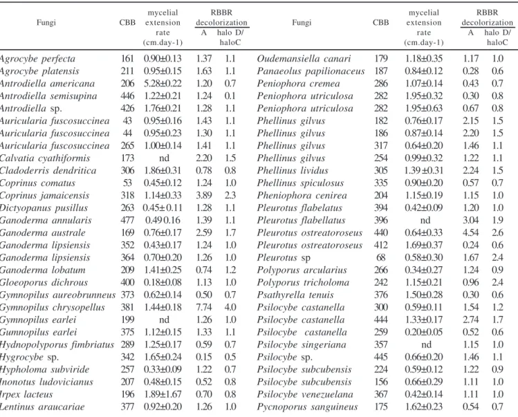
Out of the 125 fungi evaluated 106 were able to decolorize the RBBR in solid medium. Ceriporiopsis aneirina CCB261 and Oligoporus sp. CCB164 decolorized the dye without a clear decolorization halo. Fourty four fungi showed the decolorization halo bigger than the growth ring (Table 1). Collybia dryophila CCB308, Climacodon pulcherrimus CCB191, Fomitela supina CCB414, Fomitopsis spraguei CCB214, Ganoderma lucidum CCB168, Gloeophyllum striatum CCB188, Gymnopilus earlei CCB249, Lentinus crinitus, CCB212, Lentinus sp. CCB174, Oudemansiella canarii CCB241, Phellinus gilvus CCB485, P. umbrinellus CCB386, Pleurotus flabelatus CCB210 and CCB197, Psilocybe silvatica CCB443, Schizophyllum commune CCB473, Trametes
Table 1. Remazol Brilliant Blue R (RBBR) decolorization and mycelial extension rate of Basidiomycetes during growth on agar malt (2%).
mycelial RBBR mycelial RBBR
Fungi CBB extension decolorization Fungi CBB extension decolorization
rate A halo D/ rate A halo D/
(cm.day-1) haloC (cm.day-1) haloC
Agrocybe perfecta 161 0.90±0.13 1.37 1.1 Oudemansiella canari 179 1.18±0.35 1.17 1.0 Agrocybe platensis 211 0.95±0.15 1.63 1.1 Panaeolus papilionaceus 187 0.84±0.12 0.28 0.6 Antrodiella americana 206 5.28±0.22 1.20 0.7 Peniophora cremea 286 1.07±0.14 0.43 0.7 Antrodiella semisupina 446 1.22±0.21 1.24 0.1 Peniophora utriculosa 282 1.95±0.32 0.30 0.8 Antrodiella sp. 426 1.76±0.21 1.28 1.1 Peniophora utriculosa 282 1.95±0.63 0.67 0.8 Auricularia fuscosuccinea 43 0.95±0.16 1.43 1.1 Phellinus gilvus 182 0.76±0.17 2.15 1.5 Auricularia fuscosuccinea 44 0.95±0.23 1.30 1.1 Phellinus gilvus 186 0.87±0.14 2.20 1.5 Auricularia fuscosuccinea 265 1.00±0.14 1.41 1.1 Phellinus gilvus 317 0.64±0.20 1.46 1.1 Calvatia cyathiformis 173 nd 2.20 1.5 Phellinus gilvus 254 0.99±0.32 1.22 1.1 Cladoderris dendritica 306 1.86±0.31 0.78 0.8 Phellinus lividus 305 1.39 ±0.31 2.24 1.5 Coprinus comatus 53 0.45±0.12 1.24 1.0 Phellinus spiculosus 335 0.90±0.20 0.57 0.7 Coprinus jamaicensis 318 1.14±0.33 3.89 2.3 Pheniophora cenirea 204 1.15±0.19 1.15 1.0 Dictyopanus pusillus 263 0.45± 0.11 1.28 1.1 Pleurotus flabelatus 394 0.42±0.09 1.20 1.0 Ganoderma annularis 477 0.49 0.16 1.39 1.1 Pleurotus flabellatus 396 nd 3.04 1.9 Ganoderma australe 169 0.76±0.17 2.59 1.7 Pleurotus ostreatoroseus 440 0.64±0.33 4.54 2.6 Ganoderma lipsiensis 352 0.43±0.17 1.24 1.0 Pleurotus ostreatoroseus 412 1.69±0.37 0.24 0.6 Ganoderma lipsiensis 364 0.70±0.20 1.26 1.0 Pleurotus sp 68 0.58±0.30 1.67 2.4 Ganoderma lobatum 209 1.41±0.25 0.74 1.2 Polyporus arcularius 266 0.34±0.27 1.24 0.9 Gloeoporus dichrous 400 0.18±0.08 1.13 1.0 Polyporus tricholoma 242 1.15±0.21 0.96 2.4 Gymnopilus aureobrunneus 373 0.62±0.14 0.50 0.7 Psathyrella tenuis 376 1.50±0.28 0.30 0.6 Gymnopilus chrysopellus 381 1.44±0.18 7.74 4.0 Psilocybe castanella 300 0.59±0.11 1.54 1.2 Gymnopilus earlei 199 nd 1.26 1.0 Psilocybe castanella 444 1.33±0.17 2.74 1.7 Gumnopilus earlei 375 1.12±0.15 1.33 1.1 Psilocybe castanella 259 0.20±0.05 0.52 0.6 Hydnopolyporus fimbriatus 289 1.25±0.17 0.59 0.7 Psilocybe singeriana 357 nd 1.15 1.0
Hygrocybe sp. 342 1.65±0.24 0.15 0.5 Psilocybe sp. 445 0.66±0.20 1.46 1.1
Lentinus bertieri 255 1.19±0.21 1.48 1.0 Pycnoporus sanguineus 273 1.73±0.23 1.54 0.5 Lentinus bertieri 260 1.42±0.27 0.28 1.1 Pycnoporus sanguineus 277 2.53±0.53 1.28 0.6 Lentinus crinitus 356 1.33±0.11 1.20 1.0 Pycnoporus sanguineus 294 1.58±0.30 0.50 0.7 Lentinus crinitus 217 1.47±0.26 1.54 1.0 Pycnoporus sanguineus 458 1.56±0.31 0.80 0.8 Lentinus striatulus 253 1.48±0.19 1.26 1.0 Rigidoporus microporus 334 0.51±0.15 2.22 1.5 Lentinus strigellus 380 1.23±0.21 1.02 0.9 Ripartitella brasiliensis 467 1.59±0.33 1.11 1.0 Lentinus strigosus 162 1.09±0.23 3.28 2.0 Russula hygrophytica 328 1.62±0.27 0.72 0.8 Lentinus strigosus 178 1.11±0.20 1.33 1.1 Stereum ostrea 267 1.28±0.11 0.91 0.6 Lentinus strigosus 250 0.22±0.08 4.22 1.0 Trametes pubescens 166 0.81±0.17 0.63 0.8 Lentinus velutinos 268 1.31±0.24 0.33 1.1 Trametes versicolor 202 1.42±0.21 2.22 1.5 Lentinus velutinos 288 nd 1.67 1.2 Trametes versicolor 372 1.10±0.12 1.33 1.1 Lentinus villosus 271 1.43±0.19 1.43 1.2 Trametes villosa 176 1.59±0.28 1.30 1.1 Lentinus zeyheri 247 1.44±0.19 0.11 1.1 Trametes villosa 213 1.43±0.28 1.39 1.2
Lepista sp. 110 0.84±0.16 1.30 1.1 Trametes villosa 291 1.53±0.20 1.43 1.1
Marasmius cladophyllus 361 1.69±0.21 1.30 1.1 Trametes villosa 295 1.57±0.33 1.43 1.1 Marasmius niveus 455 1.67±0.34 1.26 1.0 Trametes villosa 165 1.61±0.35 1.80 1.1 Marasmius smaragdinus 470 0.35±0.07 0.65 0.8 Trichaptum bysogenum 203 1.56±0.27 1.33 1.1 Marasmius sp. 378 1.26±0.15 1.15 1.0 Trogia buccinalis 390 1.39±0.24 1.20 1.0 Melanoporia nigra 184 1.31±0.35 1.15 1.0 Tyromyces pseudolacteus 193 1.40±0.31 1.33 1.1
CCB: culture collection number; A = coefficient of decolorization (1), using Phanerochaete chrysosporium ATCC2832 as reference strain; nd:
not determined; halo D/halo C = ratio between the decolorization halo and growth halo; All values reported as means ± standard deviation for three replica cultures.
versicolor CCB158, Trichaptum biforme CCB297 and Tubaria furfuracea CCB374 were unable to decolorize the RBBR.
Ninety-six fungi showed more decolorization than P. chrysosporium (Table 1). Agaricus porosporus CCB299, Ceriporiopsis aneirina CCB261, Collybia subpruinosa CCB404, Ganoderma Lipsiense CCB321, Merulius corium CCB355, Nothopanus hygrophanus CCB216, Oligoporus sp. CCB164, Pycnoporus sanguineus CCB113, Schizophyllum commune CCB307 isolates showed decolorization activity equal or lower than P. chrysosporium.
A total of 55 fungi showed average growth velocity higher than or equal to 1cm day-1 (Table 1). The selection of fungi with high growth rates is one of the desirable characteristics in the application of bioremediation processes since this factor can make the strains more competitive with the native soil biota (6). Other facilities include the handling in laboratory and the production of spawn.
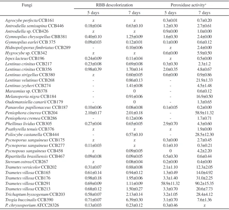
Thirty-four fungi grew in BGS solid medium and the enzymatic extracts of 30 showed RBBR decolorization and peroxidase activity (Table 2). A. perfecta, Antrodiella sp., Hygrocybe sp., Psathyrellatenuis and Pycnoporussanguineus CCB175 extracts did not show RBBR decolorization, but showed extracellular peroxidase activity.
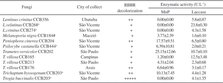
Twelve fungi with expressive RBBR decolorization capacity were tested for Mn-peroxidase and laccase activities (Table 3). Neither Lentinus species nor Trogia buccinalis showed MnP activity. Laccase activity was produced by all of the native
fungi. Until this study, the production of laccase activity was not described in Peniophora cinerea, Psilocybe castanella, Trichaptum byssogenum and Trogia buccinalis.
Table 2. Extracellular Brilliant Remazol Blue R (RBBR) decolorization and peroxidose activity produced by Basidiomycetes during growth on BGS solid medium.
Fungi RBB descolorization Peroxidase activitya 5 days 7 days 5 days 7 days
Agrocybe perfecta CCB161 z z 0.3±0.01 0.7±0.20
Antrodiella semisupina CCB446 0.18±0.04 0.63±0.10 1.2±0.30 2.7±0.61
Antrodiella sp. CCB426 z z 0.9±0.00 1.0±0.00
Gymnopilus chrysopellus CBB381 0.40±0.10 1.25±0.09 1.6±0.30 2.4±0.00
Gymnopilus earlei CCB 375 0.09±0.03 0.12±0.01 0.1±0.00 0.6±0.12
Hidnopolyporus fimbriatus CCB289 - 0.10±0.06 z 2.4±0.00
Hygrocybe sp.CCB342 z z 0.6±0.00 5.9±0.50
Irpex lacteus CCB196 0.24±0.09 0.11±0.04 z 0.5±0.00
Lentinus crinitus CCB217 0.23±0.08 0.69±0.08 0.3±0.30 2.3±1.2
Lentinus crinitus CCB356 0.98±0.39 1.70±0.14 2.0±0.35 4.8±0.67
Lentinus strigellus CCB380 z 0.60±0.05 0.6±0.00 0.9±0.86
Lentinus velutinus CCB268 - 0.86±0.13 - 21.9±1.33
Lentinus zeyheri CCB274 - 1.41±0.08 - 4.5±1.48
Marasmius sp.CCB378 - 0 - 0.6±0.12
Melanoporia nigra CCB184 - 0.91±0.06 - 16.9±0.50
Oudemansiella canarii CCB179 - 0 - 1.3±0.65
Panaeolus papilionaceus CCB187 0.10±0.06 0.08±0.08 0.1±0.05 0.2±0.00
Peniophora cinerea CCB204 2.10±0.17 2.01±0.18 - 58.9±11.32
Peniophora cremea CCB286 - 0.12±0.06 - 1.7±0.71
Phellinus lividus CCB305 0.27±0.04 0.65±0.05 2.9±0.70 4.3±0.60
Psathyrella tenuis CCB376 z z z 1.9±0.00
Psilocybe castanella CCB444 - 0.57±0.10 - 28.5±12.30
Pycnoporus sanguineus CCB175 z z 0.3±0.00 2.7±0.45
Pycnoporus sanguineus CCB277 0.11±0.03 z 0.1±0.10 0.3±0.21
Pycnopous sanguineus CCB458 z 0.09±0.05 0 4.2±2.20
Ripartitella brasiliensis CCB467 0.09±0.08 0.09±0.05 0.5±0.30 0.6±0.44
Stereum ostrea CCB267 z 0.08±0.04 0.2±0.00 0.4±0.00
Trametes versicolor CCB202 0.31±0.07 0.88±0.08 2.1±1.10 12.3±2.05
Trametes villosa CCB165 0.61±0.14 0.94±0.12 1.3±0.49 14.0±4.92
Trametes villosa CCB176 0.98±0.18 1.95±0.06 3.3±1.40 31.0±2.25
Trametes villosa CCB291 0.69±0.09 1.11±0.09 58.9±11.32 90.2±15.35
Trametes villosa CCB213 0.68±0.12 1.50±0.27 3.3±0.70 20.6±7.73
Trichaptum byssogenum CCB203 0.58±0.07 2.13±0.14 3.2±1.05 28.4±4.12
Trogia buccinalis CCB390 0.71±0.07 6.39±0.30 3.1±0.70 7.6±1.36
P. chrysosporium ATCC28326 0.13±0.03 0.23±0.12 0.3±0.46 z
- = not determined; z = no activity; a: Enzymatic units = µmol of oxidate substrate L-1, in the presence of H
2O2; All values reported as means ±
standard deviation for three replica cultures.
The enzymatic system involved in RBBR decolorization was produced by basidiomycete fungi in solid medium and in soils. This system was produced even in the absence of the RBBR and the decolorization was performed in the absence of added H2O2 showing that there are different combinations of extracellular ligninolytic enzymes produced in significant quantities under simple conditions of cultivation. Which
Table 3. Manganese peroxidase and laccase activities during Brilliant Remazol Blue R (RBBR) decolorization by rot fungi.
Fungi City of collect RBBR Enzymatic activity (U L -1) decolorization MnP Laccase
Lentinus crinitus CCB356 Ubatuba ++ 0.00±0.00 5.8±0.87
L.velutinus CCB268a São Vicente + 0.00±0.00 23.8±0.30
L.crinitus CCB274a São Vicente + 0.00±0.00 4.3±1.58
Melanoporia nigra CCB184# Maceió + 3.77±2.39 1.6±0.10
Peniophora cinerea CCB204 São Vicente ++ 17.57±9.51 6.5±0.80
Psilocybe castanella CCB444a São Vicente + 6.39±10.81 2.0±0.21
Trametes versicolor CCB202 São Paulo + 25.15±12.66 10.7±0.10
T. villosa CCB165 Campinas + 1.20±0.00 12.5±3.48
T. villosa CCB213 São Paulo + 4.31±2.04 2.3±0.68
T. villosa CCB176 Assis ++ 6.64±0.96 3.1±0.17
Trichaptum byssogenum CCB203a São Vicente ++ 10.13±7.45 4.4±1.28
Trogia buccinalis CCB203a São Paulo +++++ 0.00±0.00 4.1±1.35
a: fungi collected in places where dumps of organochlorines residues ocorred; # brown rot fungi; Enzymatic units = µmol of oxidate substrate L-1;
+++++ = 100 % of decolorization ( in relation to the highest decolorization = 6.39, T. buccinales); ++++ = 75 a 99% of decolorization; +++ =
50 a 74 % of decolorization; ++ = 25 a 49% of decolorization; + = até 24 % of decolorization; All values reported as means ± standard deviation for three replica cultures.
Figure 1. Brilliant Remazol Blue R (RBBR) decolorization by extracellular extracts of Psilocybe castanella CCB444 and Trametes villosa CCB176 during growth in organochlorine soils: () PCP-soil, () HCB-soil and (----) control-soil.
ACKNOWLEDGMENTS
This work was supported by FAPEMIG, CNPq and Rhodia do Brasil Ltda.
RESUMO
Produção de enzimas ligninolíticas e descoloração do corante azul brilhante de Remazol R por fungos
basidiomicetos tropicais brasileiros
O corante azul brilhante Remazol R (RBBR) foi usado como substrato para avaliar 125 fungos basidiomicetos isolados de ecossistemas tropicais brasileiros quanto a atividade ligninolítica. A descoloração do RBBR por extratos obtidos do crescimento de fungos em meio sólido e em solo contaminado com organoclorados foi também avaliada. Cento e seis fungos descoloriram o RBBR durante o crescimento em meio sólido (agar extrato malte); noventa e seis desses fungos mostraram crescimento micelial e descoloração superiores a uma cepa de Phanerochaete chrysosporium usada como referência. Trinta e cinco fungos foram crescidos em meio sólido contendo bagaço de cana-de-açúcar (BGS) e seus extratos foram avaliados quanto a descoloração do RBBR e a produção de atividade de peroxidases. Todos os fungos mostraram atividade de peroxidases, mas 5 foram incapazes de descolorir o RBBR. Diferentes padrões de enzimas lignolíticas foram detectados em 12 desses extratos fúngicos, porém todos mostraram atividade de laccase. Atividade de peroxidase dependente de manganês (MnP) foi produzida por Melanoporia nigra, Peniophora cinerea, Psilocybe castanella, três cepas de Trametes villosa, T. versicolor e Trichaptum byssogenum. Trogia buccinalis apresentou a maior descoloração do RBBR e não produziu MnP. Descoloração do RBBR sem produção de MnP foi também observada para as três cepas de Lentinus testadas. Altos níveis de atividades de peroxidases e laccases não mostraram relação com significativa descoloração do RBBR. Foi observada descoloração do RBBR por extratos obtidos de P. castanella, L. crinitus, P. cinerea e duas cepas de T. Villosa durante crescimento em solos contaminados com pentaclorofenol e hexaclorobenzeno. Estes fungos mostraram maior descoloração de RBBR na presença de compostos organoclorados do que em solos não contaminados.
Palavras-chave: fungos de podridão branca, peroxidases, lacase, pentaclorofenol, hexaclorobenzeno
REFERENCES
1 . Antier, P.; Minjares, A.; Roussos, S.; Raimbault, M.; Viniegra-González, G. Pectinase-hyperproducing mutants of Aspergillus niger
C28B25 for solid-state fermentation of coffee pulp. Enzyme Microbiol. Technol., 15, 254-260, 1993.
2 . Deveci, T.; Unyayara, A.; Mazmanci, M.A. Production of Remazol Brilliant Blue R decolourising oxygenase from the culture filtrate of Funalia trogii ATCC200800. J. Mol. Catal. B: Enzymatic, 30, 25-32, 2004.
3 . Field, J.A.; Jong, E.; Feijoo-Costa, G.; De Bont, J.A.M. Screening for ligninolytic fungi applicable to the biodegradation of xenobiotics. Trends Biotechnol., 11, 44-49, 1993.
4 . Glenn, J.K.; Gold, M.H. Decolorization of several polymeric dyes by the lignin-degradating basidiomycete Phanerochaete chrysosporium. Appl. Environm. Microbiol., 45, 1741-1747, 1983.
5 . Kuwahara, M.; Glenn, J.K.; Morgan, M.A.; Gold, M.H. Separation and characterization of two extracellular H2O2-dependent oxidases from ligninolytic cultures of Phanerochaete chrysosporium. Febs Lett., 169, 247-250, 1984.
6 . Lamar, R.T.; Larsen, M.J.; Kirk, T.K. Sensitivity to and degradation of pentachlorophenol by Phanerochaete spp. Appl. Environm. Microbiol., 56, 3519-3526, 1990.
7 . Machado, K.M.G.; Matheus, D.R.; Fabris, C.; Bononi, V.L.R. Efeito da adição de substrato lignocelulósico no crescimento de fungos basidiomicetos em solo com baixo teor de matéria orgânica. Workshop sobre Biodegradação, Campinas, 1996, p.243.
8 . Machado, K.M.G.; Matheus, D.R.; Monteiro, R.T.R.; Bononi, V.L.R. Biodegradation of pentachlorophenol by tropical basidiomycetes in soils contaminated with industrial residues. World J. Microbiol. Biotechnol., 21, 297-301, 2004.
9 . Matheus, D.R.; Bononi, V.L.R.; Machado, K.M.G. Biodegradation of hexachlorobenzene by basidiomycetes in soil contaminated with industrial residues, World J. Microbiol. Biotechnol., 16, 415-421, 2000.
10. Moreira, P.R.; Almeida-Vara, E.; Sena-Martins, G.; Polonia, I.; Malcata, F.X.; Duarte, J.C. Decolourisation of Remazol Brilliant Blue R via a novel Bjerkandera sp. Strain. J. Biotechnol., 89, 107-111, 2001.
11. Novotyny, C.; Svobodova, K.; Erbanova, P.; Cajthaml, T.; Kasinath, A.; Lang, E.; Sasek, V. Ligninolytic fungi in bioremediation: extracellular enzyme production and degradation rate. Soil Biol. Biochem., 36, 1545-1551, 2004.
12. Okino, L.K.; Machado, K.M.G.; Fabris, C.; Bononi, V.L.R. Ligninolytic activity of tropical rainforest basidiomycetes. World J. Microbiol. Biotechnol., 16, 889-893, 2000.
13. Shah, V.; Nerud, F. Lignin degrading system of white-rot fungi and its exploitation for dye decolorization. Review. Can. J. Microbiol., 48, 857-870, 2002.
14. Shin, K-S.; Oh, I-K.; Kim, C-J. Production and purification of remazol brilliant blue R decolorizing peroxidase from the culture filtrate of Pleurotus ostreatus. Appl. Environm. Microbiol., 63, 1744-1748, 1996. 15. Sun, Y.; Pignatello, J.J. Activation of Hydrogen Peroxide by Iron (III) Chelates for Abiotic Degradation of Herbicides and Insecticides in Water. J. Agric. Food Chem., 41, 308-312, 1993.
16. Szklarz, G.D.; Antibus, R.K.; Sinsabaugh, R.L.; Linkins, A. Production of phenol oxidases and peroxidases by wood-rotting fungi. Mycologia, 81, 234-240, 1989.
17. Thurston, C.F. The structure and function of fungal laccases. Microbiology, 140, 19-26, 1994.
18. Ulmer, D.C.; Leisola, M.S.A.; Fiechter, A. Possible induction of the ligninolytic system of Phanerochaete chrysosporium. J. Biotechnol., 1, 13-24, 1984.