Tuberculous pneumonia:
a study of 59 microbiologically confirmed cases*
,**
Pneumonia tuberculosa:
um estudo de 59 casos confirmados microbiologicamente
Jose Moreira, Jamila Belicanta Fochesatto, Ana L Moreira, Marisa Pereira, Nelson Porto, Bruno Hochhegger
Abstract
Objective: To study the clinical, epidemiological, radiographic and endoscopic features of individuals with tuberculous pneumonia. Methods: We evaluated 2,828 consecutive tuberculosis patients treated at a public health center between December of 2005 and February of 2007. Of those, 59 (2.1%) had pulmonary involvement consistent with fistula between a lymph node and a bronchus. Results: Of the 59 patients studied, 43 (73%) were between 20 and 50 years of age, 31 (53%) were male, and 28 (47%) were Black. The most common symptoms were cough (in 100%), fever (in 88%), expectoration (in 81%), and weight loss (in 40%). Comorbidities were reported in 35 cases (59%), the most common being HIV infection (in 20%) and diabetes (in 15%). On chest X-rays, consolidation was observed, predominantly in the upper lobes (in 68%). The diagnostic confirmation (identification of AFB) was made through the sputum smear microscopy in the majority of the cases and by bronchoscopy (BAL examination or bronchial biopsy) in the remainder. Bronchial lesions were clearly indicative or suggestive of fistula in three cases and five cases, respectively. Conclusions: Tuberculous pneumonia presents as acute respiratory infection, initiating with a dry cough that is followed by fever. Chest X-rays show alveolar consolidation. In most cases, tuberculous pneumonia was accompanied by at least one comorbid condition, the most common being HIV infection, and the etiological diagnosis was made through sputum smear microscopy for AFB. Bronchoscopy findings were indicative of bronchial fistula in eight cases (13%).
Keywords: Mycobacterium tuberculosis; Pneumonia; Bronchial fistula; Lymph nodes.
Resumo
Objetivo: Estudar os aspectos clínicos, epidemiológicos, radiológicos e endoscópicos encontrados em indivíduos com pneumonia tuberculosa. Métodos: Entre dezembro de 2005 e fevereiro de 2007, foram estudados 2.828 pacientes com tuberculose que foram consecutivamente atendidos em uma unidade de saúde pública. Desses, 59 (2,1%) tiveram envolvimento pulmonar compatível com fístula entre um linfonodo e um brônquio.
Resultados: Dos 59 pacientes estudados, 43 (73%) tinham entre 20 e 50 anos de idade, 31 (53%) eram do sexo masculino, e 28 (47%) eram negros. Os sintomas mais frequentes foram tosse (100%), febre (88%), expectoração (81%) e perda de peso (40%). Comorbidades foram registradas em 35 pacientes (59%), especialmente a infecção por HIV (20%) e diabetes (15%). Na radiografia de tórax, a consolidação predominou nos lobos superiores (em 68%). A confirmação diagnóstica (presença de BAAR) foi feita principalmente por baciloscopia direta do escarro, seguida por broncoscopia (LBA e biópsia brônquica). Lesões brônquicas claramente indicativas ou sugestivas de fístula foram identificadas em três casos e cinco casos, respectivamente. Conclusões: A pneumonia tuberculosa apresenta-se como uma infecção respiratória aguda, com tosse seca seguida por febre. A radiografia de tórax mostra consolidação alveolar. Na maioria dos casos, a pneumonia tuberculosa foi acompanhada por pelo menos uma comorbidade, especialmente a infecção por HIV, e a confirmação etiológica foi obtida principalmente através do exame de escarro direto para BAAR. Os achados de broncoscopia foram indicativos de fístula brônquica em oito casos (13%).
Descritores: Mycobacterium tuberculosis; Pneumonia; Fístula brônquica; Linfonodos.
* Study carried out under the auspices of the Graduate Program in Pulmonology at the Universidade Federal do Rio Grande do Sul – UFRGS, Federal University of Rio Grande do Sul – Porto Alegre, Brazil.
Correspondence to: Bruno Hochhegger. Rua João Alfredo, 558/301, Cidade Baixa, CEP 90050-230, Porto Alegre, RS, Brasil. Tel 55 51 3314-3665. E-mail: [email protected]
Financial support: None.
Submitted: 27 May 2010. Accepted, after review: 25 November 2010.
(15) At that stage, tuberculin skin test results can
be negative, as occurs in cases of tuberculous effusions.(16) The drained lymph node can remain
as a cavity in the perforated bronchial wall, later closing (leaving a fibrous scar or an area of calcification or disappearing and leaving no vestiges).(5,14,17)
After tuberculous pneumonia has become established, its further evolution will basically depend of the number of bacilli present in the caseous material aspirated from the lymph node into the lung. If few or no germs are present in the area of consolidation, resolution of the pneumonia can occur, a condition previously designated “epituberculosis”.(18,19) Otherwise, the
presence of bacilli promotes the progression of the disease, with additional necrotic cavities and dissemination to other lung regions. In addition, there can be permanent lesions, such as bronchial stricture or bronchiectasis, particularly when the focus is in the middle lobe or lingula, resulting in late complications, such as bleeding and recurrent bacterial pneumonia.(20)
The radiographic and tomographic findings in tuberculous pneumonia are typically characteristic of alveolar consolidation, whose location depends on the bronchus involved. Tree-in-bud opacities are frequently seen adjacent to the main lesion.(21,22) In some cases,
mediastinal lymphadenopathy can be observed on the affected side.(23)
Bacteriological confirmation, with identification of Mycobacterium tuberculosis, can be established by sputum smear microscopy, sputum culture, examination of BAL, bronchoscopic aspiration, or biopsy of the bronchial lesion. In two case series involving a total of 93 patients, M. tuberculosis was most often identified by sputum smear microscopy, especially when multiple examinations were conducted.(24,25)
The non-classical forms of pulmonary tuberculosis, including pneumonia, are often seen in HIV-infected individuals. In a recent case series of 231 patients with pulmonary tuberculosis, 113 were HIV-infected.(26)
The objective of this study was to analyze clinical, epidemiological, radiographic, and endoscopic findings in a cohort of adult individuals with microbiologically confirmed tuberculous pneumonia.
Introduction
Although tuberculosis can present in several forms and can affect practically any organ, it involves the lungs in approximately 80.0% of cases. The most common extrapulmonary sites are the pleura, lymph nodes, bones, genitourinary tract, and central nervous system.(1,2)
One complication of lymph node tuberculosis is fistula formation and the consequent migration of bacilli and caseous material to other structures, such as the pleural, pericardial, and peritoneal cavities, as well as the bronchus or pulmonary vein. The main radiographic feature of tuberculous pneumonia is consolidation of the pulmonary region surrounding the affected bronchus. Clinically, dry irritating cough typically occurs a few days before the onset of fever and pulmonary involvement, which emerge after the fistula is fully formed,(3) potentially evolving to
severe outcomes.(4)
According to Schwartz,(5) there are writings
regarding openings in the tracheal or bronchial wall secondary to adjacent tuberculous lymph nodes dating from ancient times. However, reports that were more precise began to appear by end of the 18th century—by Morgagni (in 1761), Laluette (in 1780), Cayol (in 1810), and others. Subsequently, there were reports, mainly based on autopsy studies, of cases in children and adults. Among 1,654 patients evaluated between 1942 and 1954,(6-9) fistulae,
of varying dimensions, between bronchi and lymph nodes were identified in 145 (8.7%). The first endoscopic observations of such fistulae were made at beginning of the 20th century, when endoscopy was reserved for use as a diagnostic tool in clinically advanced cases.
(10-12) In two case series,(5,13) involving a total
of 3,800 patients submitted to endoscopy, the authors identified 231 cases of recent bronchial fistulas with visible orifices to a lymph node and 199 cases of bronchial wall scarring indicative of previous fistula (6.1% and 5.2%, respectively). More recently, other authors have evaluated the modifications that occur during the evolution of the bronchial tuberculosis.(3,14)
Although bronchial fistulas are typically single, occurring in a lobar or segmental bronchus, multiple bronchial fistulae have been reported.(5) The infiltration of caseous material
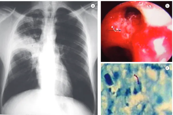
The microbiological confirmation was obtained mainly through sputum smear microscopy, which was positive for AFB in 41 (69.5%) of the 59 patients. Bronchoscopy was performed in 18 patients (30.5%). Bronchial wall lesion was identified and biopsied in 8 (44.4%) of those 18 patients. The findings were clearly indicative of fistula in 3 (16.7%) and suggestive of fistula (evidence of scarring) in 5 (27.8%). In all eight of those cases, the alterations were identified in large bronchi. In all 18 of the patients submitted to bronchoscopy, the microbiological diagnosis was made through examination of the biopsy fragment or of the BAL fluid (Table 4).
The treatment given was the rifampin-isoniazid-pyrazinamide combination in 57 patients (96.6%) and the streptomycin-isoniazid-ethambutol combination in 2 (3.4%). There was progressive clinical and radiographic improvement, and the fever subsided after 2-3 weeks of treatment. In most cases, adverse reactions were minimal, although 3 of the patients (5.1%) developed hepatitis.
Methods
We studied 2,828 consecutive tuberculosis patients treated at a public health center in the city of Porto Alegre, Brazil, between January of 2005 and February of 2007. Of those, 59 (2.1%) presented with a clinical and radiographic profile of noncavitary pulmonary consolidation and were selected for inclusion, all with microbiological confirmation (identification of AFB by sputum smear microscopy, sputum culture, BAL examination, or bronchoscopic biopsy). For sputum smear microscopy, we used the Ziehl-Neelsen method, and cultures were performed on Löwenstein-Jensen medium. Patients with other forms of tuberculosis were excluded, as were those in which there was no microbiological confirmation. Radiographic, bronchoscopic, and microbiological investigations were all performed by experienced professionals.
The data were stored and analyzed in the 2003 Microsoft Excel Program. Descriptive statistics and the chi-square test to compare proportions were used. The level of significance was set at 5%.
Results
Among the 59 patients with tuberculous pneumonia, there was a slight predominance of males and White individuals. Of the 59 patients, 43 (72.9%) were between 20 and 50 years of age (Table 1).
We found that 35 (59.3%) of the patients presented with at least one comorbidity, the most common of which were HIV infection and diabetes (Table 2). The most common symptoms were cough, which was generally dry at onset, and fever (Table 3).
In 38 (64.4%) of the patients, the white blood cell count was not significantly altered, leukocytosis with slight left shift being observed in 21 (35.6%). The tuberculin skin test with PPD (2 TU) was negative in 10 (38.5%) of the 26 patients so tested.
Pulmonary consolidation was found in the upper lobes, primarily on the right, in 40 (67.8%) of the cases, a frequency significantly greater than that observed at other locations (p < 0.001), although the middle lobe was involved in 9 cases (15.3%). Small discrete foci adjacent to the main lesion were seen in most of the patients (Figure 1).
Table 1 - Demographic characteristics of 59 patients with tuberculous pneumonia.
Characteristic n (%) Gender
Male 31 (52.5) Female 28 (47.5) Ethnicity
White 30 (50.8) Black 28 (47.5)
Other 1 (1.7)
Age (years)
< 20 2 (3.4) 20-50 43 (72.9) > 50 14 (23.7)
Table 2 - Co-morbidities present in 59 patients with tuberculous pneumonia.
Comorbidity n (%)
None 24 (40.7)
HIV infection 12 (20.3)
Diabetes 9 (15.3)
Lung disease 3 (5.1) Heart disease 3 (5.1) Depression 3 (5.1)
outpatients was 2.1%, which is lower than the 7.8% reported by Picon et al.(24) However, those
authors evaluated only patients, their sample was smaller (n = 231), and the proportion of HIV-infected patients was higher in their study.
In our study, the clinical diagnosis of tuberculous pneumonia was suggested by the presence of dry cough preceding the other respiratory symptoms and the systemic symptoms. The radiographic findings of consolidation block and small discrete adjacent foci were also indicative of the condition. Although microbiological confirmation is usually made through sputum smear microscopy, it is occasionally necessary to collect samples by bronchoscopy, as it was in the present study. Because this type of lesion often contains few germs, multiple sputum examinations can be required in order to detect AFB.
We found evidence of recent or prior fistula in 44.4% of the patients submitted to bronchoscopic examination, a proportion similar
Discussion
The onset of tuberculous pneumonia, unlike that of the classical form of pulmonary tuberculosis, typically has an acute clinical presentation, with cough, fever, and chest pain, often being confused with and treated as common bacterial pneumonia, as occurred in two of the cases evaluated here. However, as the disease evolves and the patient fails to respond to antibiotics, the correct diagnosis will be made. Therefore, in cases of pneumonia, tuberculosis must be considered, especially in regions where the disease is more prevalent and in individuals infected with HIV.(26,27)
In southern Brazil, Whites outnumber non-Whites.(28) However, in the present study, 28
(47.5%) of the cases of tuberculous pneumonia occurred in Black individuals. This finding is in agreement with those of other studies.(24,25)
We found that the prevalence of tuberculous pneumonia among consecutive tuberculosis
T cells within the large area of inflammation, as described in cases of pleural effusions in patients with tuberculosis.(16) Leukocytosis with
a left shift was observed in only 35.6% of our patients with tuberculous pneumonia, as has been previously reported,(23,24) which is obviously
different from what occurs in typical cases of bacterial pneumonia. The good response to treatment with the antituberculosis drugs used, as well as the low frequency of side effects, was as expected for the management of tuberculosis in general.
References
1. Hopewell PC. Tuberculosis and other mycobacterial diseases. In: Mason RJ, Murray JF, Broaddus VC, Nadel JA, editors. Textbook of Respiratory Medicine. V I. Philadelphia: Saunders; 2005. p. 979-1043.
2. Talavera WR, Miranda K, Lessnau L. Klapholz E. Extrapulmonary tuberculosis. In: Friedman LN, editor. Tuberculosis: current concepts and treatment. Boca Raton: CRC Press; 2001. p. 139-90.
3. Lee JH, Park SS, Lee DH, Shin DH, Yang SC, Yoo BM. Endobronchial tuberculosis. Clinical and bronchoscopic features in 121 cases. Chest. 1992;102(4):990-4. Erratum in: Chest 1993;103(5):1640.
4. Paes A, Peçanha C, Ramalho S, Nakamura L, Araujo Jr ML, Campos CF, et al. Pneumonia Tuberculosa. Pulmão RJ. 2004;13(2):127-31.
5. Schwartz P. Tuberculose Pulmonaire: Rôle des Ganglions Lymphatiques. Paris: Masson; 1959.
6. Uehlinger E. Die pathologische Anatomie der Bronchustuberkulose. Bibl Tuberk. 1942;4(1):31-55.
7. Schwartz Ph. Die lymphadenogenen Bronchialschädingungen und ihre Bedeutung für die Entwicklung der Lungenswindsucht. Beitr Klin Tuberk. 1950;103(2-3):182-91.
8. Könn G. Ueber den Einbruch tuberkulös verkäster Lymphknoten in das Bronchialsystem und seine Folgen für die Lungentuberkulose. Beitr Pathol Anat. 1953;113(1):59-89.
9. Voegtli J. Morphologie und Aetiologie der Bronchialwundnarben und ihre Beziehungen zum primären Bronchialkrebs. Pathol Bakteriol. 1954;17(2):161-76.
10. Schrötter HV. Ein seltener Fall von Tuberkulose. Wien Klin Wschr. 1905;18:1110-21.
11. Pauncz M, Winternitz AA. Beitrag zur direkten Tracheo-Bronchoskopie. Arch Laryng Rhinol. 1908;21(3):290-2. 12. Brock RC. Post-tuberculous broncho-stenosis and
bronchiectasis of the middle lobe. Thorax. 1950;5:5-39. 13. Jondot J. Bronchoscopie et séquelles endo-bronchiques
de la fistulisation ganglionnaire de primo-infection tuberculeuse [thesis]. Lyon: University of Lyon; 1953. 14. Smith LS, Schillaci RF, Sarlin RF. Endobronchial
tuberculosis. Serial fiberoptic bronchoscopy and natural history. Chest. 1987;91(5):644-7.
15. Rich AR. Pathogenesis of Tuberculosis. Springfield: CC Thomas; 1944.
16. Rossi GA, Balbi B, Manca F. Tuberculous pleural effusions. Evidence for selective presence of PPD-specific
to that reported in previous studies.(3,5,13,14) It
should be borne in mind that bronchoscopy is typically reserved for cases in which there is no prior confirmation by sputum smear microscopy or for cases that are in the more advanced stages, when the fistula may not be so easily seen.
On chest X-rays, we observed a predominance of lesions in the upper lobes, which is in agreement with the findings of other studies of pulmonary tuberculosis.(21,22) However, in cases
of tuberculous pneumonia, consolidations in middle and lower lobes are not uncommon, reflecting variations in the location of the diseased lymph node causing the fistula. Our finding of middle lobe involvement in 15.3% of the cases is lower than that found in a recent review of 85 cases.(29)
The results of the tuberculin skin test were negative in 38.5% of the patients so tested, a proportion that is significantly higher that the 14.0% reported in a series of 50 adult patients with active pulmonary tuberculosis.(30) This
elevated rate of tuberculin skin test negativity is in accordance with the findings of other studies and can be partially attributed to the relatively high number of HIV-infected patients in our sample. However, it might also be understood as resulting from the sequestration of responsive
Table 4 - Microbiological confirmation (AFB identification) in 59 cases of tuberculous pneumonia.
Test n (%)
Sputum smear microscopy 41 (69.5) BAL examination 10 (17.0) Bronchial biopsya 8 (13.5)
aAmong the 18 cases submitted to fiberoptic bronchoscopy,
the findings were clearly indicative of fistula in 3 cases (16.7%) and suggestive of fistula in 5 (27.8%).
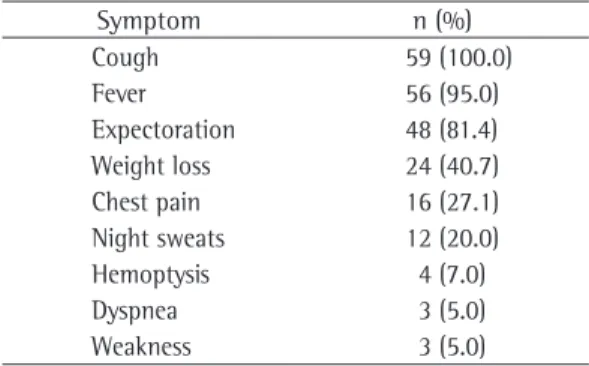
Table 3 - Clinical manifestations in 59 patients with tuberculous pneumonia.
Symptom n (%)
Cough 59 (100.0)
Fever 56 (95.0)
Expectoration 48 (81.4) Weight loss 24 (40.7) Chest pain 16 (27.1) Night sweats 12 (20.0) Hemoptysis 4 (7.0)
Dyspnea 3 (5.0)
estudo de 17 casos e revisão de literatura. Rev AMRIGS. 1994;38(4):299-303.
26. Picon PD, Caramori ML, Bassanesi SL, Jungblut S, Folgierini M, Porto Nda S, et al. Differences in the clinical and radiological presentation of intrathoracic tuberculosis in the presence or absence of HIV infection. J Bras Pneumol. 2007;33(4):429-36.
27. Daley CL, Small PM, Schecter GF, Schoolnik GK, McAdam RA, Jacobs WR Jr, et al. An outbreak of tuberculosis with accelerated progression among persons infected with the human immunodeficiency virus. An analysis using restriction-fragment-length polymorphisms. N Engl J Med. 1992;326(4):231-5.
28. IBGE [homepage on the Internet]. Brasília: Ministério do Planejamento, Orçamento e Gestão. [cited 2010 May 20]. Síntese de Indicadores Sociais 2008. Available from: http://www.ibge.gov.br/home/estatistica/ populacao/condicaodevida/indicadoresminimos/ sinteseindicsociais2008/default.shtm
29. Kala J, Sahay S, Shah A. Bronchial anthracofibrosis and tuberculosis presenting as a middle lobe syndrome. Prim Care Respir J. 2008;17(1):51-5.
30. de Araujo AC, Takeda AK, Herrero CB, de Freitas IW, Nakandakare IK, Guedes EA, et al. In vitro tuberculin reaction and cellular and humoral immune response in patients with pulmonary tuberculosis [Article in Portuguese]. Rev Saude Publica. 1983;17(2):94-111. T-lymphocytes at site of inflammation in the early phase
of the infection. Am Rev Respir Dis. 1987;136(3):575-9. 17. Canetti G. Dynamic aspects of the pathology and
bacteriology of tuberculous lesions. Am Rev Tuberc. 1956;74(2 Part 2):13-21; discussion, 22-7.
18. Eliasberg H, Neuland W. Die epituberkulose Infiltration bei tuberkülosen Säuglingen und Kindern. Jb Kinderheilk. 1920;93(1):88-93.
19. Fish RH, Pagel W. The morbid anatomy of epituberculosis. J Path Bact. 1938;47(3):593-601.
20. Graham EA, Burford TH, Mayer JH. Middle lobe syndrome. Postgrad Med. 1948;4(1):29-34.
21. Moreira JS, Porto NS, Mattos WL. Pneumonia Tuberculosa. In: Correa da Silva LC, editor: Compêndio de Pneumologia. São Paulo: Fundo Editorial Byk; 1991. p. 501-7.
22. Lee KS. Pulmonary tuberculosis. In: Müller NS, Silva CI, editors. Imaging of the chest. Philadelphia: Saunders-Elsevier; 2008. p. 322-31.
23. Leung AN. Pulmonary tuberculosis: the essentials. Radiology. 1999;210(2):307-22.
24. Picon PD, Rizzon CF, Hoeffel F JR, Porto NS, Oliveira ME. Pneumonia Tuberculosa. In: Picon PD, Rizzon CF, Ott WP, editors. Tuberculose: Epidemiologia, Diagnóstico e Tratamento em Clínica e Saúde Pública. Rio de Janeiro: Médica e Científica; 1993. p. 291-306.
25. Costa PG, Mensch MR, Oliveira MJ, Menezes JL, Gutierrez RS, Mattos WL. Pneumonia tuberculosa:
About the authors
Jose Moreira
Professor. Graduate Program in Pulmonology at the Universidade Federal do Rio Grande do Sul – UFRGS, Federal University of Rio Grande do Sul; Physician. Pereira Filho Ward, Santa Casa Hospital Complex in Porto Alegre, Porto Alegre, Brazil.
Jamila Belicanta Fochesatto
Graduate Student. Graduate Program in Pulmonology at the Universidade Federal do Rio Grande do Sul – UFRGS, Federal University of Rio Grande do Sul – Porto Alegre, Brazil.
Ana L Moreira
Professor. Federal University of Health Sciences of Porto Alegre, Porto Alegre, Brazil.
Marisa Pereira
Physician. Public Health Center no. 2, Secretaria da Saúde e Meio Ambiente do Estado do Rio Grande do Sul – SSMA-RS, Rio Grande do Sul Department of Health and the Environment – Porto Alegre, Brazil.
Nelson Porto
Radiologist. Physician. Pereira Filho Ward, Santa Casa Hospital Complex in Porto Alegre, Porto Alegre, Brazil.
Bruno Hochhegger