REGIONAL COOLING FOR REDUCING BRAIN
TEMPERATURE AND INTRACRANIAL PRESSURE
Luis Vicente Forte
1, Cássio Morano Peluso
2, Mirto Nelso Prandini
3,
Roberto Godoy
4, Salomon Soriano Ordinola Rojas
5Abstract – Objective: To evaluate the effectiveness of regional cooling for reducing brain temperature (BrTe) and intracranial pressure (ICP) in patients where conventional clinical treatment has failed. Method: Regional cooling was carried out using ice bags covering the area of the craniectomy (regional method) in 23 patients. The BrTe and ICP were determined using a fiber optic sensor. Thirteen patients (56.52%) were female. The ages ranged from 16 to 83 years (mean of 48.9). The mean APACHE II score was 25 points (11–35). The patients were submitted, on mean, to 61.7 hours (20–96) of regional cooling. Results: There was a significant reduction in mean BrTe (p<0.0001–from 37.1ºC to 35.2ºC) and mean ICP (p=0.0001–from 28 mmHg to 13 mmHg). Conclusion:
Our results suggest that mild brain hypothermia induced by regional cooling was effective in the control of ICP in patients who had previously undergone decompressive craniectomy.
KEy wORds: intracranial hypertension, intracranial pressure, cerebral hypothermia, brain edema, brain injuries.
Resfriamento cerebral regional para redução da temperatura e pressão intracraniana
Resumo – Objetivo: Avaliar a eficácia do resfriamento regional na redução da temperatura cerebral (TeCe) e pressão intracraniana (PIC) após falha das medidas clínicas convencionais de tratamento. Método: O resfriamento cerebral foi realizado com bolsas com gelo, colocadas sobre a área de craniectomia (método regional) em 23 doentes. A TeCe e PIC foram verificadas com sensor de fibra óptica. Treze (56,52%) eram do sexo feminino. A idade variou de 16 a 83 anos (média 48,96). A pontuação média no índice APACHE II foi 25 pontos (11–35). Os doentes foram submetidos, em média, a 61,7 horas (20–96) de resfriamento regional. Resultados:
Houve uma redução significativa da TeCe média (p<0,0001–de 37,1ºC para 35,2ºC) e da PIC média (p=0,0001– de 28 mmHg para 13 mmHg). Conclusão: Nossos resultados sugerem que o resfriamento regional foi eficaz no controle da PIC nos doentes submetidos, previamente, a craniectomia descompressiva.
PAlAvRAs-CHAvE: hipertensão intracraniana, pressão intracraniana, hipotermia induzida, edema encefálico, traumatismos encefálicos.
Hospital são Joaquim, Real e Benemérita sociedade Portuguesa de Beneicência de são Paulo, são Paulo sP, Brazil and Hospital Meridional, Cariacica Es, Brazil: 1Neurosurgeon, Intensive and Critical Medicine, Neurosurgical Intensive Care Unit, Hospital Meridional; 2Neurosurgeon, Neurosurgical Intensive
Care Unit, Hospital Meridional; 3Neurosurgeon, Associate Professor of department of Neurosurgery of Federal University of são Paulo (UNIFEsP), Escola
Paulista de Medicina, são Paulo sP, Brazil; 4Neurosurgeon, Head of Neurosurgery, Hospital são Joaquim; 5Intensive and Critical Medicine,
Neurosurgi-cal Intensive Care Unit, Hospital são Joaquim.
Received 14 April 2008, received in inal form 8 January 2009. Accepted 25 March 2009.
Dr. Luis Vicente Forte – Avenida Estudante José Júlio de Souza 2650 / 1402 - 29102-010 Vila Velha ES - Brasil. E-mail: [email protected]
Intracranial hypertension (ICH), associated with var-ious neurologic diseases, is responsible for high morbid-mortality1. The ideal treatment for ICH is focused on a
combined analysis of the hemodynamic and cerebral me-tabolism. However, there is no single system of treatment that can be used for all patients.
At the beginning of the 1990s, the works of Clifton et al.2, Marion et al.3 and shiozaki et al.4 demonstrated the
safety and possible beneits of mild to moderate
hypo-thermia in the treatment of patients with severe traumat-ic brain injury (TBI).
since that time, mild to moderate hypothermia has been one of the measures adopted for the treatment of acute ICH, particularly in patients where other conven-tional measures have failed5. However Clifton et al.6 did
present, hypothermia for the treatment of various acute cerebral disorders is based on the individual experience7,8.
despite the progressive consolidation of hypothermia as a therapy, a number of questions still need to be answered. we therefore present our results with the use of brain hypothermia induced by regional cooling for the control of intracranial pressure (ICP).
METHOD
The Research Project was approved by the Research Ethics Committee of the Hospital são Joaquim da Real e Beneméri-ta sociedade Portuguesa de Beneicência de são Paulo (CEPesp 236/04) and the Research Ethics Committee of the Hospital são Paulo–Federal University of são Paulo–Escola Paulista de Me-dicina (CEP 0421/06).
The study included patients with acute ICH refractory to clinical treatment (Table 1) and decompressive craniectomy; who have been submitted to at least 12 hours of regional cooling (re-stricted to the cephalic segment), in an intensive care unit (ICU). The ICP and brain temperature (BrTe) were continually mon-itored in all the patients, using a iber optic catheter. The seda-tion schemes (Table 2) were adjusted to attain and maintain lev-el 6 on the Ramsay scale.
Cerebral cooling was achieved using ice bags covering the area of the craniectomy (regional method). The total cerebral cooling time was dependant on the improvement demonstrat-ed in the Ct scans and the stabilization of the ICP during the re-warming phase. The patients were submitted, on mean, to 61.7 hours (20–96 hours) of regional cooling.
The following variables were evaluated in the “pre” and “post” hypothermia periods: cardiac frequency (CF), mean ar-terial pressure (MAP), central venous pressure (CvP), ICP, cere-bral perfusion pressure (CPP), PaCO2, sataO2 and BrTe. The start
time of the cerebral cooling was used as the reference for the study of “pre” and “post” variables. The PaCO2 was not
correct-ed according to temperature (“a-stat” strategy).
The “pre” variables corresponded with the last records, for each item, determined prior to the start of cooling. The “post” variables consisted of the mean for the values determined, for each item, in the 48 hours following the start of hypothermia. The effectiveness of hypothermia in the control of ICH was eval-uated by the variation in “pre” and “post” ICP values.
The evolution of the patients was classiied according to the Glasgow Outcome scale (GOs) at the moment of discharge from the ICU. No subsequent evaluation was carried out in the pres-ent study. The APACHE II scores, time of cerebral cooling and length of stay in the ICU were calculated.
Casuistic
Cerebral hypothermia was induced in 23 patients during the period of July 1997 to december 2003. Thirteen patients (56.52%) were female. The ages ranged from 16 to 83 years (mean 48.9). Regarding to the cause of the brain lesion, six patients were vic-tims of severe TBI (26.09%), ten presented complications follow-ing subarachnoid hemorrhage (sAH) (43,48%), four had suffered severe acute ischemic stroke (17.39%), two presented exacerba-tion of tumor edema (,69%) and one presented extensive intrac-erebral hemorrhage (4.34%). The interval between neurological deterioration and the beginning of hypothermia was, on mean, 21.4 hours (2–106 hours).
Statistical analysis
The quantitative variables were presented descriptively, in tables containing mean, standard deviation, median, minimum and maximum values. The pre and posthypothermia means were compared with the paired student-t test.
values of p<0.05 were considered statistically signiicant and signed with an asterisk.
RESULTS
Table 3 shows the values of the variables for 23 pa-tients in the pre and post hypothermia period. Table 4
Table 1. Clinical treatments of the intracranial hypertension.
1. General measures–respiratory, hemodynamic, biochemical and temperature control. 2. Postural care–lying position, elevated to 30 degrees, with centered head position. 3. sedation and analgesia + Neuromuscular blocker.
4. Optimized ventilation.
5. Hyperosmolar substance – Manitol 20% (0.25 g to 0.5 g/kg/dose). Hypertonic saline solutions were not used in the present study. 6. Increase in arterial pressure to maintain the pressure of cerebral perfusion – Noradrenalin (0.05 to 2.0 μg/kg/min).
Table 2. Medications most used in each pharmacological group.
1. Hypnotic = Propofol 1% (initially 1–2 mg/Kg and continuous infusion 0.3–4 mg/Kg/h).
2. Benzodiazepines = Midazolam (initially 0.05-0.1 mg/Kg and continuous infusion 0.5–1.5 μg/Kg/min). 3. Opioid = Fentanyl (initially 1.0–3.0 μg/Kg and continuous infusion 2.0–6.0 μg/Kg/h).
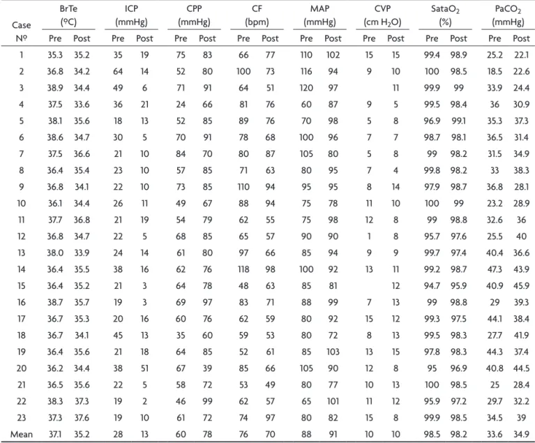
Table 3. Values of the variables studied, for each patient, in the pre and post regional cooling period.
Case Nº
BrTe (ºC)
ICP (mmHg)
CPP (mmHg)
CF (bpm)
MAP (mmHg)
CvP (cm H2O)
sataO2
(%)
PaCO2
(mmHg)
Pre Post Pre Post Pre Post Pre Post Pre Post Pre Post Pre Post Pre Post
1 35.3 35.2 35 19 75 83 66 77 110 102 15 15 99.4 98.9 25.2 22.1
2 36.8 34.2 64 14 52 80 100 73 116 94 9 10 100 98.5 18.5 22.6
3 38.9 34.4 49 6 71 91 64 51 120 97 11 99.9 99 33.9 24.4
4 37.5 33.6 36 21 24 66 81 76 60 87 9 5 99.5 98.4 36 30.9
5 38.1 35.6 18 13 52 85 89 76 70 98 5 8 96.9 99.1 35.3 37.3
6 38.6 34.7 30 5 70 91 78 68 100 96 7 7 98.7 98.1 36.5 31.4
7 37.5 36.6 21 10 84 70 80 87 105 80 5 8 99 98.2 31.5 34.9
8 36.4 35.4 23 10 57 85 71 63 80 95 7 4 99.8 98.2 33 38.3
9 36.8 34.1 22 10 73 85 110 94 95 95 8 14 97.9 98.7 36.8 28.1
10 36.1 34.4 26 11 49 67 88 94 75 78 11 10 100 99 23.2 28.9
11 37.7 36.8 21 19 54 79 62 55 75 98 12 8 99 98.8 32.6 36
12 36.8 34.7 22 5 68 85 65 57 90 90 1 8 95.7 97.6 25.5 40
13 38.0 33.9 24 14 61 80 97 66 85 94 9 9 99.7 97.4 40.4 36.6
14 36.4 35.5 38 16 62 76 118 98 100 92 13 11 99.2 98.7 47.3 43.9
15 36.4 35.2 21 3 64 78 48 63 85 81 12 94.7 95.9 40.9 45.9
16 38.7 35.7 19 3 69 97 83 71 88 99 7 13 99 98.8 29 39.3
17 36.7 35.3 20 16 60 76 62 59 80 92 15 12 99.3 97.5 44.1 38.4
18 36.7 34.1 45 13 35 60 59 53 80 72 8 13 99.5 98.3 27.7 41.9
19 36.4 35.6 21 18 64 85 52 61 85 103 13 15 97.8 98.3 44.3 37.4
20 36.2 34.4 38 51 67 39 85 66 105 90 12 8 95 96.9 40.8 44.5
21 36.5 35.6 22 5 58 72 53 49 80 77 10 13 100 98.5 25 28.4
22 38.3 37.3 19 2 46 99 62 57 65 101 11 12 95.9 97.2 29.7 32.2
23 37.3 37.6 19 10 61 72 74 97 80 82 15 8 99.9 98.5 34.5 39
Mean 37.1 35.2 28 13 60 78 76 70 88 91 10 10 98.5 98.2 33.6 34.9
CF: cardiac frequency; CPP: cerebral perfusion pressure; CvP: central venous pressure; ICP: intracranianal pressure; MAP: mean arterial pressure; PaCO2: parcial pression CO2; sataO2: arterial saturation O2; BrTe: brain temperature.
shows the mean, standard deviation, median, minimum and maximum value of the variables studied in the pre and posthypothermia period, as well as the results of the paired student-t test.
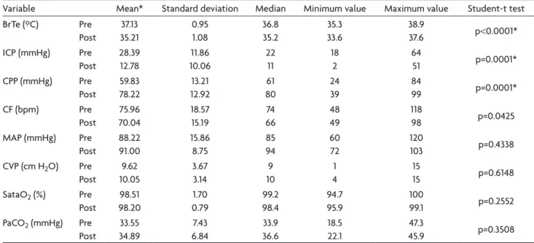
There was a signiicant reduction (p<0.0001) in mean BrTe, from 37.1ºC (35.3ºC–38.9ºC), prior to cooling, to 35.2ºC (33.6ºC–37.6ºC) in the post-hypothermia period (Table 4, Fig 1).
There was a signiicant drop (p=0.0001) in mean ICP from 28 mmHg (18–64 mmHg), in the pre-cooling period, to 13 mmHg (2 mmHg–51 mmHg) in the postcooling peri-od (Table 4, Fig 2). during the pre-cooling periperi-od, 19 of the 23 (82.60%) patients presented ICP higher than or equal to 20 mmHg and only two patients (8.69%) maintained an ICP over 20 mmHg after cooling.
There was a signiicant increase (p=0.0001) in mean
CPP, from 60 mmHg (24–84 mmHg) in the pre-cooling pe-riod, to 78 mmHg (2 mmHg–39 mmHg) in the post-cooling period (Table 4, Fig 3). In the pre-cooling period, 18 of the 23 (78.26%) patients presented CPP lower than or equal to 70 mmHg, while only four patients (17.39%) maintained CPP below 70 mmHg after cooling.
The control variables (CF, MAP, CPv, sataO2 and
Pa-CO2) did not present statistically signiicant alterations between the pre and post-hypothermia periods (Table 4).
DISCUSSION
The pathogenics alterations caused by primary brain lesion are extensive and complex. In general, they deter-mine a series of biochemical reactions which result in the: liberation of excitatory neurotransmitters and production of inlammatory agents, cytokine, metabolites of arachi-donic acid, nitric oxide, free radicals (reperfusion) and activity of the intracellular pathways which cause pro-grammed cell death (apoptosis).
Hypothermia inluences virtually all biochemical reac-tions caused by the primary brain lesion9. The
biochem-ical action can be explained by the physbiochem-ical properties involved. Biological reactions are catalyzed by enzymes and the characteristics of the environment (pH,
temper-Table 4. Measure of the variables in the pre and post regional cooling periods.
variable Mean* standard deviation Median Minimum value Maximum value student-t test
BrTe (ºC) Pre
Post
37.13 35.21
0.95 1.08
36.8 35.2
35.3 33.6
38.9
37.6 p<0.0001*
ICP (mmHg) Pre
Post
28.39 12.78
11.86 10.06
22 11
18 2
64
51 p=0.0001*
CPP (mmHg) Pre
Post
59.83 78.22
13.21 12.92
61 80
24 39
84
99 p=0.0001*
CF (bpm) Pre
Post
75.96 70.04
18.57 15.19
74 66
48 49
118
98 p=0.0425
MAP (mmHg) Pre
Post
88.22 91.00
15.86 8.75
85 94
60 72
120
103 p=0.4338
CvP (cm H2O) Pre
Post
9.62 10.05
3.67 3.14
9 10
1 4
15
15 p=0.6148
sataO2 (%) Pre
Post
98.51 98.20
1.70 0.79
99.2 98.4
94.7 95.9
100
99.1 p=0.2552
PaCO2 (mmHg) Pre
Post
33.55 34.89
7.43 6.84
33.9 36.6
18.5 22.1
47.3
45.9 p=0.3508
CF: cardiac frequency; MAP: mean arterial pressure; CPP: cerebral perfusion pressure; PaCO2: parcial pression CO2; CvP: central venous pressure; sataO2: arterial saturation O2; ICP: intracranianal pressure; BrTe: brain temperature.
Fig 1. Graph showing the mean brain temperature (ºC) and standard deviation in the pre and post regional cooling periods.
Fig 2. Graph showing the mean intracranial pressure (mmHg) and standard deviation in the pre and post regional cooling periods.
ature, pressure, among others) directly inluence the en-zyme activity.
The enzymes demonstrate an adequate performance within a narrow temperature range around 37ºC. The in-crease or reduction in temperature directly affects the speed of the biological reactions. This variation is deter-mined by the temperature coeficient (Q10), i.e. the Q10 of
the brain is approximately 2.3, therefore an alteration of 10º C in the temperature will lead to a 2.3-fold increase or decrease in brain metabolism10.
In the central nervous system, around 70% of the en-ergy produced in the form of ATP is used in the processes of active transport and nervous excitation. The lack of en-ergy compounds compromises the function of the pumps and ionic pathways which control the eflux of potassi-um ions (K+) and inlux of calcium ions (Ca2+), sodium ions (Na+) and water. Thus, cellular edema, activation of phos-pholipases and proteases result in irreversible lesion of the membranes, and death of the cell.
Hypothermia can stabilize the intracellular environ-ment, through the adequate functioning of the ionic
pumps and pathways. Cooling causes reduction in ener-gy demand and a relative increase in the concentration of energy compounds (ATP) which are probably used in the maintenance of active transport.
The ability to maintain the potential of the cell mem-brane reduces the ionic permeability, and the activity of the Na+/K+ and Na+/Ca2+ ion pumps determine the func-tional evidences of the strategy of saving energy during the cooling11. Thus, hypothermia hinders the inlux of Ca2+
and the consequent activation of proteolytic enzymes. The clinical application of cerebral hypothermia be-gan with the pioneering work of neurosurgeon Temple Fay, in 1938, but the program was abandoned during the second world war12.
From the 1950s, studies on profound cerebral hypo-thermia beginning. despite the promising results in ex-perimental models13, the practical application led to
var-ious complications (hemodynamic and hydroelectrolytic disturbances, alterations in coagulation and increased in infections) and was rapidly suspended.
In the 1980s, experimental studies demonstrated the
Table 5. APACHE II score, risk of death and evolution of each patient.
Case Nº APACHE II Risk of death (%) stay in ICU (days) GOs
1 31 69,5 8 1
2 31 69,5 6 1
3 22 34,1 31 5
4 32 72,5 7 1
5 26 52,3 19 3
6 22 38 26 3
7 19 28,3 11 4
8 29 80,8 16 1
9 27 56 78 2
10 18 25,5 29 3
11 29 63 39 3
12 30 66,3 44 1
13 24 45,1 17 3
14 28 59,5 5 1
15 35 80,3 48 1
16 19 28,3 18 3
17 19 28,3 16 4
18 20 31,4 98 3
19 33 75,3 8 1
20 28 59,5 12 1
21 24 45,1 18 3
22 28 59,5 11 1
23 11 10,9 20 5
Mean 25 51,3 25,4 –
beneficial effects of mild to moderate cerebral hypo-thermia in the treatment of acute brain lesions, and en-abled the application of new cerebral cooling techniques. Prandini et al.14 used surface cooling, with ice packs
ap-plied over the area of the craniectomy to induce cere-bral hypothermia in provoked ischemic lesion in rabbits brain. The experimental results suggested that the induc-tion of mild hypothermia (around 34ºC) exerts a neuro-protective and therapeutic effect, following severe acute ischemic brain lesion.
At the beginning of the 1990s, three isolated clini-cal trails demonstrated the beneits of mild to moderate therapeutic hypothermia in severe TBI2-4.
Following these trials, systemic hypothermia began to be used in others clinical situations: severe acute ischemic stroke15-17, subarachnoid hemorrhage8, fulminating
hepati-tis and anoxia-ischemic encephalopathy8,18. Our study was
also comprised of patients with refractory ICH, with dif-ferent causes, and a predominance of traumatic and vas-cular lesions.
However, the results of the further multicentric study did not conirm the initial indings. Clifton et al. carried out a randomized, prospective multicentric controlled study of 392 patients with severe TBI (GCs≤8). They did not ind any signiicant difference in mortality between the group submitted to hypothermia and the group submitted to normothermia, after six months of TBI. They recorded a signiicant reduction in ICP with the hypothermia group, but associated with a higher tendency to infections6.
A possible explanation for the disassociation between the results is the lack of standardization of the method19. In
recent years, different techniques, objectives and evalua-tion criteria have been used and compared, indiscriminately. Thus, we should consider the following aspects of the technique, which are important for analyzing the results of the therapeutic hypothermia: intensity of cooling; the cooling method used; the site at which the temperature is determined; the start and time of maintenance, and the rewarming strategy.
Intensity of cooling
For every 1ºC drop in temperature there is a reduction in the cerebral metabolism of around 6%10. Meanwhile, an
increase in temperature of just 1ºC to 2ºC will worsen the primary lesion and increase tissue necrosis20.
Induced hypothermia is classiied according the tem-perature achieved. The accentuated body cooling, for pro-longed periods, is associated with high rates of morbid-mortality.
The majority of authors adopt a body core tempera-ture between 32ºC and 33ºC, which characterizes the hy-pothermia as moderate and increases the risk of complica-tions. This choice was made because the initial
experimen-tal and clinical trials indicate that a potent neuroprotec-tive action is obtained at this temperature. As Tokutomi et al.21 we opted for mild cerebral hypothermia (up to 34.0ºC)
as the target temperature in our treatment scheme.
Cooling method
The choice of cooling method was another important technical aspect. This decision directly inluenced the in-tensity and speed of hypothermia induction.
The techniques are divided into: (1) surface cooling
(a) Blankets2,3,6,15,16,21
(b) Ice packs or immersion in cold water15,22
(c) Cooling helmet7,22
(2) deep cooling
(a) with cold solutions • Intravenous infusion8,23
• Gastric washing3,6
• Peritoneal washing24
• Nasopharyngeal cooling22
(b) Closed intravascular system16
The cooling techniques were associated, in the majori-ty of instances, with the circulation of cooled air leading to a decrease in environmental temperature to around 18ºC.
However, the brain temperature is not constant. The cerebral metabolism is responsible for the production of heat, and the loss occurs through conduction, convec-tion and radiaconvec-tion.
The conduction of heat occurs through the tissues. In normal conditions, this mechanism contributes shortly to temperature control, as the intact cranium is an important barrier (thermal isolator). In convection, heat loss occurs as a result of the CBF and arterial blood temperature.
Gentilello25 describes in detail, the physical mechanisms
of heat loss or transfer. Heat conduction occurs though contact between the two masses. The transfer rate depends on the temperature gradient, interface, size of the contact area and thermal conductivity of the material. The transfer is also affected by the distance that the heat needs to trav-el, i.e. the thickness of the skin and subcutaneous tissue. Heat can also be transferred to the environment in the form of electromagnetic waves, which do not require con-tact with any mass or luid. Radiation is proportional to the fourth power of the temperature gradient, surface area of the body and characteristics of the heat emission itself.
Site of temperature determination
The body temperature luctuates throughout the day, and varies according to regions. disease can exacerbate the temperature gradients. Therefore, the body temper-ature observed in different regions does not relect the intracranial temperature. The sites for determining tem-perature can be divided into:
(1) surface: axiliary region26
(2) deep
(a) Core: esophagus27, urinary bladder3,15,16, internal
jugular vein28, lower cava vein16, pulmonary artery18,
rectum5,21,22 and tympanic membrane27,18
(b) Intracranial: lateral ventricle22, brain3,5,7,21,26 and
epidural space22
The determination of body core temperature is used generally for the control of patients admitted to the ICU. A difference between core and brain temperature (brain-core temperature gradient) was observed in patients with acute brain lesion. The BrTe tends to be 1ºC higher than the core temperature.
Rumana et al.28 demonstrated that this difference in
temperature tends to increase in situations that compro-mise the CPP. They observed that in episodes where there is a drop in CPP, to levels of 20 mmHg a 50 mmHg, the brain-body core gradient can reach 2.1ºC. Henker et al.29,
also observed that the difference between brain and core temperature can be as high as 2.0ºC.
The BrTe should act as a guide for therapeutic inter-ventions in patients with acute brain lesion, and serve as a control for cooling until the desired temperature is reached. various different sensors were developed to mon-itor BrTe, the majority of which are coupled to ICP sensors.
Start and duration
The best moment to induce hypothermia depends on the objectives to be reached. It is likely that therapeutic window remains only for a few hours, in order to obtain the neuroprotective action of the cerebral hypotherm-ia30. In this situation, cooling may be used as a temporary
strategy (a bridge) until the deinitive treatment can be carried out8. In our study, the regional cooling was
main-tained for around 61.7 hours (20–96 hours) for the con-trol de ICP. In the majority of the studies reviewed, the maintenance span of hypothermia was 24 to 48 hours. de-spite these differences, we believe that our favorable re-sults are related to the rapid reduction in BrTe, provided by the regional cooling, and the more prolonged mainte-nance of the hypothermia.
Strategy in the rewarming phase
The rewarming phase consisted of a new challenge specially for patients submitted to systemic hypothermia. Two strategies may be used: passive or active rewarming.
The passive form is based on the endogenous generation of heat, resulting in a gradual increase in central tempera-ture. However, this strategy prolongs the total time of hy-pothermia and can, in theory, increase the harmful effects. Active rewarming was used in the majority of studies. The temperature was raised using a blanket, lamps, and heated solutions. The speed of rewarming varied between 0.5ºC every two hours3 and 1.0ºC per day for three days4.
The increase in ICP is the most feared complication in the rewarming phase, but it is not the only one15. we
should remember that hyperthermia increases the neu-ronal lesion and worsens the development of patients with severe brain lesion.
To avoid these complications, schwab et al.15
recom-mend the use of slow rewarming for the control of the ICP and CPP, in patients with extensive acute ischemic stroke submitted to moderate hypothermia.
Our technique enabled gradual and passive rewarm-ing of the brain, with the intermittent application of ice packs to the area of the craniectomy (regional method), to avoid an abrupt elevation in BrTe.
In conclusion, despite the limited number of patients, our results suggest that mild cerebral hypothermia, in-duced by regional cooling, was effective in the control of intracranial pressure in patients who had previously un-dergone decompressive craniectomy. However, cerebral hypothermia, for the treatment of various acute cerebral lesions, continues to be used based on the experience of each author, and on the results of isolated clinical series. In our understanding, the best explanation for the dis-crepancy in the results observed in the literature is the lack of clearly-deined criteria for the indication, induc-tion, maintenance and suspension of cerebral hypotherm-ia, which led to a comparison of the results obtained by different methods.
REFERENCES
1. Narayan RK, Greenberg RP, Miller JD, et al. Improved coni
-dence of outcome prediction in severe head injury. A compara-tive analysis of the clinical examination, multimodality evoked potentials, CT scanning, and intracranial pressure. J Neurosurg 1981;54(6):751-762.
2. Clifton GL, Allen S, Barrodale P, et al. A phase II study of mod-erate hypothermia in severe brain injury. J Neurotrauma 1993; 10:263-271.
3. Marion DW, Obrist WD, Carlier PM, et al. The use of moder-ate therapeutic hypothermia for patients with severe head in-juries: a preliminary report. J Neurosurg 1993;79:354-362. 4. Shiozaki T, Sugimoto H, Taneda M, et al. Effect of mild
hypo-thermia on uncontrollable intracranial hypertension after se-vere head injury. J Neurosurg 1993;79:363-368.
rela-tionship to intracranial pressure, cerebral perfusion pressure, cerebral blood flow, and outcome. J Neurotrauma 2002;19: 559-571.
6. Clifton GL, Miller ER, Choi SC, et al. Lack of effect of induc-tion of hypothermia after acute brain injury. N Engl J Med 2001;344:556-563.
7. Wang H, Olivero W, Lanzino G, et al. Rapid and selective ce-rebral hypothermia achieved using a cooling helmet. J Neu-rosurg 2004;100:272-277.
8. Polderman KH, Rijnsburger ER, Peerdeman SM, et al. Induc-tion of hypothermia in patients with various types of neuro-logic injury with use of large volumes of ice-cold intravenous
luid. Crit Care Med 2005;33:2744-2751.
9. Gunn AJ, Gunn TR. The ‘pharmacology’ of neuronal rescue with cerebral hypothermia. Early Hum Dev 1998;53:19-35. 10. McCullough JN, Zhang N, Reich DL, et al. Cerebral metabolic
suppression during hypothermic circulatory arrest in humans. Ann Thorac Surg 1999;67:1895-1921.
11. Boutilier RG. Mechanisms of cell survival in hypoxia and hy-pothermia. J Exp Biol 2001;204:3171-3181.
12. Wang H, Olivero W, Wang D, et al. Cold as a therapeutic agent. Acta Neurochir (Wien) 2006;148:565-570.
13. Rosomoff HL. Hipothermia and cerebral vascular lesions. I. Experimental interruption of the middle cerebral artery dur-ing hypothermia. J Neurosurg 1956;13:332-343.
14. Prandini MN, Lacanna SN, Valente PR, et al. Regional mild hypothermia in the protection of the ischemic brain. Acta Cir Bras 2002;17:232.
15. Schwab S, Georgiadis D, Berrouschot J, et al. Feasibility and safety of moderate hypothermia after massive hemispheric in-farction. Stroke 2001;32:2033-2035.
16. Georgiadis D, Schwarz S, Aschoff A, et al. Hemicraniectomy and moderate hypothermia in patients with severe ischemic stroke. Stroke 2002;33:1584-1588.
17. Els T, Oehm E, Voigt S, et al. Safety and therapeutical beneit
of hemicraniectomy combined with mild hypothermia in com-parison with hemicraniectomy alone in patients with malig-nant ischemic stroke. Cerebrovasc Dis 2006;21:79-85. 18. Zeiner A, Holzer M, Sterz F, et al. Mild resuscitative
hypo-thermia to improve neurological outcome after cardiac arrest.
A clinical feasibility trial. Hypothermia After Cardiac Arrest (HACA) Study Group. Stroke 2000;31:86-94.
19. Clifton GL, Choi SC, Miller ER, et al. Intercenter variance in clinical trials of head trauma-experience of the National Acute Brain Injury Study: hypothermia. J Neurosurg 2001;95:751-755. 20. Ginsberg MD, Busto R. Combating hyperthermia in acute
stroke: a signiicant clinical concern. Stroke 1998;29:529-534.
21. Tokutomi T, Morimoto K, Miyagi T, et al. Optimal temperature for the management of severe traumatic brain injury: effect of hypothermia on intracranial pressure, systemic and intracranial hemodynamics, and metabolism. Neurosurgery 2003;52:102-111. 22. Mellergard P. Changes in human intracerebral temperature in
response to different methods of brain cooling. Neurosurgery 1992;31:671-677.
23. Baumgardner JE, Baranov D, Smith DS, et al. The
effective-ness of rapidly infused intravenous luids for inducing mod
-erate hypothermia in neurosurgical patients. Anesth Analg 1999;89:163-169.
24. Cancio LC, Wortham WG, Zimba F. Peritoneal dialysis to in-duce hypothermia in a head-injured patient: case report. Surg Neurol 1994;42:303-307.
25. Gentilello LM. Practical approaches to hypothermia. In Ad-vances in trauma and critical care 1994,9:39-79.
26. Yoo DS, Kim DS, Park CK, et al. Signiicance of temperature
difference between cerebral cortex and axilla in patients un-der hypothermia management. Acta Neurochir 2002;81(Suppl): S85-S87.
27. Schuhmann MU, Suhr DF, v Gosseln HH, et al. Local brain surface temperature compared to temperatures measured at standard extracranial monitoring sites during posterior fossa surgery. J Neurosurg Anesthesiol 1999;11:90-95.
28. Rumana CS, Gopinath SP, Uzura M, et al. Brain temperature exceeds systemic temperature in head-injured patients.Crit Care Med 1998;26:562-567.
29. Henker RA, Brown SD, Marion DW. Comparison of brain tem-perature with bladder and rectal temtem-peratures in adults with severe head injury. Neurosurgery 1998;42:1071-1075. 30. Markarian GZ, Lee JH, Stein DJ, et al. Mild hypothermia: