GASTROENTEROLOGIA EXPERIMENT
AL / EXPERIMENT
AL
GASTROENTEROLOGY
.031)*/&%0&4/05130.05&
&401)"(&"-$"3$*/0(&/&4*4
*/3"54&9104&%50
%*&5):-/*5304".*/&
Carlos Frota
DILLENBURG
1, Cleber Dario Pinto
KRUEL
1, Carlos Thadeu
CERSKI
2,
Maria Isabel
EDELWEISS
2, Tiago Luís Dedavid e
SILVA
3and André Silvio
SCHIER
3ABSTRACT –Background - The high incidence of esophageal cancer in the north of Iran has been associated to the consumption of opium
and exposure to nitrosamines. Diethylnitrosamine has an established potential of producing experimental cancer in the esophagus and liver. Aim - To evaluate by histopathology the effect of oral administration of morphine and diethylnitrosamine during 23 weeks on the hepatic and esophageal carcinogenesis on 176 rats. Methods - We divided the rats into the following groups: Morph: morphine; Den: diethylnitrosamine; Den+morph: Den and morphine in the same solution; Den/morph: Den and morphine in different solutions and days. Results - Morphine did not promote neoplasias. The highest neoplastic incidents were found: a) in the esophagus, Den in relation to Den/morph and Den+morph (71.1%, 55.8%, and 50.0%); b) in the liver, Den and Den/morph in relation to Den+morph (73.8%, 81.4%, and 40.9%); c) higher incident of hepatic neoplasia than esophageal in Den/morph (81.4% and 55.8%). Different doses of diethylnitrosamine were ingested among the groups Den, Den/morph, and Den+morph, respectively 2.9, 2.8, and 2.3 mg/kg/day. Conclusions - These results show that the morphine did not promote esophageal carcinogenesis and may have stimulated the hepatic metabolism of the first pass of the carcinogen.
HEADINGS – Esophageal neoplasms. Carcinoma, squamous cell. Morphine. Diethylnitrosamine. Rats.
*/530%6$5*0/
Esophageal cancer is among the 10 most frequent in the world and if we consider the social impact on the endemic regions, the problem becomes a severe scourge for those populations. An intimate correlation has been seen between sickness and regional habits, meaning the way people live(43),
and it has been shown that the origin of esophageal cancer is associated with different factors in each region(16, 26).
The high incidence of esophageal cancer has been associated with the consumption of opium habit in high-incidence regions in the north of Iran(15, 19, 24) and with the exposure to
nitrosamines in China, mainly through their diet(8, 21, 22, 41).
The reasons for the association between opium and cancer of the esophagus are not known, but trials show that a dose of morphine sulfate, the main alkaloid of opium, caused an alkylation increase in the DNA in the esophagus and a decrease in the liver in rats that received diethylnitrosamine (DEN) after the morphine. DEN, a nitrosamine, is considered one of the substances with the highest potential for producing cancer in the esophagus and liver of rats and mice(20, 31), and the alkylation of
the DNA constitutes a known alteration precursory to the carcinogenesis provoked by DEN in these organs.
This way the morphine would cause a lowering of the hepatic carcinogenesis and an increase of the esophageal by influencing the hepatic metabolism of DEN. Once suggested that the observations made in rats could be imputed to man(31), these would be possible metabolic
evidences, in an acute trial, for the association between opium and cancer of the esophagus in rats and therefore, by extension, in humans.
In the present study, we offered DEN in the rat’s drinking water for 3 days during each one of the 23 weeks, and compared with the groups that consumed morphine simultaneously or not with DEN. We evaluated the effect of the chronic administration of morphine and DEN on the hepatic and esophageal carcinogenesis.
.&5)0%4
We purchased the DEN from SIGMA (St. Louis, MO, USA): N-0756, density – 0.95 g/mL, and the morphine sulfate from CRISTÁLIA (Itapira, SP, Brazil): DCB 0856.03-7, density - 10 mg/mL. We obtained the 176 Wistar rats, ranging from 185-215 g, from Bioterio of the State Foundation of Health Protection and Research in Rio Grande do Sul, Porto Alegre, RS, Brazil. Water,
Departments of 1Surgery and 2Pathology, Federal University of Rio Grande do Sul 3School of Medicine, Porto Alegre, RS, Brazil.
food (Nuvilab CR1, based on criteria from the National Research Council and National Institute of Health – USA), and the diluted substances were changed 3 times a week at which time we measured the quantities ingested of each solution. All of the animals received human care and the protocols were approved by the Scientific Commission and the Health Research and Ethics Commission of GPPG-HCPA (the Research and Post-Graduate Group of “Hospital de Clínicas” of Porto Alegre, RS, Brazil).
In a Comparative Study of Multiple Groups, we divided 176 rats into groups with 44 animals and they ingested:Morph:
morphine;Den: DEN;Den+morph: DEN + morphine in the same solution;Den/morph: DEN and morphine in different solutions and days. The rats ingested morphine solution during 4 days a week in theDen/morph group and 3 in the other groups, while the DEN was ingested during 3 days a week in the respective groups. We used the estimated doses of 5 mg/kg/day for morphine and DEN. The animals were weighed in the beginning, at 3 months, and before euthanasia, which occurred at 161 days (23 weeks). We dried esophagi and livers and immediately placed them in 10% buffered formalin until analysis.
We examined the esophagus in its entire extension since it wraps around itself (like a jellyroll) and removed three samples to represent the liver. Pathologists that were unaware of the group of origin of the specimen, examined the sections in hematoxylin-eosin (HE) under a common light microscope. We considered always the lesion of higher grade of alteration of each piece.
We classified the esophagi as(32):1) normal histology, 2)
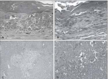
hyperplasia, 3) esophagitis, 4) papilloma, 5) low-grade dysplasia, 6) high-grade dysplasia (including carcinoma in situ), and 7) invasive carcinoma: of the mucosa, of the muscularis of mucosa, and of the sub-mucosa (Figure 1A and 1B). We considered malignant neoplasias the high-grade dysplasia and invasive carcinoma, and called them neoplastic esophageal lesions or esophageal CA.
We classified the livers as(5) 1) normal histology, 2) focus
of clear cells, 3) neoplastic nodules, and 4) hepatocellular carcinoma (Figure 1C and 1D). We considered malignant neoplasias the focus of clear cells, the neoplastic nodules, and the hepatocellular carcinoma, and called them neoplastic hepatic lesions or hepatic CA.
The quantitative variables was analysed by ANOVA with a criterion of classification and the differences localized by the Tukey Test. Among the categorical variables, we compared the groups by chi-square with the differences located among the groups by the post-hoc procedure proposed by Zar. The significance level adopted wasD = 0.05.
3&46-54
We did not observe changes in conduct of the rats subordinate to the ingestion of morphine. There were two non-programmed deaths at 31 and 74 days of the trial, both of them in the
Den group. Histopathologic analysis was not carried out in 13 esophageal specimens and in 3 hepatic: 2 due to non-programmed deaths and the others due to lack of material for the analysis. Therefore, we submitted 163 esophaguses and 173 livers to the histological exam.
The morphine ingested in isolated form (((Morph) did not induce significant carcinogenesis in any of the organs analyzed (Table 1).
The incidence of neoplastic esophageal lesions was larger in theDen group (71.1%) in relation to the other groups (P<0.001). This incidence was similar between the two groups that ingested DEN and morphine (((Den+morph 50.0% and Den/morph 55.8%) (Table 1).
The incidence of neoplastic hepatic lesions was less in the group that ingested DEN and morphine simultaneously in the same solution (((Den+morph 40.9%) than in the others that
FIGURE 1.(A) HE 200x, high-grade dysplasia in esophagus of animal in theDen+morph group, dyskaryotic cells affecting more than half of the squamous epithelium, without invading the base membrane; (B) HE 200x, invasive epidermoid carcinoma that affects the muscularis of mucosa in the esophagus of animal in theDen+morph group; (C) HE 40x, focus of clear cells in liver of animal in theDen group; (D) HE 100x, hepatocelullar carcinoma in animal of theDen group
TABLE 1.Neoplastic incidences and doses of the substances broken down by treatment group
Morph Den Den+morph Den/morph P
Esophageal CA n = 40
0 (0.0) a
n = 38 27 (71.1) c
n = 42 21 (50.0) b
n = 43 24 (55.8) b
< 0.001
Hepatic CA n = 44
1 (2.3) a
n = 42 31 (73.8) c
n = 44 18 (40.9) b
n = 43 35 (81.4) c
< 0.001
DEN dose - 2.9 ± 0.3a 2.3 ± 0.1b 2.8 ± 0.1c < 0.001
Morphine dose 2.5 ± 0.2a - 2.3 ± 0.1 b 2.4 ± 0.1a < 0.001
Data is expressed in numbers (percentages) of positive cases or as an average ± standard deviation of mg/kg/day. Distinct letters-index represent statistically significant differences among the groups.Morph group: ingested morphine;
ingested the carcinogen (Den/morph 81.4% and Den 73.8%) (P<0.001) (Table 1).
The possible effect of the morphine on the metabolism of first hepatic pass of the DEN may be observed in Table 1. The Den and Den+morph presented a similar incidence of esophageal and hepatic lesions while the Den/morph presented a higher percentage of hepatic lesions than esophageal ones (P = 0.013).
The esophageal histology was normal for the vast majority of the animals of the Morph group (95.0%), in contrast with the groups that ingested DEN, which were normal in only 2.6% of the Den, 16.7% of the Den+morph, and 14.0% of the Den/ morph. The Den group caused a greater incidence of alterations, with a tendency to lesions of greater epithelial destructuring than the groups treated also with morphine (Den+morph and Den/morph) (Table 2).
Almost all of the animals of the Morph group presented a normal hepatic histology (97.7%), which did not occur with the groups that ingested DEN. In these, the histology was normal in 26.2% of the Den, 59.1% of the Den+morph, and 18.2% of the Den/morph. Though the Den/morph group represented greater incidence of changes, the three cases of hepatocellular carcinoma occurred in the Den group (Table 3).
The dose ingested was based on the quantity of weekly solution ingested, on the animal’s weight, and on the concentration of the substances in the liquid offered the animals. The average dose ingested was inferior to the estimate of 5 mg/kg/day in all the groups.
Among the groups exposed to the carcinogen, they all ingested different doses of DEN among themselves (Den: 2.9 mg/kg/ day, Den+morph: 2.3 mg/kg/day, Den/morph: 2.8 mg/kg/day) (P<0.001). From the groups that ingested morphine (Morph: 2.5 mg/kg/day; Den+morph: 2.3 mg/kg/day; and Den/morph: 2.4 mg/kg/day), the Den+morph group ingested less morphine than the other groups (P<0.001) (Table 1).
%*4$644*0/
The nitrate compounds are presented in the diet or in the environment in doses much below those necessary to develop cancer, and for this reason the biggest concern is with chronic exposure. The carcinogenic nitrates are present in the diet (mainly in food preservatives, colorings, and flavorings), in tobacco and alcoholic beverages (beer, whisky, and liquors), through occupational exposure (rubber, pesticide, cosmetic, and leather tanning industries), and in personal and domestic hygiene products (cosmetics, shampoos, and detergents)(9, 13, 20, 36). Their
hepatotoxic and carcinogenic potential has been established both in humans as well as in animals(6, 27, 35) and described that
the nitrosamines produce the alkylation of the DNA in human tissues in vitro(20). Based on these studies, it has been suggested
that nitrosamines could be related to cancer of the esophagus in man(2, 14, 21, 22, 23).
The discovery that nitrosamines present a well-defined organotropism allowed the development of study models of cancer in various organs. Many authors used the model of esophageal and hepatic carcinogenesis induced by DEN in mice and rats and showed various levels of neoplastic incidence depending on the dose and time of duration of their studies(4, 17, 18, 29, 30, 32, 33, 35). The
knowledge that the N-nitroso compounds show similar biological activity in animal and human tissues would suggest that the observations made in rats could be extrapolated to man(2, 31).
The incidence of cancer of the esophagus is influenced by different factors in the various endemic areas of the world. In Europe and United States it is related with the exposure to alcohol and tobacco, associated or not(7, 40), in the south of Brazil with
the consumption of hot matte tea(11), and in the north of Iran it
has been associated by epidemiologists with the smoking of opium and the ingestion of opium pipe residues (sukhteh)(19).
TABLE 2. Relative frequency of the results from the esophageal microscopy broken down by treatment groups
Morph
(n = 40)
Den
(n= 38)
Den+morph
(n = 42)
Den/morph
(n = 43) P
Normal 38 (95.0) 1 (2.6) 7 (16.7) 6 (14.0) < 0.001
Hyperplasia 0 (0.0) 2 (5.3) 4 (9.5) 1 (2.3) 0.166
Esophagitis 0 (0.0) 2 (5.3) 4 (9.5) 3 (7.0) 0.283
Papilloma 1 (2.5) 0 (0.0) 1 (2.4) 0 (0.0) 0.572
Low-grade dysplasia 1 (2.5) 6 (15.8) 5 (11.9) 9 (20.9) 0.084
High-grade dysplasia 0 (0.0) 19 (50.1) 19 (45.2) 18 (41.9) < 0.001
Invasive carcinoma 0 (0.0) 8 (21.0) 2 (4.8) 6 (14.0) < 0.001
Mucous 0 (0.0) 6 (15.8) 2 (4.8) 4 (9.3) 0.051
Muscle of mucous 0 (0.0) 1 (2.6) 0 (0.0) 2 (4.7) 0.314
Sub-mucous 0 (0.0) 1 (2.6) 0 (0.0) 0 (0.0) 0.346
Data is expressed in numbers (percentage) of cases. Morph Group: ingested morphine; Den: diethylnitrosamine (DEN); Den+morph: DEN and morphine in the same solution; Den/morph: DEN and morphine in different solutions and days
TABLE 3. Relative frequency of the results from the hepatic microscopy broken down by treatment groups
Morph
(n = 44)
Den
(n= 42)
Den+morph
(n = 44)
Den/morph
(n = 43) P
Normal 43 (97.7) 11 (26.2) 26 (59.1) 8 (18.6) < 0.001
Focus of clear cells 1 (2.3) 25 (59.5) 8 (18.2) 28 (65.1) < 0.001
Neoplastic nodules 0 (0.0) 3 (7.1) 10 (22.7) 7 (16.3) 0.005
Hepatocellular carcinoma 0 (0.0) 3 (7.1) 0 (0.0) 0 (0.0) 0.023
Experiments show that ethanol alters the pharmacokinetics of the nitrosamines(1, 37, 38, 39). It causes higher exposure of
nitrosamines to the esophageal tissue and capable of inducing cancer of the esophagus in animals(1, 3, 37, 38, 39). RIBEIRO-PINTO
and SWANN(31) showed that morphine, opium’s main alkaloid,
also changes the pharmacokinetics and distribution of DEN in a way similar to ethanol. This reinforces the hypothesis that these two substances have a common base in their influence on esophageal carcinogenesis: their effects on pharmacokinetics from the nitrosamines to which man is exposed.
The dose of 5 mg/kg/day of DEN used in this study during 3 days a week was based on the studies of RUBIO et al(33) and
RIBEIRO-PINTO and SWANN(31). The first author administered
DEN in the concentration of 0.04 mL/L of drinking water (7 mg/kg/day) during 3 days a week, which promoted esophageal tumors after periods of 4 and 6 months in mice (1 and 3 tumors/ cm of esophageal mucous, respectively), with low mortality. RIBEIRO-PINTO and SWANN(31) demonstrated the alkylation
of the hepatic and esophageal DNA with 3 mg/kg/dose of DEN. Other authors confirm the carcinogenic power of these doses: GIBEL(17) provoked esophageal cancer in 30% and 56% of the
rats with the respective doses of 2.5 and 10 mg/kg/day, and SCHMÄHL et al(35) provoked hepatocellular carcinomas in 92%
of the rats of his trial in a period of 138 ± 10 days using doses between 5 and 7.5 mg/kg/day of DEN, ingested daily.
In relation to morphine, it has been demonstrated that a single dose of 5 mg/kg increased the alkylation of the esophageal DNA of rats by 90% and lowered the hepatic by 10% when administrated 45 minutes previously to a single dose of DEN. These results do not change significantly with the increase of the doses of morphine sulfate from 10 and 20 mg/kg(31). In the
present study, we used 5 mg/kg/day of morphine sulfate, which would not cause changes in the activity of the animals and provoke the pharmacokinetic changes referred to above(31).
The dose ingested by the animals in this study was substantially lower to the dose estimated. Though the esophageal and hepatic carcinogenesis are known to be dependent on the dose of DEN, RUBIO et al.(33, 34) described that the time elapsed
would also be of great importance in the formation of tumors in the esophagus of mice. According to these authors, clones of esophageal cells would be “programmed” for carcinogenesis in early stages of the treatment with DEN, and that a large number of tumors would occur in longer intervals even after only a few doses of DEN. While animals treated for 3 months presented a tumor index (TI) = 0.9, animals treated for 3 months and maintained alive for 4 additional months, with a carcinogen-free diet, presented a TI that was 5 times higher (TI = 4.6)(34). In this present study, a percentage of animals
affected by esophageal neoplastic lesions higher than that estimated in the beginning of the study was developed in the Den group (obtained = 71%; estimated = 30%), despite the doses ingested were only 59% of that expected in this group. Based on this, it can be inferred that both the time elapsed of 23 weeks as well as the doses actually ingested of DEN were enough to provoke the carcinogenic effects expected in the two organs evaluated with practically no mortality.
The dose ingested of morphine varied between 2.3 and 2.5 mg/kg/day, which is only 48% of the estimated 5 mg/kg/day. These doses are equivalent to 170 mg/day for man, which are similar to the levels used in analgesics in occidental medicine and making them comparable to this population of chronic users of morphine. On the other hand, the dose is possibly inferior to those reported in the population of addicts in the north of Iran, who received 3 g of opium daily(19). For this reason, it most likely
does not apply to this population where the relation between opium and cancer of the esophagus was first described(12).
With the ethical intention of lowering the unnecessary euthanasia of animals, a control group with eight rats was created, which ingested only water. This group presented a normal macro and microscopic analysis in the present study, the same of hundreds of animals that used the same source of water in other research from this Institution(18). This group was excluded from the analysis
since the number of eight animals caused a strong asymmetry in the data, jeopardizing an adequate statistical analysis.
Among the animals exposed only to DEN, 71% presented esophageal neoplastic lesions and 73% hepatic, which are incidents similar to the other authors(17, 29, 25, 35). Among the groups exposed
only to morphine, esophageal neoplasias were not found, but only one pre-neoplastic hepatic lesion (2.3%) was developed in one animal group of Morph, a focus of clear cells. BANNASCH et al.(5) affirm that although these lesions can occur in control
animals, the incidence is low, as is the incidence of naturally occurring hepatocellular carcinoma.
There was a higher incidence of esophageal neoplastic lesions in the animals that ingested only the carcinogenic (71.1%) (P<0.001) in relation to the two groups that ingested DEN and morphine (Den+morph and Den/morph). These two groups presented a neoplastic incidence similar between themselves (50% and 55.8%) (Table 1). Even though the doses of DEN were different among the groups of Den, Den/morph, and Den+morph (2.9, 2.8, and 2.3 mg/kg/day) (P<0.001), a relative correspondence could be seen between them and the incidence of esophageal neoplasias: the group that ingested more carcinogen present a greater incidence of neoplasia. These results suggest that the morphine does not exert an inductive carcinogenic effect on the esophageal mucosa.
Dillenburg CF, Kruel CDP, Cerski CT, Edelweiss MI, Silva TLD, Schier AS. Morfina não promove carcinogênese esofágica em ratos expostos à dietilnitrosamina.Morfina não promove carcinogênese esofágica em ratos expostos à dietilnitrosamina. Arq Gastroenterol. 2007;45(1):87-92.
RESUMO –Racional - A alta incidência de câncer esofagiano no norte do Irã foi associada ao consumo de ópio e exposição às nitrosaminas. A dietilnitrosamina possui potencial estabelecido de produzir câncer experimental em esôfago e fígado. Objetivo - Avaliar por histopatologia o efeito da administração oral de morfina e de dietilnitrosamina na carcinogênese esofágica e hepática em ratos. Métodos - Durante 23 semanas, 176 ratos ingeriram diferentes soluções, sendo
divididos em grupos: Morf: morfina; Den: dietilnitrosamina; Den+morf: dietilnitrosamina e morfina numa mesma solução;Den/morf: dietilnitrosamina e morfina em diferentes soluções e dias. Resultados - Morf não promoveu neoplasias. Encontraram-se maiores incidências neoplásicas: a) no esôfago, Den em relação àDen/morf e Den+morf(71,1%, 55,8% e 50,0%); b) no fígado, Den e Den/morfem relação àDen+morf(73,8%, 81,4% e 40,9%); c) maior incidência de neoplasia hepática do que esofágica em Den/morf(81,4% e 55,8%). Diferentes doses de dietilnitrosamina foram ingeridas entre os grupos Den, Den/morfeDen+morf, respectivamente 2,9, 2,8 e 2,3 mg/kg/dia. Conclusões - A morfina não promoveu a carcinogênese esofágica e pode ter estimulado o metabolismo hepático de primeira passagem do carcinógeno.
DESCRITORES – Neoplasias esofágicas. Carcinoma de células escamosas. Morfina. Dietilnitrosamina. Ratos. The mechanism of changes produced by the morphine is not
clear, but the participation of the enzymatic cytochrome P450 system is probable(31). Though chronic or sub-acute treatment
of adult male rats with morphine have lowered the levels of some P450s, elevated doses of morphine between 5 and 20 mg/kg/day administered to rats for 4 or more days induced other P450s such as sub-groups 1A2, 2B1, and 2E1(28). The P450 2E1
carries out a substantial part of the hepatic metabolism of the DEN in the rat(42). The morphine is not only metabolized by the
hepatic P450(28, 31), but it also acts as an inductive agent of this
P450 and could have an influence on the metabolism of the DEN’s first hepatic pass, which would stimulate and attenuate the activation of the carcinogen. Morphine, when administered in an isolated, non-continual (3 days a week), and chronic way, could induce weekly the 2E1 sub-group of the hepatic enzyme P450, maintaining it active and free for the moment when the carcinogen is ingested. This way, the DEN would be mostly metabolized in the liver, causing it to be locally active and capable of provoking a high incidence of carcinoma in this organ. If it is mostly metabolized in the liver, there would be less hematic bioavailability for other organs that also have P450 such as the esophagus, and consequently a lower local activation and capacity to form carcinomas in this organ. Doses of DEN known to be carcinogenic such as those offered to the Den/morph group could have been utilized by this metabolic mechanism of pre-activation by the morphine and promote a significantly higher percentage of hepatic neoplasias than esophageal, respectively 81.4 and 55.8% (Table 1).
When offered DEN and morphine simultaneously, the induction and utilization of the hepatic P450 2E1 by the morphine could occur, keeping this P450 “occupied” at the time in which the carcinogen does its metabolism of the first pass, thus characterizing a competitive phenomenon between the two substances. Depending on the level of competition at the hepatic level, the hematic
bioavailability of DEN would be unaltered or even increased and consequently an unaltered or increased metabolization and carcinogenesis in the esophagus. This could have been the metabolic phenomenon that occurred in the Den+morph group when statistically similar results were seen of 40.9% of hepatic neoplasias and 50% of esophageal neoplasias (Table 1). These results suggest that the morphine could stimulate the hepatic metabolism of DEN in chronic exposure when the substances are ingested in an interspersed way. This hypothesis is based on indirect results of neoplastic incidence in histological results and needs new studies with direct metabolic measurement in order to reach more definite conclusions.
We concluded that morphine did not present an inductive effect on the esophageal carcinogenesis induced by the ingestion of diethylnitrosamine in this experimental model. We suppose that the morphine may have stimulated the hepatic metabolism of the first pass of the carcinogen.
"$,/08-&%(.&/54
• The State Foundation of Health Protection and Research – FEPPS, for them making the biotério available as well as for the animals used in this study.
• The Post-Graduate Research Group of the “Hospital de Clínicas” in Porto Alegre through its Research Support Fund and the Post-Graduate Program in Medicine: Surgery, from the Federal University of Rio Grande do Sul, for them making the financial resources available to carry out this study. • Claudia Helena Werlang Dillenburg, Pathological Physician
of the Histolab Laboratory, Novo Hamburgo, RS, Brazil, for the participation on pathological analysis.
3&'&3&/$&4
1. Anderson LM, Chhabra SK, Nerurkar PV, Souliotis VL, Kyrtopoulos SA. Alcohol-Anderson LM, Chhabra SK, Nerurkar PV, Souliotis VL, Kyrtopoulos SA. Alcohol- Alcohol-related cancer risk: a toxicokinetic hypothesis. Alcohol. 1995;12:97-104. 2. Archer MC. Mechanisms of action of N-nitroso compounds. Cancer Surv. 1989;8:241-50. 3. Aze Y, Toyoda K, Furukawa F, Mitsumori K, Takahashi M. Enhancing effect of
ethanol on esophageal tumor development in rats by initiation of diethylnitrosamine. Carcinogenesis. 1993;14:37-40.
4. Baker JR, Mason MM, Yerganian G, Weisburger EK, Weisburger JH. Induction of tumors of the stomach and esophagus in inbred Chinese hamsters by oral diethylnitrosamine. Proc Soc Exp Biol Med. 1974;146:291-3.
5. Bannasch P, Becker FF, Busey W, Farber E, Firminger HI, Garner FM, Gössner W, et al. Report of a workshop on classification of specific hepatocellular lesions in rats. Cancer Res. 1975;35:3214-23.
6. Bartsch H, Ohshima H, Pignatelli B, Calmels S. Human exposure to endogenous N-nitroso compounds: quantitative estimates in subjects at high risk for cancer of the oral cavity, oesophagus, stomach and urinary bladder. Cancer Surv. 1989;8:335-62.Cancer Surv. 1989;8:335-62. 7. Blot WJ. Esophageal cancer trends and risk factors. Semin Oncol. 1994;21:403-10. 8. Cheng KK, Day NE, Duffy SW, Lam TH, Fok M, Wong J. Pickled vegetables in the
aetiology of oesophageal cancer in Hong Kong Chinese. Lancet. 1992;339:1314-8. 9. Cheng KK. The etiology of esophageal cancer in Chinese. Semin Oncol. 1994;21:411-5. 10. Cochin J, Axelrod J. Biochemical and pharmacological changes in the rat following
chronic administration of morphine, nalorphine and normorphine. J Pharmacol Exp Ther. 1959;125:105-10.
11. De Barros SG, Ghisolfi ES, Luz LP, Barlem GG, Vidal RM, Wolff FH, Magno VA. [High temperature “mate” infusion drinking in a population at risk for squamous cell carcinoma of the esophagus]. Arq Gastroenterol. 2000;37:25-30.
12. Dowlatshahi K, Miller RJ. Role of opium in esophageal cancer: a hypothesis. CancerCancer Res. 1985;45:1906-7.
13. Ender F, Havre GN, Madsen R, Ceh L, Helgebostad A. Studies on conditions under which N-nitrosodimethylamine is formed in herring meal produced from nitrite-preserved herring. The risk of using nitrite uncritically as a preservative agent. Z Tierphysiol Tierernahr Futtermittelkd. 1967;22:181-9.
14. Forman D. Are nitrates a significant risk factor in human cancer? Cancer Surv. 1989;8:443-58.
15. Ghadirian P, Stein GF, Gorodetzky C, Roberfroid MB, Mahon GA, Bartsch H, Day NE. Oesophageal cancer studies in the Caspian littoral of Iran: some residual results, including opium use as a risk factor. Int J Cancer. 1985;35:593-7.
16. Ghadirian P, Vobecky J, Vobecky JS. Factors associated with cancer of the oesophagus: an overview. Cancer Detect Prev. 1988;11:225-34.
17. Gibel W. Experimentelle untersuchungen zur zynkarzinogenese beim ösophaguskarzinom. Arch Geschwulstforsch. 1967;30:181-9.
18. Gurski RR, Schirmer CC, Kruel CR, Komlos F, Kruel CD, Edelweiss MI. Induction of esophageal carcinogenesis by diethylnitrosamine and assessment of the promoting effect of ethanol and N-nitrosonornicotine: experimental model in mice. Dis Esophagus. 1999;12:99-105.
19. Hewer T, Rose E, Ghadirian P, Castegnaro M, Malaveille C, Bartsch H, Day NE.Hewer T, Rose E, Ghadirian P, Castegnaro M, Malaveille C, Bartsch H, Day NE., Day NE.. Ingested mutagens from opium and tobacco pyrolysis products and cancer of the oesophagus. Lancet. 1978;2:494-6.
20. Lijinsky W. Chemistry and biology of N-nitroso compounds. Cambridge: Cambridge University Press; 1992. (Cambridge Monographs on Cancer Research).
21. Lin K, Shen ZY, Lu SH, Wu YN. Intake of volatile N-nitrosamines and their ability to exogenously synthesize in the diet of inhabitants from high-risk area of esophageal cancer in southern China. Biomed Environ Sci. 2002;15:277-82.
22. Lin K, Shen W, Shen Z, Wu Y, Lu S. Dietary exposure and urinary excretion of total N-nitroso compounds, nitrosamine acids and volatile nitrosamine in inhabitants of high- and low-risk areas for esophageal cancer in southern China. Int J Cancer. 2002;102:207-11.
23. Lu SH, Chui SX, Yang WX, Hu XN, Guo LP, Li FM. Relevance of N-nitrosamines to oesophageal cancer in China. IARC Sci Publ. 1991;105:11-7.
24. Malaveille C, Friesen M, Camus AM, Garren L, Hautefeuille A, Béréziat JC, Ghadirian P, Day NE, Bartsch H. Mutagens produced by the pyrolysis of opium and its alkaloids as possible risk factors in cancer of the bladder and oesophagus. Carcinogenesis 1982;3:577-85.
25. Mandard AM, Marnay J, Herlin P, Elie H, Tuyns AJ, Le Talaer JY. Cancer of the esophagus induced in the Wistar rat by ethyl-N-butyl-nitrosamine. Bull Cancer. 1984;71:419-24.
26. Montesano R, Hollstein M, Hainaut P. Genetic alterations in esophageal cancer and their relevance to etiology and pathogenesis: a review. Int J Cancer. 1996;69:225-35.
27. Pérez YS, Legleu CC, Cuellar CG, Carreón JP, García SH, Neyoy CG. OxidativeOxidative stress in carcinogenesis. Correlation between lipid peroxidation and induction of preneoplastic lesions in rat hepatocarcinogenesis. Cancer Lett. 2005;217:25-32. 28. Rane A, Liu Z, Henderson CJ, Wolf CR. Divergent regulation of cytochrome P450
enzymes by morphine and pethidine: a neuroendocrine mechanism? Mol Pharmacol. 1995;47:57-64.
29. Reuber MD. Carcinomas of the esophagus in rats ingesting diethylnitrosamine. Eur J Cancer. 1975;11:97-9.
30. Reuber MD. Histopathology of preneoplastic and neoplastic lesions of the esophagus in BUF rats ingesting diethylnitrosamine. J Natl Cancer Inst. 1977;58:313-21. 31. Ribeiro-Pinto LF, Swann PF. Opium and oesophageal cancer: effect of morphine and
opium on the metabolism of N-nitrosodimethylamine and N-nitrosodiethylamine in the rat. Carcinogenesis. 1997;18:365-9.
32. Rubio CA. Epithelial lesions antedating oesophageal carcinoma. I. Histologic study in mice. Pathol Res Pract. 1983;176:269-75.
33. Rubio CA, Liu FS, Chejfec G, Sveander M. The induction of esophageal tumors in mice: dose and time dependency. In Vivo. 1987;1:35-8.
34. Rubio CA, Munck-Wikland E, Fagerberg J, Strander H, Kuylenstierna R, Kruel C.Rubio CA, Munck-Wikland E, Fagerberg J, Strander H, Kuylenstierna R, Kruel C.A, Munck-Wikland E, Fagerberg J, Strander H, Kuylenstierna R, Kruel C., Munck-Wikland E, Fagerberg J, Strander H, Kuylenstierna R, Kruel C. Further studies on the carcinogenic-free interval following exposure in experimental esophageal tumorigenesis. In Vivo. 1993;7:81-4.
35. Schmähl D, Preussmann R, Hamperl H. Leberkrebs-erzeugende wirkung von diäthylnitrosamin nach oraler gabe bei ratten. Naturwissenchaften. 1960;47:89. 36. Siddiqi MA, Tricker AR, Kumar R, Fazili Z, Preussmann R. Dietary sources of
N-nitrosamines in a high-risk area for oesophageal cancer - Kashmir, India. IARC Sci Publ. 1991;105:210-3.
37. Swann PF. Effect of ethanol on nitrosamine metabolism and distribution. Implications for the role of nitrosamines in human cancer and for the influence of alcohol consumption on cancer incidence. IARC Sci Publ. 1984;57:501-12.
38. Swann PF. The possible role of nitrosamines in the link between alcohol consumption and esophageal cancer in man. Toxicol Pathol. 1984;12:357-60.
39. Swann PF, Coe AM, Mace R. Ethanol and dimethylnitrosamine and diethylnitrosamine metabolism and disposition in the rat. Possible relevance to the influence of ethanol on human cancer incidence. Carcinogenesis. 1984;5:1337-43.
40. Tuyns AJ, Castegnaro M, Toussaint G, Walker EA, Griciute LL, Le Talaer JY. Research on the etiological factors of oesophageal cancer in the West of France. Bull Cancer. 1980;67:15-28.
41. Wu Y, Chen J, Ohshima H, Pignatelli B, Boreham J, Li J, Campbell TC, Peto R, Bartsch H. Geographic association between urinary excretion of N-nitroso compounds and oesophageal cancer mortality in China. Int J Cancer. 1993;54:713-9.
42. Yoo JS, Ishizaki H, Yang CS. Roles of cytochrome P450IIE1 in the dealkylation and denitrosation of N-nitrosodimethylamine and N-nitrosodiethylamine in rat liver microsomes. Carcinogenesis. 1990;11:2239-43.
43. Zhu JQ, Xiao Y, Liu ZQ, Chen JS, Guo ZL. The effects of Chinese tea on the methylation of DNA by the esophageal carcinogen N-nitrosomethylbenzylamine. Biomed Environ Sci. 1991;4:225-31.