Arquivos Brasileiros de Cardiologia - Volume 86, Nº 4, April 2006
305
Image
Image
Real Time Tridimensional Echocardiography in a
Patient with Hypertrophic Cardiomyopathy
Marcelo Luiz Campos Vieira*, Prasad Maddukuri **, Natesa G. Pandian**,
Wilson Mathias Jr.*, José A. F. Ramires*
*Instituto do Coração do Hospital das Clínicas – FMUSP and ** Tufts University – New England Medical Center - São Paulo, SP, Brazil - Boston, MA, USAMailing Address: Marcelo Luiz Campos Vieira • Rua Cardoso de Melo, 463/21 - 04548-002 – São Paulo, SP - Brazil E-mail: [email protected] Received on 10/09/05 • Accepted on 10/17/05
Real time tridimensional echocardiography (3D echo) allows cardiac structural visualization based on the combination of multiple observation planes, thus providing new morphological and functional information 1,2. The case of the 32-year old, male patient, diagnosed
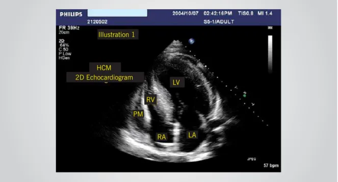
with hypertrophic cardiomyopathy is here described. The bidimensional echocardiography analysis (2D echo) showed hypertrophy at medium and basal segments of interventricular septum (16 mm) and the observation of pacemaker electrode in right chambers (Figure 1). Left atrium measurement (anteroposterior plane) showed a
3.2 cm diameter (normal value < 4 cm). The real time echocardiographic tridimensional analysis confi rmed 2D echo fi ndings (Figures 2A, 2B), and added information regarding left atrial anatomy, showing atrial enlargement in its elevation plane (depth plane), with left atrial volume of 85 mL (reference value in normal volunteers: 42 + 17 (18-79 ) mL. The 3D echo added structural information to this hypertrophic cardiomyopathy case. In the future, cavity measures may be provided in regard to volume rather than being associated to measures of one single observation plane.
Fig. 1 - Transthoracic bidimensional echocardiogram (2D) (4-chamber apical projection) of patient, showing hypertrophy at medium and basal segments of interventricular septum. LV- left ventricle; LA- left atrium; RV- right ventricle; RA- right atrium; PM- pacemaker electrode in right chambers; HCM- hypertrophic cardiomyopathy
Illustration 1
HCM 2D Echocardiogram
LV
RV
PM
Arquivos Brasileiros de Cardiologia - Volume 86, Nº 4, April 2006
306
R
EFERENCESREAL TIME TRIDIMENSIONAL ECHOCARDIOGRAPHY IN A PATIENT WITH HYPERTROPHIC CARDIOMYOPATHY
1. Ahmad M. Real-time three-dimensional echocardiography in assessment of heart disease. Echocardiography 2001; 18(1): 73-7.
2. Kisslo J, Firek B, Takahiro O, Kang DH, Fleishman CE, Stetten G et al. Real-time volumetric echocardiography: the technology and the possibilities. Echocardiography 2000; 17: 773-9.
Fig. 2 - A) Real time transthoracic tridimensional echocardiogram (3D) (apical projection) of patient. Blue circle (arrow)- reference point for depth plane. LA- left atrium. B) Real time transthoracic tridimensional echocardiogram (3D) (apical projection). LV - left ventricle
Illustration 2A Left Atrium 3D Echocardiogram
LA
LV Illustration 2B
Left Ventricle 3D Echocardiogram