Central actions of glucocorticoids in the
control of body fluid homeostasis: Review
S.G. Ruginsk
1, A. Lopes da Silva
1, R.R. Ventura
2, L.L.K. Elias
1and
J. Antunes-Rodrigues
11
Departamento de Fisiologia, Faculdade de Medicina de Ribeirão Preto, Universidade de São Paulo,
Ribeirão Preto, SP, Brasil
2
Departamento de Fisiologia, Universidade Federal do Paraná, Curitiba, PR, Brasil
Correspondence to: J. Antunes Rodrigues, Departamento de Fisiologia, FMRP, USP, Av. Bandeirantes,
3900, 14049-900 Ribeirão Preto, SP, Brasil
Fax: +55-16-3633-0017. E-mail: [email protected]
The involvement of the hypothalamic-pituitary-adrenal axis in the control of body fluid homeostasis has been extensively investigated in the past few years. In the present study, we reviewed the recent results obtained using different approaches to investigate the effects of glucocorticoids on the mechanisms of oxytocin and vasopressin synthesis and secretion in response to acute and chronic plasma volume and osmolality changes. The data presented here suggest that glucocorticoids are not only involved in the mechanisms underlying the fast release but also in the transcriptional events that lead to decreased synthesis and secretion of these neuropeptides, particularly oxytocin, under diverse experimental conditions of altered fluid volume and tonicity. The endocannabinoid system, through its effects on glutamatergic neurotransmission within the hypothalamus and the nuclear factor κB-mediated transcriptional activity, seems to be also involved in the specific mechanisms by which
glucocorti-coids exert their central effects on neurohypophyseal hormone synthesis and secretion.
Key words: Corticosterone; Vasopressin; Oxytocin; Endocannabinoid; Glutamate; NFκB
Presented at the IV Miguel R. Covian Symposium, Ribeirão Preto, SP, Brazil, May 23-25, 2008.
Publication supported by FAPESP.
Received July 4, 2008. Accepted November 18, 2008
Introduction
The control of body fluid homeostasis involves com-plex mechanisms that regulate water and electrolyte in-gestion and excretion. It is well established that plasma osmolality is mainly regulated by water balance, while sodium, which is one of the major components of the extracellular compartment, is a determinant factor for the maintenance of blood volume. Specific areas in the central nervous system (CNS) implicated in the control of hydro-mineral homeostasis receive information from specialized systems that detect peripheral changes in both volume and tonicity of extracellular fluid. The baroreceptors, lo-cated in the cardiovascular system, are activated in re-sponse to changes in blood volume or blood pressure and
the nucleus of the solitary tract (NTS) plays a key role in the integrative responses that reach the forebrain, particularly the hypothalamus. On the other hand, the osmosensitive areas are located in the circumventricular organs and the presence of both osmo- and sodium-sensitive neurons in these nuclei has been well characterized. Increases of 1-2% in plasma osmolality stimulate water drinking and also enhance neurohypophyseal secretion of vasopressin (AVP) and oxytocin (OT). Conversely, a 10% decrease in blood volume is known to increase AVP secretion.
depend-ent on the integrity of the third vdepend-entricle anterovdepend-entral region, which comprises the organum vasculosum of lamina terminalis, the ventral portion of the median preoptic nucle-us (MnPO) and the subfornical organ (SFO). These struc-tures, once stimulated, can determine responses that in-volve 1) the behavioral induction of thirst or salt appetite, or both, 2) changes in sympathetic and renal nerve activity, 3) activation of the renin-angiotensin-aldosterone system, or 4) secretion of AVP and OT from the neurohypophysis, and 5) secretion of natriuretic peptides from the heart.
Both AVP and OT are produced by magnocellular neu-rons of the paraventricular (PVN) and supraoptic (SON) nuclei of the hypothalamus and stored in terminals located in the neurohypophysis. AVP is mostly known for its antidi-uretic effects, but it is also involved in the thermoregulatory and cardiovascular responses (1). The activation of the V2
vasopressinergic receptors in the kidneys leads to increased permeability to water and consequent fluid reabsorption along the distal nephron. A stimulatory action of AVP on atrial natriuretic peptide (ANP) release has also been pro-posed (2). OT was shown to act either directly on the kidneys to promote sodium excretion or by an indirect pathway, stimulating ANP release from the heart (3,4). The participa-tion of the natriuretic peptidergic system in response to hypervolemia and hyperosmolality has been well estab-lished also in the brain (5, for a review, see Ref. 6).
The prototype of the natriuretic peptides is the circulat-ing peptide containcirculat-ing 28 amino acids (ANP 99-126 amino acids), which is processed from the atrial prohormone (1-126 amino acids). Other members of the natriuretic peptide family are BNP, CNP and urodilatin. The natriuretic pep-tides act at the cell membrane level through three types of receptors (A, B, and C). A and NPR-B, but not NPR-C, have an intracellular guanylate cyclase domain that generates cyclic guanosine monophosphate (cGMP) from guanosine triphosphate, which in turn acti-vates protein kinase G (7). In contrast, NPR-C is likely to act as a clearance receptor and to remove natriuretic peptides from the circulation (8). Recently, it has also been shown to have physiological effects on the heart and vasculature (9). The expression of NPR-A, analyzed by RT-PCR, was detected in all kidney layers (6).
The natriuretic effects of OT occur through a dual mechanism: generation of nitric oxide (NO) in the kidney, leading to increased cGMP and, at higher doses, induction of ANP release from the heart that, in turn, also increases cGMP. Oxytocin-induced natriuresis occurs mainly through a cGMP-mediated decrease in tubular Na+ reabsorption. In
contrast to ANP, which increases cGMP in the renal ves-sels as well as in the tubules, OT acts through its receptors located in NOergic cells identified in the macula densa and
proximal tubules, increasing cGMP production and closing Na channels. Thus, both ANP- and OT-induced natriuresis and kaliuresis appear to be mediated by cGMP (6).
The renin-angiotensin-aldosterone system also plays an important role in the regulation of body fluid homeosta-sis. An interaction between the angiotensinergic pathways and neurohypophyseal secretion has been described (6). Lauand et al. (10) recently showed that intracerebroven-tricular administration of angiotensin II (ANG II) increases AVP and OT secretion and also activates neurons in hypothalamic areas related to the control of fluid homeo-stasis. Several hormones (ANG II, AVP, OT, ANP, and mineralocorticoids) injected into the anterior hypothala-mus of the rat modify neuronal activity and appear to be involved in the regulation of fluid and electrolyte balance. ANG II induces a delayed sodium appetite following water intake (11). It has been suggested that an active inhibitory system may exist to restrain NaCl intake. Peptides and hormones with the opposite effect to that of ANG II on fluid and electrolyte balance, such as ANP, may attenuate ANG II-induced salt appetite (6). It has also been demonstrated that pre-treatment with an OT receptor antagonist increases NaCl intake induced by intracerebroventricular injection of ANG II, without significant changes in water intake, sug-gesting an inhibitory action of OT on salt appetite (12). Taken together, these data provided a link between the central mechanisms controlling body fluid homeostasis and the peripheral adjustments of renal and cardiovascu-lar systems in response to acute increases in plasma volume and osmolality.
Significant progress has been made to identify the neural circuits involved in the physiological and behavioral osmoregulatory responses. There is growing evidence for the participation of the hypothalamic-pituitary-adrenal (HPA) axis in the adaptive responses following changes in blood volume and osmolality. The presence of the cytoplasmic glucocorticoid receptor (GR) has already been reported in the SON (13) and also in the parvocellular portion of the PVN, where this receptor is predominantly co-expressed with corticotrophin-releasing factor (CRF) (14). Further-more, AVP and OT are weak secretagogues of adrenocor-ticotropic hormone (15-17), strongly suggesting an inter-action between the HPA axis and the secretion of neurohy-pophyseal hormones.
Glucocorticoids - general aspects
cytoso-lic GR, which, in the absence of the endogenous ligand, is assembled into a multiprotein complex (20). Although re-tained in the cytoplasm, this conformation enables high-affinity ligand binding.
Produced by the adrenal glands under the stimulation of the HPA axis, the glucocorticoids act as ligand-depend-ent transcription factors that positively regulate genes through interaction with DNA enhancer sequences, called glucocorticoid response elements (GREs) (21-23). The activated GR was also shown to negatively regulate the expression of inflammatory genes through direct protein-protein interaction with proinflammatory transcription fac-tors, without DNA binding (24,25).
The genomic and long-term mechanisms that follow glucocorticoid binding to the cytoplasmic GR and nuclear entry are also known as the classic pathway, but recent evidence suggests that fast glucocorticoid actions are mediated by membrane receptors and activation of nonge-nomic signaling events. In fact, glucocorticoids have been reported to bind specifically to cell membrane sites and to induce electrolytic movement changes (26,27). Although the molecular characterization of these receptors remains to be established in humans, a functional corticosterone membrane-associated receptor was identified in the am-phibian brain (28).
The expression of GR in CRF-expressing neurons in the parvocellular PVN reinforces the direct feedback ac-tion of glucocorticoid in the control of CRF synthesis and release (14,29). Co-localization of mineralocorticoid re-ceptor (MR) and GR was also observed in the parvocellu-lar region, but not in the magnocelluparvocellu-lar region of the PVN (30). These results suggest that MR and GR may interact in the control of HPA axis activity.
There are several lines of evidence showing that glucorticoid action is modulated by the presence of 11ß-hydroxysteroid dehydrogenases (11ß-HSDs) in several tis-sues (31). 11ß-HSD1 activates cortisone to cortisol to facili-tate GR-mediated action. In addition, 11ß-HSD2 plays an important role in aldosterone target tissues where it cata-lyzes the opposite reaction (i.e., inactivation of cortisol to cortisone) to prevent activation of MR by cortisol. Therefore, 11ß-HSD activity allows aldosterone occupancy of MR by inactivating endogenous glucocorticoids. In the kidney, 11ß-HSD2 is mainly expressed in collecting ducts, where it is co-localized with MR. Inhibition of 11ß-HSD2 can result in glucocorticoid-dependent mineralocorticoid excess and hy-pertension. This enzyme is also found in several tissues that are not classic targets for mineralocorticoids, such as peri-and circumventricular regions peri-and NTS. Therefore, tissue-specific glucorticoid metabolism may be involved in the control of fluid balance and blood pressure.
Effects of glucocorticoids on neurohypophyseal
hormone secretion
Several studies from our laboratory have demonstrated that isotonic (0.9% NaCl) blood volume expansion (BVE) increases plasma concentrations of OT and ANP and decreases plasma AVP levels, resulting in increased wa-ter and sodium excretion. On the other hand, rats submit-ted to hypertonic (1.8% NaCl) BVE presensubmit-ted increased plasma levels of AVP, OT and ANP in order to promote water reabsorption and renal sodium excretion (32-34).
In 2004, Durlo et al. (33) reported the first evidence for the participation of the HPA axis in hormone secretion induced by BVE in rats. These investigators reported that both hypervolemia and hyperosmolality increased plasma corticosterone concentrations, an event also known to occur in response to other types of stress, such as forced swimming, immobilization and hypoglycemia (35). This study also demonstrated that pre-treatment with dexa-methasone, a synthetic glucocorticoid, blunted OT, but not ANP secretion, induced by BVE. These results were con-firmed by Ruginsk et al. (34), who also showed that previ-ous administration of dexamethasone did not alter AVP secretion in response to isotonic and hypertonic BVE. Pre-treatment with dexamethasone also decreased the secre-tion of OT but not AVP induced by central angiotensinergic and cholinergic stimulation (10).
Taken together, these results suggest that, besides their genomic action on the modulation of transcriptional events, glucocorticoids are likely to present a fast negative modulation of OT but not AVP release from neurohypophy-seal stores. In fact, Limbourg and Liao (36) reported that glucocorticoids could exert short-term effects on NO pro-duction in the heart, contributing to vasodilatatory responses during ischemic conditions. These data suggested that glucocorticoids may have actions that depend on non-genomic mechanisms, since the effects are rapidly ob-served.
Additionally, the HPA axis also seems to be involved in the mechanisms underlying chronic adjustments to os-motic challenges. Berghorn et al. (37) reported that long-term hyperosmolality induced an increase in the number of receptors for glucocorticoids in magnocellular vasopressi-nergic cells. Furthermore, increased plasma corticoster-one levels were also reported in response to salt loading (38-40). These investigators also suggested that the HPA responsiveness to either CRF or AVP seems to be im-paired after long-term osmotic stimulation.
ap-proaches has been generating conflicting results. Ventura et al. (40) demonstrated that the increase in plasma OT and AVP induced by salt loading was accompanied by an increased NO synthase activity in the SON and PVN, although the previous administration of NO synthase in-hibitor produced different effects on the secretion of these peptides. These data indicate that NO differentially modu-lates the secretion of neurohypophyseal hormones in re-sponse to chronic salt loading.
Effects of glucocorticoids on hypothalamic
neuronal activation
The detection of immediate-early gene products, such as Fos nuclear protein, has been used extensively as a marker of neuronal activation (41). The number of Fos-positive neurons in the PVN and SON was increased in rats submitted to hypertonic BVE and this effect was observed in parallel with enhanced neurohypophyseal secretion of AVP and OT (34). Furthermore, the use of specific antibodies has also allowed the identification of neuron phenotype, which is of interest to clarify the particular cellular mechanisms in-volved in response to a specific stimulus. Data obtained by Godino et al. (42) and extended by results from our labora-tory (34) have shown that hypertonic BVE induced increased numbers of vasopressinergic and oxytocinergic neurons activated in both the PVN and SON. On the other hand, the number of oxytocinergic, but not vasopressinergic, magno-cellular neurons activated in these nuclei was increased in rats submitted to isotonic BVE.
The responsiveness of magnocellular neurosecretory cells to plasma hyperosmolality is subject to inputs from the osmosensitive neurons (43), which are also activated by administration of hypertonic solution. A significant number of these neurons project to the hypothalamic magnocellular nuclei and other areas involved in the regulation of body fluid homeostasis, since hypertonicity also increases the number of Fos-positive neurons in the SFO, central nucleus of the amygdala, parabrachial nucleus, locus coeruleus, ventrolat-eral medulla, NTS, and area postrema (42). The activation of these areas depends on intact osmosensory pathways and mediates the autonomic, endocrine and behavioral responses involved in the osmoregulation (44).
The glucocorticoids seem to be also involved in the mechanisms underlying magnocellular neuronal activa-tion following BVE. Ruginsk et al. (34) showed that, be-sides inhibiting hormone secretion, pre-treatment with dexa-methasone decreased the number of oxytocinergic neu-rons activated in response to both isotonic and hypertonic BVE. This inhibitory effect induced by dexamethasone was observed on vasopressinergic neurons following
hy-pertonic but not isotonic BVE. We also demonstrated that Fos expression and plasma AVP levels were both in-creased by osmotic, cholinergic and angiotensinergic cen-tral stimulation. However, dexamethasone pre-treatment induced a decrease of Fos expression in the MnPO, PVN and SON, but did not affect AVP secretion. Therefore, it appears that Fos expression and AVP secretion were parallel events that occurred in response to a given stimu-lus, rather than interacting with one another (10). The apparent divergence between unaltered AVP secretion and decreased neuronal activity observed in dexametha-sone-pretreated rats subjected to hypertonic BVE or os-motic, cholinergic and angiotensinergic central stimulation suggests that the glucocorticoids may differentially modu-late the AVP release from neurohypophyseal storages and the de novo hormone synthesis in the PVN and SON.
According to some other studies, glucocorticoid ac-tions on magnocellular hypothalamic neurons seem to be predominantly inhibitory. Di et al. (45,46) proposed that corticosterone could act on transmembrane G-coupled receptors and stimulate the release of endocannabinoids in the hypothalamus. According to these investigators, the endocannabinoids would act as retrograde messengers and not only inhibit the release of glutamate, the main excitatory neurotransmitter in the CNS, but also stimulate the release of gamma-aminobutyric acid (GABA), contrib-uting to the decreased activity of both parvocellular and magnocellular hypothalamic neurons.
Results obtained in our laboratory indicate that there is an increase in total glutamate content in the PVN and SON of rats submitted to BVE and this response is not altered by dexamethasone pre-treatment, suggesting that the bal-ance between neurotransmitter production and release would not be affected in this condition (47). These results confirm the crucial role of glutamate in mediating the responses to hypervolemia and hyperosmolality.
Lopes da Silva et al. (48) also suggested that dexameth-asone is likely to inhibit the expression of AVP and OT mRNA in the hypothalamic PVN and SON of rats submitted to water restriction and salt loading. The participation of the nuclear factor κB (NFκB) in the mediation of glucocorticoid
Discussion
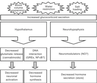
In the experimental models of BVE, water restriction and salt loading, the autonomic, neuroendocrine, cardio-vascular, renal and behavioral systems are recruited to restore body fluid homeostasis (6,49). A better under-standing of these mechanisms has been obtained with the recent contributions of molecular studies, but many of the events at the ultrastructural level are still unknown. The participation of the HPA axis, particularly the glucocorti-coids, in mediating the responses to plasma volume and osmolality changes seems to involve diverse mechanisms ranging from the control of molecular aspects within the CNS to systemic modulation of hormone release, as sum-marized in Figure 1.
The glucocorticoid interaction with DNA binding sites has been well characterized and may also be activated after the acute stimulus or under prolonged exposure to stress. A GRE was identified in target genes (50) and by DNA sequence analysis, several elements upstream to the transcriptional start point of the rat OT gene were identi-fied, matching the consensus sequence of enhancers inducible by glucocorticoids (51). Therefore, the observed effect of prolonged dexamethasone pre-treatment, reduc-ing OT mRNA content and the consequent hormone se-cretion, could be due to an inhibition of OT gene transcrip-tion. The presence of GREs is speculated in the AVP gene, although no relevant result has been reported thus far.
The participation of the NFκB cascade in the
transcrip-tional events evoked by glucocorticoids is also a promising area of study, although many pathways are known to converge to this common point. Additionally, the involve-ment of the nitrergic system in the mediation of AVP and OT release represents a growing area of investigation, since NO seems to differentially regulate the release of both neuropeptides.
The study of the pharmacological benefits of cannabis-like substances was described very early in medical sci-ences, but it was only in 1988 that the cannabinoid CB1
receptor was described in the CNS. The main endogenous ligands for this type of receptor are anandamide and 2-arachidonoylglycerol and a clear relationship between the predominant anatomical localization of CB1 receptors in
the CNS and the cognitive, affective and motor systems was then established (52). Since then, the endocannab-inoids have been extensively studied in the mediation of homeostatic responses, such as those related to energy balance, pain and behavior.
Hartman et al. (53) reported the first evidence for the in vivo participation of the cannabinoid system in the control of body fluid homeostasis. This study demonstrated that previous administration of naloxone, an opioid antagonist, to adult rats enhanced OT but not AVP secretion induced by hypertonic saline injection. Additionally, these investi-gators also showed that the central content of OT but not AVP was depleted in naloxone-pretreated mature rats. The most recent link between the endocannabinoids and the neuroendocrine system that controls body fluid ho-meostasis was obtained from in vitro manipulations of CB1
receptors. The inhibition of the glutamatergic neurotrans-mission within the hypothalamus by endocannabinoids, as reported by Di et al. (45,46), provides a new insight into the molecular mechanisms that may be recruited in the pres-ence of specific disturbances in body fluid homeostasis.
In conclusion, it has been suggested that the HPA axis might be involved in the integrative control of body fluid homeostasis, modulating neurohypophyseal hormone se-cretion, supporting the idea that the hypothalamus, par-ticularly the PVN, contains a complex network with specific and integrative pathways. The advance of new techniques, such as interference RNA, blocking specific proteins, pep-tides or neurotransmitters, as well as the data on electro-physiological studies will provide in the near future useful tools for the understanding of mechanisms of glucocorti-coid control of body fluid homeostasis.
Decreased glutamate release
(cannabinoids)
DNA interaction (GREs, NFκB?)
Neuromodulators (NO?) Neurohypophysis Hypothalamus
Increased glucocorticoid secretion Blood
volume expansion
Water
restriction loadingSalt
Decreased neuronal activation
Decreased hormone synthesis
Decreased hormone secretion (store)
Figure 1. Figure 1. Figure 1. Figure 1.
References
1. Steiner AA, Branco LG. Hypoxia-induced anapyrexia: impli-cations and putative mediators. Annu Rev Physiol 2002; 64: 263-288.
2. Itoh H, Nakao K, Yamada T, Morii N, Shiono S, Sugawara A, et al. Modulatory role of vasopressin in secretion of atrial natriuretic polypeptide in conscious rats. Endocrinology 1987; 120: 2186-2188.
3. Soares TJ, Coimbra TM, Martins AR, Pereira AG, Carnio EC, Branco LG, et al. Atrial natriuretic peptide and oxytocin induce natriuresis by release of cGMP. Proc Natl Acad Sci U S A 1999; 96: 278-283.
4. Jankowski M, Hajjar F, Kawas SA, Mukaddam-Daher S, Hoffman G, McCann SM, et al. Rat heart: a site of oxytocin production and action. Proc Natl Acad Sci U S A 1998; 95: 14558-14563.
5. Zamir N, Skofitsch G, Eskay RL, Jacobowitz DM. Distribu-tion of immunoreactive atrial natriuretic peptides in the cen-tral nervous system of the rat. Brain Res 1986; 365: 105-111.
6. Antunes-Rodrigues J, de Castro M, Elias LL, Valenca MM, McCann SM. Neuroendocrine control of body fluid metabo-lism. Physiol Rev 2004; 84: 169-208.
7. Woodard GE, Rosado JA. Natriuretic peptides in vascular physiology and pathology. Int Rev Cell Mol Biol 2008; 268: 59-93.
8. Maack T, Suzuki M, Almeida FA, Nussenzveig D, Scarbor-ough RM, McEnroe GA, et al. Physiological role of silent receptors of atrial natriuretic factor. Science 1987; 238: 675-678.
9. Rose RA, Giles WR. Natriuretic peptide C receptor signal-ling in the heart and vasculature. J Physiol 2008; 586: 353-366.
10. Lauand F, Ruginsk SG, Rodrigues HL, Reis WL, de Castro M, Elias LL, et al. Glucocorticoid modulation of atrial natri-uretic peptide, oxytocin, vasopressin and Fos expression in response to osmotic, angiotensinergic and cholinergic stim-ulation. Neuroscience 2007; 147: 247-257.
11. Fitzsimons JT. Angiotensin, thirst, and sodium appetite. Physiol Rev 1998; 78: 583-686.
12. Blackburn RE, Demko AD, Hoffman GE, Stricker EM, Verbalis JG. Central oxytocin inhibition of angiotensin-in-duced salt appetite in rats. Am J Physiol 1992; 263: R1347-R1353.
13. Kiss JZ, van Eekelen JA, Reul JM, Westphal HM, de Kloet ER. Glucocorticoid receptor in magnocellular neurosecre-tory cells. Endocrinology 1988; 122: 444-449.
14. Liposits Z, Uht RM, Harrison RW, Gibbs FP, Paull WK, Bohn MC. Ultrastructural localization of glucocorticoid receptor (GR) in hypothalamic paraventricular neurons synthesizing corticotropin releasing factor (CRF). Histochemistry 1987; 87: 407-412.
15. Gillies GE, Linton EA, Lowry PJ. Corticotropin releasing activity of the new CRF is potentiated several times by vasopressin. Nature 1982; 299: 355-357.
16. Schwartz J, Vale W. Dissociation of the adrenocorticotropin secretory responses to corticotropin-releasing factor (CRF) and vasopressin or oxytocin by using a specific cytotoxic analog of CRF. Endocrinology 1988; 122: 1695-1700. 17. Antoni FA. Hypothalamic control of adrenocorticotropin
se-cretion: advances since the discovery of 41-residue cortico-tropin-releasing factor. Endocr Rev 1986; 7: 351-378. 18. Tronche F, Kellendonk C, Reichardt HM, Schutz G. Genetic
dissection of glucocorticoid receptor function in mice. Curr Opin Genet Dev 1998; 8: 532-538.
19. McEwen BS. Protective and damaging effects of stress mediators. N Engl J Med 1998; 338: 171-179.
20. Lu NZ, Cidlowski JA. Glucocorticoid receptor isoforms gen-erate transcription specificity. Trends Cell Biol 2006; 16: 301-307.
21. Giguere V, Hollenberg SM, Rosenfeld MG, Evans RM. Func-tional domains of the human glucocorticoid receptor. Cell 1986; 46: 645-652.
22. Hollenberg SM, Evans RM. Multiple and cooperative trans-activation domains of the human glucocorticoid receptor. Cell 1988; 55: 899-906.
23. Beato M, Herrlich P, Schutz G. Steroid hormone receptors: many actors in search of a plot. Cell 1995; 83: 851-857. 24. Jonat C, Rahmsdorf HJ, Park KK, Cato AC, Gebel S, Ponta
H, et al. Antitumor promotion and antiinflammation: down-modulation of AP-1 (Fos/Jun) activity by glucocorticoid hor-mone. Cell 1990; 62: 1189-1204.
25. Karin M. New twists in gene regulation by glucocorticoid receptor: is DNA binding dispensable? Cell 1998; 93: 487-490.
26. Suyemitsu T, Terayama H. Specific binding sites for natural glucocorticoids in plasma membranes of rat liver. Endocri-nology 1975; 96: 1499-1508.
27. Avanzino GL, Ermirio R, Ruggeri P, Cogo CE. Effects of corticosterone on neurons of reticular formation in rats. Am J Physiol 1987; 253: R25-R30.
28. Evans SJ, Murray TF, Moore FL. Partial purification and biochemical characterization of a membrane glucocorticoid receptor from an amphibian brain. J Steroid Biochem Mol Biol 2000; 72: 209-221.
29. Uht RM, McKelvy JF, Harrison RW, Bohn MC. Demonstra-tion of glucocorticoid receptor-like immunoreactivity in glu-cocorticoid-sensitive vasopressin and corticotropin-releas-ing factor neurons in the hypothalamic paraventricular nu-cleus. J Neurosci Res 1988; 19: 405-409.
30. Han F, Ozawa H, Matsuda K, Nishi M, Kawata M. Colocali-zation of mineralocorticoid receptor and glucocorticoid re-ceptor in the hippocampus and hypothalamus. Neurosci Res 2005; 51: 371-381.
31. Seckl JR. 11Beta-hydroxysteroid dehydrogenases: chang-ing glucocorticoid action. Curr Opin Pharmacol 2004; 4: 597-602.
32. Haanwinckel MA, Elias LK, Favaretto AL, Gutkowska J, McCann SM, Antunes-Rodrigues J. Oxytocin mediates atrial natriuretic peptide release and natriuresis after volume ex-pansion in the rat. Proc Natl Acad Sci U S A 1995; 92: 7902-7906.
33. Durlo FV, Castro M, Elias LL, Antunes-Rodrigues J. Interac-tion of prolactin, ANPergic, oxytocinergic and adrenal sys-tems in response to extracellular volume expansion in rats. Exp Physiol 2004; 89: 541-548.
vol-ume expansion. Exp Neurol 2007; 206: 192-200.
35. Lang RE, Heil JW, Ganten D, Hermann K, Unger T, Rascher W. Oxytocin unlike vasopressin is a stress hormone in the rat. Neuroendocrinology 1983; 37: 314-316.
36. Limbourg FP, Liao JK. Nontranscriptional actions of the glucocorticoid receptor. J Mol Med 2003; 81: 168-174. 37. Berghorn KA, Knapp LT, Hoffman GE, Sherman TG.
Induc-tion of glucocorticoid receptor expression in hypothalamic magnocellular vasopressin neurons during chronic hypoos-molality. Endocrinology 1995; 136: 804-807.
38. Dohanics J, Verbalis JG. Naloxone disinhibits magnocellu-lar responses to osmotic and volemic stimuli in chronically hypoosmolar rats. J Neuroendocrinol 1995; 7: 57-62. 39. Elias LL, Dorival CA, Moreira AC. The opposite effects of
short- and long-term salt loading on pituitary adrenal axis activity in rats. Horm Metab Res 2002; 34: 207-211. 40. Ventura RR, Gomes DA, Reis WL, Elias LL, Castro M,
Valenca MM, et al. Nitrergic modulation of vasopressin, oxytocin and atrial natriuretic peptide secretion in response to sodium intake and hypertonic blood volume expansion. Braz J Med Biol Res 2002; 35: 1101-1109.
41. Hoffman GE, Smith MS, Verbalis JG. c-Fos and related immediate early gene products as markers of activity in neuroendocrine systems. Front Neuroendocrinol 1993; 14: 173-213.
42. Godino A, Giusti-Paiva A, Antunes-Rodrigues J, Vivas L. Neurochemical brain groups activated after an isotonic blood volume expansion in rats. Neuroscience 2005; 133: 493-505.
43. Bourque CW. Central mechanisms of osmosensation and systemic osmoregulation. Nat Rev Neurosci 2008; 9: 519-531.
44. Gardiner TW, Verbalis JG, Stricker EM. Impaired secretion of vasopressin and oxytocin in rats after lesions of nucleus medianus. Am J Physiol 1985; 249: R681-R688.
45. Di S, Malcher-Lopes R, Halmos KC, Tasker JG. Nongeno-mic glucocorticoid inhibition via endocannabinoid release in the hypothalamus: a fast feedback mechanism. J Neurosci 2003; 23: 4850-4857.
46. Di S, Malcher-Lopes R, Marcheselli VL, Bazan NG, Tasker JG. Rapid glucocorticoid-mediated endocannabinoid re-lease and opposing regulation of glutamate and gamma-aminobutyric acid inputs to hypothalamic magnocellular neurons. Endocrinology 2005; 146: 4292-4301.
47. Ruginsk SG, Oliveira FR, Elias LL, Antunes-Rodrigues J. Effects of glucocorticoids on neuronal activity, hormone secretion and glutamate content after blood volume expan-sion (BVE). Proc Aust Neurosc Soc 2008; 18: 31.
48. Lopes da Silva A, Elias LL, Antunes-Rodrigues J. Role of glucocorticoids on oxytocin and vasopressin mRNA expres-sion in two models of osmotic stimulation, presented in a poster session during. The Endocrine Society’s 2008 An-nual Meeting, held in San Francisco (CA, USA) from June 15 to 18; 2008.
49. McKinley MJ, Johnson AK. The physiological regulation of thirst and fluid intake. News Physiol Sci 2004; 19: 1-6. 50. Lan NC, Karin M, Nguyen T, Weisz A, Birnbaum MJ,
Eber-hardt NL, et al. Mechanisms of glucocorticoid hormone action. J Steroid Biochem 1984; 20: 77-88.
51. Mohr E, Schmitz E. Functional characterization of estrogen and glucocorticoid responsive elements in the rat oxytocin gene. Brain Res Mol Brain Res 1991; 9: 293-298.
52. Freund TF, Katona I, Piomelli D. Role of endogenous can-nabinoids in synaptic signaling. Physiol Rev 2003; 83: 1017-1066.