Effects of stress on catecholamine
stores in central and peripheral tissues
of long-term socially isolated rats
Institute of Nuclear Sciences “Vin…a”, Laboratory of Molecular Biology and Endocrinology, Belgrade, Serbia and Montenegro
S. Dronjak and L. Gavrilovic
Abstract
Both the peripheral sympatho-adrenomedullary and central catecho-laminergic systems are activated by various psycho-social and physi-cal stressors. Catecholamine stores in the hypothalamus, hippocam-pus, adrenal glands, and heart auricles of long-term socially isolated (21 days) and control 3-month-old male Wistar rats, as well as their response to immobilization of all 4 limbs and head fixed for 2 h and cold stress (4ºC, 2 h), were studied. A simultaneous single isotope radioenzymatic assay based on the conversion of catecholamines to the corresponding O-methylated derivatives by catechol-O-methyl-transferase in the presence of S-adenosyl-l-(3H-methyl)-methionine
was used. The O-methylated derivatives were oxidized to 3H-vanilline
and the radioactivity measured. Social isolation produced depletion of hypothalamic norepinephrine (about 18%) and hippocampal dopa-mine (about 20%) stores and no changes in peripheral tissues. Immo-bilization decreased catecholamine stores (approximately 39%) in central and peripheral tissues of control animals. However, in socially isolated rats, these reductions were observed only in the hippocampus and peripheral tissues. Cold did not affect hypothalamic catechol-amine stores but reduced hippocampal dopcatechol-amine (about 20%) as well as norepinephrine stores in peripheral tissues both in control and socially isolated rats, while epinephrine levels were unchanged. Thus, immobilization was more efficient in reducing catecholamine stores in control and chronically isolated rats compared to cold stress. The differences in rearing conditions appear to influence the response of adult animals to additional stress. In addition, the influence of previ-ous exposure to a stressor on catecholaminergic activity in the brain-stem depends on both the particular catecholaminergic area studied and the properties of additional acute stress. Therefore, the sensitivity of the catecholaminergic system to habituation appears to be tissue-specific.
Correspondence
S. Dronjak
Laboratory of Molecular Biology and Endocrinology
Institute of Nuclear Sciences “Vin…a”
P.O. Box 522-090 11001 Belgrade Serbia and Montenegro Fax: +38-11-1245-5561 E-mail: [email protected]
Research supported by the Ministry for Science, Technology and Environmental Protection of Serbia (Contract #1953).
Received July 18, 2005 Accepted February 6, 2006
Key words
•Cold stress
Introduction
Stress disturbs homeostasis and may in-duce various disorders. Both the peripheral sympatho-adrenomedullary and central monoaminergic systems are activated by various psycho-social and physical stres-sors. Limbic circuits connecting, e.g., the hippocampus, amygdala and prefrontal cor-tex are sensitive to stressors such as re-straint, fear or exposure to a novel environ-ment. In contrast, physiological threats such as exposure to ether result in the activation of efferent visceral pathways that are di-rectly relayed to the paraventricular nucleus of the hypothalamus (1). Activation of lim-bic and hypothalamic brain structures is a major component of the stress reaction that integrates neuroendocrine and emotional components and thus determines the magni-tude and duration of the hormonal and neu-ral stress response. Recent reports have sug-gested that different types of stress may sometimes produce qualitatively different patterns of effects on both behavior and physiology. Using an in vivo microdialysis
method, Tanaka et al. (2,3) reported that the same immobilization stress led to a different time course of changes in norepinephrine (NE) levels in the amygdala when compared to the hypothalamus, suggesting regional differences in release due to functional dif-ferences in the stress response. Shibasaki et al. (4) showed that the response pattern of NE release to repeated stress in the hypo-thalamus was also dependent on the type of stressor employed, since restraint stress, but not tail-pinching, promotes desensitization of hypothalamic NE neurons. Animals ex-posed to prior stress exhibit an enhanced, reduced or equivalent hypothalamic-pitui-tary-adrenal response to a subsequent acute stressor (5-7). Social isolation produced by individual housing affects brain monoamine and adrenal gland functions in experimental animals (8-11). Social isolation and acute environmental changes are risk factors
in human depression, and represent a lack of the social stimuli necessary to modulate adaptive responses to new situations (12). The differences in animal rearing conditions appear to influence the responses to acute stress. Almost all reported studies have ex-amined the effects of isolation during the weaning period. However, Miura et al. (13) suggested that animals suffering isolation in adulthood also show behavioral changes, indicating that isolation can alter the central nervous system activity of adult animals previously reared in a group.
We investigated the influence of long-term social isolation on catecholamine stores in central and peripheral tissues in response to two different types of additional stressors, i.e., immobilization and cold.
Material and Methods
Three-month-old male Wistar rats weigh-ing 300-340 g maintained under standard laboratory conditions with water and food
ad libitum were used. Before being
sub-jected to stress the animals were housed in groups of four individuals. The animals were divided into two groups, a control group consisting of four animals per cage and a group of animals that were housed individu-ally for the 21-day study period. Both groups were exposed to short-term (2 h) immobili-zation or cold stress. The animals were treated as humanely as possible according to the recommendations of the “Vin…a” Institute which are based on the Guide for Care and Use of Laboratory Animals of the National Institutes of Health, Bethesda, MD, USA.
decapitated and the hippocampi, hypo-thalami, heart auricles, and adrenal glands were quickly dissected and immersed in cold 0.1 N HClO4 (0.3 µg tissue per 30 µL 0.1 N
HClO4 at 4ºC). The tissues were then
ho-mogenized in a motor-driven homogenizer and centrifuged at 10,000 rpm for 20 min at 4ºC in a K24 Janetzky centrifuge (Berlin, Germany). The resulting supernatant solu-tions (30-µL aliquots) were stored at -70oC
and later used for the analyses.
Catecholamines in the tissues were de-termined using the single isotope radioenzy-matic assay of Peuler and Johnson (15) based on the conversion of catecholamines to the corresponding O-methylated derivatives by purified catechol-O-methyl-transferase in the presence of S-adenosyl-l-(3
H-methyl)-me-thionine. The O-methylated derivatives were oxidized to 3H-vanilline. Radioactivity was
measured with a toluene-based scintillation liquid and with an LKB-Wallac model 1219 scintillation counter (Stockholm, Sweden) at 40% efficiency for tritium. The range of measurement is Window 1 5-320,
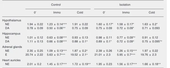
sensitivi-Table 1. Changes in catecholamine stores due to social isolation.
Control Isolation
0’ Immo Cold 0’ Immo Cold
Hypothalamus
NE 1.94 ± 0.22 1.23 ± 0.14+++ 1.91 ± 0.22 1.60 ± 0.17* 1.58 ± 0.17* 1.63 ± 0.2*
DA 0.78 ± 0.09 0.50 ± 0.06++ 0.75 ± 0.09 0.75 ± 0.09 0.72 ± 0.09* 0.71 ± 0.093
Hippocampus
NE 1.01 ± 0.12 0.63 ± 0.08+++ 0.93 ± 0.13 0.98 ± 0.11 0.77 ± 0.09*+ 0.91 ± 0.12
DA 1.11 ± 0.13 0.66 ± 0.08+++ 0.88 ± 0.1+ 0.89 ± 0.1* 0.72 ± 0.09+ 0.75 ± 0.095*+
Adrenal glands
NE 2.35 ± 0.25 1.09 ± 0.13+++ 1.87 ± 0.2+ 2.39 ± 0.26 1.26 ± 0.15+++ 1.97 ± 0.22
E 20.74 ± 2.22 5.63 ± 0.7+++ 19.52 ± 2.1+ 21.01 ± 2.3 5.95 ± 0.7+++ 19.76 ± 2.3
Heart auricles
NE 2.01 ± 0.2 1.45 ± 0.17+++ 1.72 ± 0.19++ 1.95 ± 0.23 1.56 ± 0.17+++ 1.66 ± 0.18++
Catecholamine stores were measured by the single isotope radioenzymatic method of Peuler and Johnson (15). The means and SEM for each of the two central and two peripheral tissues were calculated for each group of 6 animals. Social isolation was compared with naive control. Immobilization and cold stress were compared with their zero point. NE = norepinephrine; E = epinephrine; DA = dopamine; Immo = immobilization; cold stress = 0' to 2 h.
*P < 0.05 for 21-day social isolation vs naive control (two-way factorial ANOVA). +P < 0.05; ++P < 0.01; +++P < 0.001 for
immobilization and cold stress compared to their zero point (two-way factorial ANOVA).
ty is 20 CPM and interassay is less than 10%. Data were analyzed statistically by two-way ANOVA, with the level of significance set at P < 0.05.
Results
The results presented in Table 1 show that when compared with naive control, long-term isolation produced a significant deple-tion of only NE stores (about 18%, P < 0.05) in the hypothalamus, while in the hippocam-pus a significant decrease was observed only in dopamine (DA) content (about 20%). So-cial isolation produced no significant changes in epinephrine (E) or NE levels in adrenal glands, or in heart auricle NE levels.
immobiliza-tion had no effect on the level of these cat-echolamines in the hypothalamus of animals previously submitted to long-term social iso-lation. Immobilization stress produced a highly significant decrease (P < 0.001) in the adrenal gland content of E (about 72%) and NE (about 50%) of both naive controls and rats previously exposed to long-term social isolation compared with 0’. Also, immobili-zation led to a decrease of heart auricle NE stores in both the control (about 28%) and long-term isolated rats (about 29%).
Cold stress did not reduce hypothalamic catecholamine stores in either control or long-term isolated rats compared with 0’. It also did not affect the hippocampal NE stores, while a statistically significant decrease (P < 0.05) of DA stores was observed in this brain region in the controls (about 21%) and long-term isolated rats (about 16%) compared with 0’. Likewise, cold stress induced a re-duction (P < 0.05) of NE in the adrenal glands (about 20%) and in heart auricles (about 14%), but did not affect E stores in the adrenal glands of naive controls or the rats previously exposed to long-term isola-tion compared with 0’.
Discussion
Several studies have demonstrated that animals reared in isolation show some be-havioral and neurochemical changes (16,17). The results of almost all earlier studies con-cerning the effects of social isolation were obtained by applying isolation at the begin-ning of the weabegin-ning period when the isola-tion condiisola-tions could influence the growth and development of the central nervous sys-tem. To eliminate this possibility, in the present study adult rats were subjected to 21-day isolation. The present data show that long-term social isolation led to a significant depletion of hypothalamic NE stores, with-out affecting hippocampal NE stores. The hypothalamus is the brain structure that in-fluences all limbic structures and
long-term social isolation resulted in the reduc-tion of DA stores in the hippocampus but not in the hypothalamus. Baffi and Palkovits (23) have reported similar results regarding emotional changes, with exposure to a stress situation increasing DA metabolite levels in the hippocampus. The results obtained in the present study showed that the influence of previous exposure to a stressor on catechola-minergic activity in the brainstem depends on the particular catecholaminergic area of the brainstem studied and on the properties of additional acute stress. Thus, the sensitiv-ity of the catecholaminergic system to ha-bituation appears to be region-specific.
Long-term social isolation produced no significant changes in the catecholamine lev-els of peripheral tissues, suggesting adapta-tion of the sympathetic adrenomedullary sys-tem to long-term isolation. It has been re-ported that exposure of animals to chronic intermittent stress results in a significant increase of catecholamine biosynthesis and storage capacity in peripheral tissues com-pared to unstressed controls (24,25). Ani-mals exposed to repeated immobilization stress (41 days) showed permanently in-creased adrenal tyrosine hydroxylase mRNA levels, tyrosine hydroxylase activity and pro-tein levels (26). Our results showed that
additional immobilization stress produced a highly significant decrease in the amount of E and NE in the adrenal gland of long-term isolated rats. This could mean that these animals increasingly mobilized high stores of tissue catecholamines after exposure to an additional stressor. Cold stress induced only a reduction of NE stores in the adrenal glands and heart auricles of both naive control and long-term isolated rats. These results are in agreement with previous data obtained from our laboratory, that demonstrated an in-creased plasma level of NE upon exposure of rats to cold stress, while plasma E content remained unchanged (27).
The results of the present study showed that additional immobilization stress pro-duced a greater reduction of catecholamine stores in all tissues examined in both control and chronically stressed rats compared to that observed after exposure of animals to a novel additional cold stress. Therefore, we conclude that the properties of the additional stressor play a role in the intensity of the response of peripheral and central noradre-nergic neurons. In addition, the differences in rearing conditions of adult animals appear to influence their responses to additional stress.
References
1. Fuchs E, Flugge G. Chronic social stress: effects on limbic brain structures. Physiol Behav 2003; 79: 417-427.
2. Tanaka M, Kohno Y, Nakagawa R, Ida Y, Takeda S, Nagasaki N, et al. Regional characteristics of stress-induced increases in brain noradrenaline release in rats. Pharmacol Biochem Behav 1983; 19: 543-547.
3. Tanaka T, Yokoo H, Mizoguchi K, Yoshida M, Tsuda A, Tanaka M. Noradrenaline release in the rat amygdala is increased by stress: studies with intracerebral microdialysis. Brain Res 1991; 544: 174-176.
4. Shibasaki T, Tsumori C, Hotta M, Imaki T, Yamada K, Demura H. The response pattern of noradrenaline release to repeated stress in the hypothalamic paraventricular nucleus differs according to the form of stress in rats. Brain Res 1995; 670: 169-172.
5. Simpkiss JL, Devine DP. Responses of the HPA axis after chronic variable stress: effects of novel and familiar stressors. Neuro
Endocrinol Lett 2003; 24: 97-103.
6. Stam R, Bruijnzeel AW, Wiegant VM. Long-lasting stress sensitisa-tion. Eur J Pharmacol 2000; 405: 217-224.
7. Tan Z, Nagata S. Superimposed cold stress-induced hypothalamic-pituitary-adrenal response during long-duration restraint stress. J UOEH 2002; 24: 361-373.
8. Fone KC, Shalders K, Fox ZD, Arthur R, Marsden CA. Increased 5-HT2C receptor responsiveness occurs on rearing rats in social isolation. Psychopharmacology 1996; 123: 346-352.
9. Fulford AJ, Butler S, Heal DJ, Kendall DA, Marsden CA. Evidence for altered alpha 2-adrenoceptor function following isolation-rearing in the rat. Psychopharmacology 1994; 116: 183-190.
10. Fulford AJ, Marsden CA. Social isolation in the rat enhances alpha-2-autoreceptor function in the hippocampus in vivo. Neuroscience
1997; 77: 57-64.
release in rat hypothalamus and hippocampus in vitro. Brain Res
1997; 748: 93-99.
12. Ishida H, Mitsui K, Nukaya H, Matsumoto K, Tsuji K. Study of active substances involved in skin dysfunction induced by crowding stress. I. Effect of crowding and isolation on some physiological variables, skin function and skin blood perfusion in hairless mice. Biol Pharm Bull 2003; 26: 170-181.
13. Miura H, Qiao H, Ohta T. Attenuating effects of the isolated rearing condition on increased brain serotonin and dopamine turnover elic-ited by novelty stress. Brain Res 2002; 926: 10-17.
14. Kvetnansky R, Mikulaj L. Adrenal and urinary catecholamines in rats during adaptation to repeated immobilization stress. Endocrinology
1970; 87: 738-743.
15. Peuler JD, Johnson GA. Simultaneous single isotope radioenzy-matic assay of plasma norepinephrine, epinephrine and dopamine.
Life Sci 1977; 21: 625-636.
16. Sanchez MM, Aguado F, Sanchez-Toscano F, Saphier D. Neuroen-docrine and immunocytochemical demonstrations of decreased hy-pothalamo-pituitary-adrenal axis responsiveness to restraint stress after long-term social isolation. Endocrinology 1998; 139: 579-587. 17. Schrijver NC, Bahr NI, Weiss IC, Wurbel H. Dissociable effects of
isolation rearing and environmental enrichment on exploration, spa-tial learning and HPA activity in adult rats. Pharmacol Biochem Behav 2002; 73: 209-224.
18. Gamaro GD, Manoli LP, Torres IL, Silveira R, Dalmaz C. Effects of chronic variate stress on feeding behavior and on monoamine levels in different rat brain structures. Neurochem Int 2003; 42: 107-114. 19. Torres IL, Gamaro GD, Vasconcellos AP, Silveira R, Dalmaz C.
Effects of chronic restraint stress on feeding behavior and on mono-amine levels in different brain structures in rats. Neurochem Res
2002; 27: 519-525.
20. Jedema HP, Sved AF, Zigmond MJ, Finlay JM. Sensitization of norepinephrine release in medial prefrontal cortex: effect of different chronic stress protocols. Brain Res 1999; 830: 211-217.
21. Gilinskii MA, Petrakova GM, Amstislavskaia TG, Maslova LN, Bulygina VV. Hypothalamic monoamines in the cold stress during changes in activity of nitric oxide system. Ross Fiziol Zh Im I M Sechenova 2003; 89: 795-802.
22. Dayas CV, Buller KM, Crane JW, Xu Y, Day TA. Stressor categori-zation: acute physical and psychological stressors elicit distinctive recruitment patterns in the amygdala and in medullary noradrener-gic cell groups. Eur J Neurosci 2001; 14: 1143-1152.
23. Baffi JS, Palkovits M. Fine topography of brain areas activated by cold stress. A fos immunohistochemical study in rats. Neuroendocri-nology 2000; 72: 102-113.
24. Kvetnansky R. Transsynaptic and humoral regulation of adrenal catecholamines biosynthesis in stress. In: Usdin E, Snyder SH (Editors), Frontier in Catecholamines Research. New York: Perga-mon Press, 1973, 223-229.
25. Sanchez A, Pereira OC. Acute swimming stress induces changes in noradrenergic mechanisms in order to maintain the response pat-tern of the rat vas deferens to norepinephrine. Pharmacol Res 2002; 46: 55-60.
26. Kvetnansky R, Rusnak M, Dronjak S, Krizanova O, Sabban EL. Effect of novel stressors on tyrosine hydroxylase gene expression in the adrenal medulla of repeatedly immobilized rats. Neurochem Res
2003; 28: 625-630.