Pseudomonas syringae
pv.
tabaci
in papaya seedlings
Luís Otávio S. Beriam1, Irene M.G. Almeida, Suzete A.L. Destéfano, Eunice Grabert, Denise M. Balani, Mariana Ferreira, Júlio Rodrigues Neto
Instituto Biológico, Caixa Postal 70, 13001-970, Campinas, SP, Brazil 1Correponding author <[email protected]>
Data de chegada: 01/09/04. Aceito para publicação: 23/06/05.
1112
ABSTRACT
The natural occurrence of Pseudomonas syringae pv. tabaci causing leaf spot symptoms in papaya seedlings is reported. The pathogen was identified through biochemical, physiological, serological, and molecular assays and artificial inoculations in papaya plants. It was also shown that the strains were pathogenic to bean and tobacco plants. The restriction patterns obtained with Afa I, Alu I, Dde I, Hae
RESUMO
É relatada a ocorrência natural de Pseudomonas syringae pv. tabaci causando sintomas de lesões foliares em plântulas de mamoeiro. O patógeno foi identificado por meio de testes bioquímicos, fisiológicos, serológicos e moleculares, além de ensaios de patogenicidade em plan-tas de mamoeiro, feijoeiro e fumo. Os padrões de restrição obtidos com as enzimas Afa I, Alu I, Dde I, Hae III, Hpa II, Hinf I, Sal 3A I e Taq I, utilizando-se a técnica de PCR-RLFP da região espaçadora 16S-23S do
DNA ribossômico, foram idênticos àqueles apresentados para P. s. pv. tabaci. Primers correspondentes ao gene hrpL de P. syringae foram também testados e os resultados obtidos permitiram agrupar as linha-gens isoladas de mamão com P. s. pv. tabaci. Linhagens bacterianas estão depositadas na coleção de culturas IBSBF, Instituto Biológico, Campinas, sob n. 1687 e 1822.
Beriam, L.O.S.; Almeida, I.M.G.; Destéfano, S.A.L.; Grabert, E.; Balani, D.M.; Ferreira, M.; Rodrigues Neto, J. Pseudomonas syringae pv. tabaci in papaya seedlings. Summa Phytopathologica, v.32, p. 21-26, 2006.
Beriam, L.O.S.; Almeida, I.M.G.; Destéfano, S.A.L.; Grabert, E.; Balani, D.M.; Ferreira, M.; Rodrigues Neto, J. Pseudomonas syringae pv. tabaci em plântulas de mamoeiro. Summa Phytopathologica, v.32, p. 21-26, 2006.
Palavras-chave adicionais: bacteriose do mamoeiro, 16S-23S DNAr.
III, Hpa II, Hinf I, Sau 3A I and Taq I of the PCR-RFLP of 16S-23S DNAr were identical to the P. s. pv. tabaci patterns. Primers corres-ponding to hrpL gene of P. syringae were also tested and the results grouped the papaya strains with P s. pv. tabaci. Bacterial strains were deposited at Coleção de Culturas IBSBF, Instituto Biológico, Campinas, Brazil, under access numbers 1687 and 1822.
Brazil is the most important producer of fresh papaya (
Cari-ca papaya L.) in the world, with a planted area of approximately
30,000 ha and is responsible for an annual production of 1,6 millions of ton of fresh papaya. The main region of exportation is located at the State of Espírito Santo which represents 87.6% of the total exported fruits (24).
Papaya production may be affected by many factors such as the occurrence of phytopathogenic agents, like bacterial disea-ses, which could causes serious losses. Several bacterial speci-es could infect papaya plants. In 1956, Robbs (19) reported a
bacterial disease in Brazil causing symptoms of water soaked and angular spots on papaya leaves, naming the pathogen as
Pseudomonas caricapapayae. Nelson & Alvarez (13)
descri-bed in 1976 a disease causing symptoms of “purple stain” in papaya fruits in Hawaii, which causal agent was identified as
Erwinia herbicola. In 1979, Erwinia cypripedii causing black
rot on seedlings, trees and fruits of papaya was observed in
Taiwan (10). Two other bacterial diseases caused by Erwinia
spp. and called “erwinia mushy canker” and “erwinia decline” occurring respectively in trees and seedlings of papaya were
reported by Trujillo & Schroth (23) in Hawaii. Other diseases
induced by Erwinia sp. were also described by Webb (25) in
Saint Croix, U.S. Virgin Islands, causing canker on papaya trees and by Frossard et al. (5) who reported a papaya disease caused by Erwinia belonging to the “amylovora” group.
An Erwinia strain associated with Papaya Ringspot Virus
inducing symptoms of bud rot and causing severe damage on papaya plants was described by Robbs et al. (20) in the South of Brazil and further investigations identified it as E. carotovora
subsp. atroseptica1.
Recently, the causal agents of the diseases described by Tru-jillo & Schroth (23), Web (25) and Frossard et al. (5) were iden-tified as a new bacterial species named Erwinia papayae (6).
Enterobacter cloacae was another bacterial species
descri-bed in papaya, inducing symptoms of internal yellowing of fruits in Hawaii (14). Strains of E. cloacae were also isolated from
pa-paya fruits in Brazil1.
In September 2001 seedlings of papaya cv. Golden showing symptoms of brown colored leaf spots, with, sometimes surroun-ded by a diffuse chlorotic halo, that may progress to large necro-tic areas, were observed in commercial nurseries located at
Li-nhares county, State of Espírito Santo, Brazil. Bacterial strains
that belong to Pseudomonas syringae group (LOPAT Ia) were
isolated from these necrotic lesions in a preliminary study (2). The objective of the present study was to identify these pa-paya strains at pathovar level through biochemical, serological, pathological, and molecular tests.
MATERIAL AND METHODS
Pathogen isolation
Small pieces of diseased leaf tissues were excised from the lesions and macerated in sterile distilled water. The resultant sus-pension was streaked on plates containing Nutrient Agar (NA)
(11) or King’s B (9) media and then incubated at 280C for 48h.
Individual colonies were cultured and used in hypersensitivity tests on tobacco leaves.
Bacterial strains
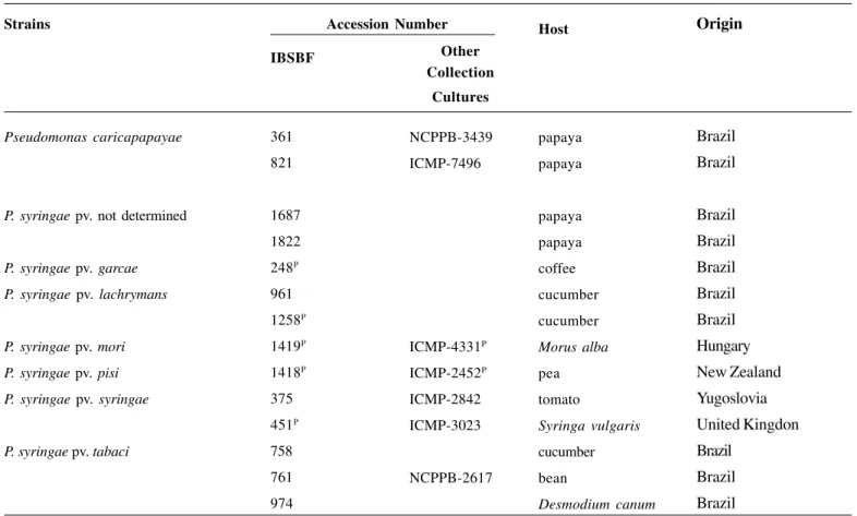
Besides the papaya isolates, other bacterial strains were in-cluded in this study for comparative purposes (Table 1). The strains were recovered from freeze-dried cultures and grown on NA at 280C for 48h.
1 Robbs,.C.F. Data not published.
Table 1. Bacterial strains used in this study
P Pathovar reference strain
IBSBF- Phytobacteria Culture Collection of Instituto Biológico, Campinas, SP, Brazil ICMP - International Collection of Micro-organisms from Plants, Auckland, New Zealand NCPPB - National Collection of Plant Pathogenic Bacteria, Harpenden, England
Host
papaya papaya
papaya papaya coffee cucumber cucumber
Morus alba
pea tomato
Syringa vulgaris
cucumber bean
Desmodium canum
Strains Origin
Brazil Brazil
Brazil Brazil Brazil Brazil Brazil Hungary New Zealand Yugoslovia United Kingdon Brazil
Brazil Brazil
Other Collection
Cultures
NCPPB-3439 ICMP-7496
ICMP-4331P ICMP-2452P ICMP-2842 ICMP-3023
NCPPB-2617
IBSBF
361 821
1687 1822 248P 961 1258P 1419P 1418P 375 451P 758 761 974
Accession Number
Pseudomonas caricapapayae
P. syringae pv. not determined
P. syringae pv. garcae P. syringae pv. lachrymans
P. syringae pv. mori P. syringae pv. pisi P. syringae pv. syringae
Pathogenicity assays
Papaya seedlings cv. Golden, tobacco (Nicotiana tabacum L.),
bean (Phaseolus vulgaris L.) and poinsettia (Euphorbia
pulche-rima Willd.) plants were inoculated by infiltration with bacterial
cell suspensions of papaya strain IBSBF 1687 containing ca.
108cfu/mL from 48-72h-old NA cultures, under moisture
cham-ber conditions. Besides leaves, fruits, stems and flowers of pa-paya were also inoculated. Negative controls were inoculated with sterile distilled water. All the inoculated and controls were maintained in a greenhouse (25 - 30°C) and examined daily for
disease development. Papaya plants were also inoculated with P.
caricapapayae strain IBSBF 361 for comparative purposes.
Biochemical and physiological assays
Biochemical tests for the identification at pathovar level were carried out according to Young & Triggs (27) and Schaad et al. (21).
Serological assays
Bacterial suspensions (ca. 109 cfu/mL) obtained from 48
h-old NA cultures as well as membrane protein complex (MPC) (22) were used as antigens. Microscopy slides for double diffusi-on tests were prepared with 3 mL of 1% purified agar in phos-phate buffered saline 0.1M, pH 7 with 200 ppm sodium azide. Papaya bacterial strains were tested with antisera against P. syrin-gae strains [P.s pv. syringae (AS-375), P. s. pv. tabaci (AS-761), P. savastanoi pv. phaseolicola (AS-736) and P. s. pv.
lachry-mans (AS-961)] obtained from the Antisera Collection of the
Laboratório de Bacteriologia Vegetal (LBV), Instituto Biológico, Campinas, SP, Brazil. All the antigen fractions were also tested against normal serum.
DNA extraction and amplification
Genomic DNA from papaya strain (IBSBF-1687), P.
syrin-gae pv. garcae (IBSBF 248P), P. s. pv. lachrymans (IBSBF 1258P), P. s. pv mori (IBSBF 1419P), P. s. pv. pisi (IBSBF 1418P), P. s.
pv. syringae (IBSBF 451P), P. s. pv. tabaci (IBSBF 758) and P. caricapapayae (IBSBF-361) were extracted (16)and the
con-centrations were estimated by comparison of the intensity of flu-orescence emitted by known concentrations of the bacteriopha-ge lambda DNA in an ethidium bromide-stained 0.6% agarose gel. Amplification of the 16S-23S spacer region was carried out using the primers pHr (12) and p23Suni322-anti (8). All PCR re-actions were performed in a total volume of 25 μL using 100 ng
of genomic DNA, 1.0 U Taq polymerase (Amersham
Bioscien-ces), 1 X Taq buffer, 200 μM dNTPs mixture, and 0.4 μM each
primer. The PCR protocol consisted of a denaturating cycle of 95oC for 2 min, followed by 25 cycles at 94oC for 1 min, 60oC for 30
s and 72oC for 3 min, and a final extension of 72oC for 5 min, in a
thermocycler (GeneAmp PCR system 9700; Perkin-Elmer Corpo-ration, Norwalk, Conn).
The primer set pshrp1F/2R, corresponding to hrpL gene of P. syringae pathovars morsprunorum, pisi and syringae (4) was
also tested. PCR was performed under the same conditions of the spacer regions and the amplifications were carried out by using
an initial denaturation step of 95oC for 2 min, followed by 25
cycles at 94oC for 1 min, 55oC for 30 s and 72oC for 1 min, and a
final extension period of 72oC for 3 min. The amplification
frag-ments were observed by electrophoresis in 1% agarose gels in 1X
TAE buffer (0.04M tris-acetate, 0.001M EDTA). The gels were stained with 0.1μg/μL of ethidium bromide and photographed under UV light using the Alpha Innotech 2200 Digital System.
PCR-RLFP of the 16S-23S spacer region and hrpL gene
PCR products (5 μL) were digested individually with each of the following restriction endonucleases Afa I, Alu I, Dde I, Hae
III, Hpa II, Hinf I, Sau 3A I and Taq I under conditions specified
by the manufacturer (Amersham Biosciences) and the restriction fragments separated by electrophoresis in 3% agarose gels using 1X TAE buffer. The gels were stained with ethidium bromide and visualized under UV. The molecular weights of the fragments were determined by comparison with a 100 bp DNA ladder (Amershan Biosciences).
RESULTS AND DISCUSSION
Cultural, morphological, physiological and biochemical tests previously carried out by Beriam et al.(2) showed that the papaya
strains belong to Pseudomonas syringae species. In this study,
these strains were investigated in order to classify them at the pathovar level.
P. syringae causes diseases in a large number of plants and
according to Young et al. (26) this species includes more than 50 pathovars, circumscribed on the basis of distinct host range. In addition, Young & Triggs (27) showed that physiological and biochemical determinative tests could be used to differentiate
Pseudomonas syringae at the pathovar level.
In this study, the papaya strains were pathogenic to tobacco, bean, and poinsettia. Bean and tobacco are natural hosts of P.s.
pv. syringae and P. s. pv. tabaci, but only P. s. pv. tabaci was also
described causing disease in poinsettia (18), suggesting that the papaya strains could be allocated as P. syringae pv. tabaci. The
determinative tests described in the literature (27) for identifica-tion at the pathovar level were very useful in this study (Table 2), corroborating the results of pathogenicity assays.
Another evidence that reinforced the identification of the pa-paya strains as P. s. pv. tabaci was the presence of precipitin
ban-ds only between the papaya isolates and P. s. pv. tabaci antisera
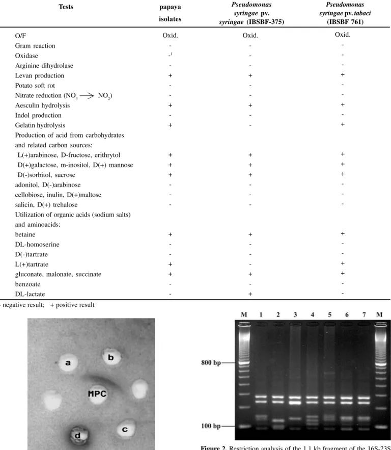
in the results of the serological assays (Figure 1).
Besides biochemical, serological and pathological results, the molecular tests also confirmed the papaya strains as P. s. pv. ta-baci. The amplification of the 16S-23S spacer region of different
pathovars of P. syringae (P.s. pv. garcae, P. s. pv. lachrymans, P.s.
pv. mori, P. s. pv. pisi, P. s.pv. syringae and P. s. pv. tabaci)
resulted in a single product for all strains. The size of the pro-duct was approximately 1.1 kilobase (kb). Only fragments ran-ging from 90 to 1100 base pairs (bp) obtained from restriction endonucleases experiments were considered for analysis. Seve-ral restriction enzymes were tested, but only Dde I yielded
dis-tinct profiles for each pathovar tested, which allowed to group the papaya strains with P. s. pv. tabaci (Figure 2 and Table 3).
The amplifications with the pshrp 1F/2R primers set were carried out with P.s. pv. syringae (IBSBF 451T), P.s. pv.tabaci
(IBSBF 758 and 974) and the papaya strain (IBSBF 1687) and yielded a fragment about 450 bp. No amplification was observed with P. caricapapayae strains. In the Alu I, Hae III, Hpa II, Hinf I,
and Taq I digestions, P.s. pv. syringae could clearly be
differenti-ated from P. s. pv. tabaci while the papaya strains showed
Figure 2. Restriction analysis of the 1.1 kb fragment of the 16S-23S
rDNA spacer regions from different pathovars of Pseudomonas syringae.
(M) 100 bp Marker (Amersham Biosciences); (1)P.syringae pv. garcae (IBSBF 248P); (2) P.syringae pv. lachrymans (IBSBF 1258P); (3)
P.syringae pv. mori (IBSBF 1419P); (4) P.syringae pv. pisi (IBSBF
1418P); (5) P.syringae pv. syringae (IBSBF 451T); (6) P.syringae pv.
tabaci (IBSBF 758); (7) papaya strain (IBSBF 1687).
Figure 1. Serological relationship between antisera against
Pseudomonas syringae pv. lachrymans (a) P.s. pv. tabaci (b), P. s. pv. syringae (d) and P. savastanoi pv. phaseolicola (c) strains and Mem-brane Complex Protein (MCP) of P. syringae from papaya.
Tests
Table 2. Determinative tests for the papaya isolates and some Pseudomonas pathovars
papaya isolates
Pseudomonas syringae pv. syringae (IBSBF-375)
O/F
Gram reaction Oxidase
Arginine dihydrolase Levan production Potato soft rot
Nitrate reduction (NO3 NO2) Aesculin hydrolysis
Indol production Gelatin hydrolysis
Production of acid from carbohydrates and related carbon sources:
L(+)arabinose, D-fructose, erithrytol D(+)galactose, m-inositol, D(+) mannose D(-)sorbitol, sucrose
adonitol, D(-)arabinose cellobiose, inulin, D(+)maltose salicin, D(+) trehalose
Utilization of organic acids (sodium salts) and aminoacids:
betaine DL-homoserine D(-)tartrate L(+)tartrate
gluconate, malonate, succinate benzoate
DL-lactate
Oxid. --1
-+ -+ -+
+ + +
-+ -+ +
-Oxid. -+ -+
-+ + +
-+ -+ -+
Oxid. -+ -+ -+
+ + +
-+ -+ + -1 - negative result; + positive result
Pseudomonas syringae pv. tabaci
Specific primers have been widely used as a rapid method
for identification of phytopathogenic bacteria like Erwinia
amylovora (1), P. savastanoi pv. phaseolicola (17), Xanthomo-nas albilineans (15), X. axonopodis pv. citri (7), and others. In
this study, the pshrp 1F/2R primer set exhibited specificity, dis-criminating P.s. pv. syringae and P.s. pv. tabaci from P. carica-papayae. Although the amplification has occurred for both P.s.
pv. syringae and P.s. pv. tabaci, the restriction profiles clearly
differentiated these pathovars, confirming the identification of the papaya strains as P.s. pv. tabaci.
According to Bradbury (3), strains of P. s. pv. tabaci can be
transmitted by seeds. Denardin2 isolated P.s. pv. tabaci from
pa-paya seed lots. Herein, the seed infection probably could be the source of primary inoculum since the papaya seedlings showed cotyledonary leaf lesions, suggesting bacterial seed transmissi-on, which represents an important vehicle of dissemination of the disease over considerable distances.
Table 3. PCR-RFLP profiles of the spacer region 16S-23S from
different pathovars of P. syringae produced by Dde I digestion.
Strains
P.s. pv. garcae (IBSBF 248P) P.s. pv. lachrymans (IBSBF 1258P) P.s. pv. mori (IBSBF 1419P) P.s. pv. pisi (IBSBF 1418P) P.s. pv. syringae (IBSBF 451T) P.s. pv. tabaci (IBSBF 758) Papaya strain (IBSBF 1687)
Fragments (bp)
290, 240, 170, 160, 110, 90 290, 240, 130, 100, 90 290, 240, 160, 90
290, 240, 170, 140, 110, 90 290, 240, 180, 160, 110, 90 290, 240, 160, 150, 110, 90 290, 240, 160, 150, 110, 90
Figure 3. Restriction patterns of the 450 bp fragment of the hrpL gene from (1) Pseudomonas syringae. pv. syringae (IBSBF 451P), (2) P.s. pv. tabaci (IBSBF 758), (3) P.s. pv. tabaci (IBSBF 974), (4) papaya strain (IBSBF-1687) digested with Hae III, Hinf I and Taq I. (M) 100 bp Marker.
Table 4. PCR-RFLP profiles produced by digestions of the hrp L gene with different restriction enzymes.
Enzymes
Afa I
Alu I
Dde I
Hae III
Hpa II
Hinf I
Mbo I
Taq I
Fragments (bp)
450
200, 180 250, 200
460
290, 190 380, 80
400 380
250, 150, 90 440
320, 150
170, 50 280, 150, < 50
Strains
P.s. pv. syringae (IBSBF 451T), P.s. pv. tabaci (IBSBF 758 and 974), papaya strain (IBSBF 1687)
P.s. pv. syringae (IBSBF 451T) P.s. pv. tabaci (IBSBF 758 and 974), papaya strain (IBSBF 1687)
P.s. pv. syringae (IBSBF 451T), P.s. pv. tabaci (IBSBF 758 and 974), papaya strain (IBSBF 1687)
P.s. pv. syringae (IBSBF 451T) P.s. pv. tabaci (IBSBF 758 and 974), papaya strain (IBSBF 1687)
P.s. pv. syringae (IBSBF 451T) P.s. pv. tabaci (IBSBF 758 and 974), papaya strain (IBSBF 1687)
P.s. pv. syringae (IBSBF 451T) P.s. pv. tabaci (IBSBF 758 and 974), papaya strain (IBSBF 1687)
P.s. pv. syringae (IBSBF 451T), P.s. pv. tabaci (IBSBF 758 and 974), papaya strain (IBSBF 1687)
P.s. pv. syringae (IBSBF 451T) P.s. pv. tabaci (IBSBF 758 and 974), papaya strain (IBSBF 1687)
REFERENCES
1. Bereswill, S.; Bugert, P.; Bruchmuller, I.; Geider K. Identification of the fire blight pathogen, Erwinia amylovora, by PCR assays with chromossomal DNA. Applied and Environmental Microbiolo-gy,Washington, v. 61, p. 2636-2642, 1995.
2. Beriam, L.O.S.; Almeida, I.M.G.; Ferrari, J.T.; Grabert, E.; Bar-bosa, A.F.; BarBar-bosa, I.; Louzeiro, I.M. Bacteriose em mamoeiro (Carica papaya L) causada por patovar de Pseudomonas syrin-gae. Summa Phytopathologica, Botucatu, v.28, n.1, p.95, 2002
(Resumo).
3. Bradbury, J.F. Pseudomonas tabaci. C.M.I. Descriptions of patho-genic fungi and bacteria, n. 129, p.1-2, 1967.
4. Destéfano, S.A.L. Detecção e identificação de bactérias fitopato-gênicas através da utilização de primers específicos. Summa Phytopathologica, Jaboticabal, v.26, n.1, p.158-160, 2000. 5. Frossard, P.; Hugon, R.; Verniere, C. Un dépérissement du
pa-payer aux Antilles françaises associé à un Erwinia sp. du groupe amylovora. Fruits, Paris, v.40, n.5, p.583-594, 1985.
6. Gardan, L.; Christen, R.; Achouak, W.; Prior, P. Erwinia papayae sp. nov., a pathogen of papaya (Carica papaya). International
nal of Systematic and Evolutionary Microbiology, Washington,
v.54, n.1, p.107-113, 2004.
7. Hartung, J.S.; Daniel, J.F.; Pruvost, O.P. Detection of Xanthomo-nas campestris pv. citri by the polymerase chain reaction me-thod. Applied and Environmental Microbiology, Washington, v.59, n.4, p.1143-1148, 1993.
8. Honeycut, R.J.; Sobral, B.W.S.; McClelland, M. tRNA intergenic spacers reveal polymorphism diagnostic of Xanthomonas albili-neans. Microbiology, Reading, v.141, n.12, p.3229-3239. 1995.
9. King, E.O.; Ward, M.K.; Raney, E.D. Two simple media for the demonstration of pyocianin and fluorescin. Journal of Labora-tory and Clinical Medicine, Milwaukee, v.44, p.301-307, 1954.
10. Leu, L.S.; Lee, C.C.; Huang, T.C. Papaya black rot caused by Erwinia cypripedii. Plant Protection Bulletin, Taichung, v.22,
n.4, p.377-384, 1980.
11. Levine, M. An introduction to laboratory technique in bacte-riology. New York: The Macmillan, 1954. p.68-79.
12. Massol-Deya, A.A.; Odelson, D.A.; Hickey, R.F.; Tiedje, J.M. Bacterial community fingerprinting of amplified 16S and 16S 23S ribossomal DNA gene sequences and restriction endonuclease analysis (ARDRA). In: Akkermans, A.D.L.; van Elsas, J.D.; de Brujin, F.D. (Ed.) Molecular microbial ecology manual.
Dor-drecht: Kluwer Academic Publishers, 1995. p. 3.3.2/1-3.3.2./8. 13. Nelson, M.N.; Alvarez, A.M. Purple stain of Carica papaya. Plant
Disease, St. Paul, v.64, n.1, p.93-95, 1980.
14. Nishijima, K.A.; Couey, H.M.; Alvarez, A.M. Internal yellowing, a bacterial disease of papaya fruits caused by Enterobacter cloa-cae. Plant Disease, St. Paul, v.71, n.11, p.1029-1034, 1987. 15. Pan, Y.-B.; Grisham, M.P.; Burner, D.M. A polymerase chain
reaction protocol for the detection of Xanthomonas albilineans, the causal agent of sugarcane leaf scald disease. Plant Disease,
St. Paul,v.81, n.2, p.189-194, 1987.
16. Pitcher, D.G.; Saunders, N.A.; Owen, R.J. Rapid extraction of bacterial genomic DNA with guanidium thiocyanate. Letters in Applied Microbiology, Oxford, v.8, p.151-156, 1989.
17. Prosen, D.; Hatziloukas, E.; Schaad, N.W.; Panapoulos, N.J. Speci-fic detection of Pseudomonas syringae pv. phaseolicola DNA in
bean seed by PCR-based amplification of a phaseolotoxin gene region. Phytopathology, St. Paul,v.83, n.8, p. 965-970, 1993. 18. Ribeiro, R.L.D.; Pimentel, J.P.; Kimura, O.; Robbs, C.F.; Akiba, F.
Caracterização da bactéria incitante do “fogo selvagem” da poinset-tia (Euphorbia pulcherrima) no Est. Rio de Janeiro. Fitopatologia Brasileira, Brasília, v.5, n.3, p.450-451, 1980.
19. Robbs, C.F. Uma nova doença bacteriana no mamoeiro (Carica papaya L). Boletim da Sociedade Brasileira de Agronomia, Rio de Janeiro, v.12, n.1/2, p.73-76, 1956.
20. Robbs, C.F.; Rodrigues Neto, J.; Malavolta Jr., V.A.; Vega, J. Uma podridão bacteriana do topo do mamoeiro causada por Erwi-nia sp. associada à plantas afetadas pelo vírus do mosaico, no Rio Grande do Sul. Fitopatologia Brasileira, Brasília, v.13, n.2,
p.107, 1988.
21. Schaad, N.W.; Jones, J.B.; Chun, N. Laboratory guide for iden-tification of plant pathogenic bacteria. St. Paul: APS Press, 2001. 373p.
22. Thaveechai, N.; Schaad, N.W. Immunochemical characterizati-on of a subspecies-specific antigenic determinant of a membrane protein extract of Xanthomonas campestris pv. campestris. Phyto-pathology, St. Paul, v.76, n.2, p.148-153, 1986.
23. Trujillo, E.E.; Schrot, M.N. Two bacterial diseases of papaya trees caused by Erwinia species in the Northern Mariana Island.
Plant Disease, St. Paul, v.66, n.2, p.116-120, 1982.
24. Ventura, J.A.; Costa, H.; Tatagiba, J.S.; Martins,D.S. Manejo de doenças e produção integrada de frutas tropicais. Fitopatologia Brasileira, Brasília, v.28, supl., p. 57-61, 2003.
25. Webb, R.R. Epidemiology and control of bacterial canker of pa-paya caused by an Erwinia sp. on St. Croix, U.S. Virgin Island.
Plant Disease, St. Paul, v.69, n.4, p.305-309, 1985.
26. Young, J.M.; Saddler, G.S.; Takikawa, Y.; De Boer, S.H.; Vaute-rin, L.; Gardan, L.; Gvozdyak, R.I.; Stead, D.E. Names of plant pathogenic bacteria 1864-1995. Review of Plant Pathology,
Wallingford, v.75, n.9, p.721-763, 1996.