Characterization and Chemical Stability of Hydrophilic and Hydrophobic Magnetic
Nanoparticles
Natália Cristina Candian Lobato a, Marcelo Borges Mansur a,c,d, Angela de Mello Ferreira b,d*
Received: September 22, 2016; Revised: March 06, 2017; Accepted: March 11, 2017
Magnetic nanoparticles can improve the eiciency of phase separation time in multi-stage operations when a magnetic ield is present. As such operations involve contact with aqueous and/or organic solutions, hydrophilic magnetic nanoparticles synthesized through the co-precipitation method were functionalized with oleic acid to attain hydrophobic magnetic nanoparticles. Both nanoparticles were characterized morphologically, chemically and magnetically. The results revealed that the particles (size ≈ 10 nm) consisted of an iron oxide mixture of magnetite and maghemite. The functionalization with oleic acid was efective in converting them into hydrophobic nanoparticles. Both particles were ferro/ferrimagnetic and the presence of oleic acid did not interfere signiicantly in the saturation magnetization value. The chemical stability of both nanoparticles were also evaluated, as an attempt of simulating broad industrial conditions to which the nanoparticles may be subjected; the hydrophilic nanoparticles were resistant at pH ≥ 4, while the hydrophobic nanoparticles were stable at pH ≥ 3.
Keywords: Magnetic Nanoparticles, Chemical Stability, Oleic Acid, Hydrophilic Nanoparticles, Hydrophobic Nanoparticles
* e-mail: angelamello@des.cefetmg.br
1. Introduction
The use of magnetic nanoparticles to develop and/or improve separation techniques has been widely studied in recent years. In general, the separation methods with nanoparticles have proven to be fast, easily automatable, reversible, and selective, in addition to being able to reach high eiciency levels 1–3. Magnetic nanoparticles have been used as contrast agents in diagnostic applications, drug release, magnetic refrigeration, magnetic separation, among others 4–6. In the ield of mineral processing, the application of nanoparticles has been studied in multi-stage operations, such as solvent extraction 1,7–10, adsorption11–15,
and ion exchange 16,17.
In solvent extraction, the application of magnetic nanoparticles is intended to magnetize the organic solution to make the separation between the aqueous and the organic phases quick and eicient when in the presence of a magnetic ield 7,18. To obtain a magnetized organic solution, hydrophobic
superparamagnetic nanoparticles are dispersed in an organic solvent containing an extractant, responsible to selectively react with the metal in the aqueous phase. After contact with the two phases, and the metal thus being transferred from
the aqueous phase to the organic phase, a magnetic ield is applied to accelerate the phase disengagement step 1,7–10.
In fact, the results of phase disengagement with magnetic nanoparticles was faster, but the values varied signiicantly depending on the operating conditions and other speciication of the system, such as the type and the concentration of the extractant, aqueous composition, concentration of the nanoparticles, etc. According to Palyska and Chmielewski8, the
use of magnetic separation in solvent extraction can achieve a separation rate 160 times faster than using the gravitational ield alone; Vatta, Koch, and Sole 9 reported that the time
separation when using the magnetic system is between 48% and 86% when compared to the traditional system time; and Lobato, Ferreira, and Mansur 10 veriied that magnetic
separation is 3-5 times faster when using magnetic luid. The superparamagnetic nanoparticles are also being studied for metal ion adsorption from an aqueous solution
11–15. Similar to in solvent extraction, the use of magnetic
properties in the adsorbent material is intended to improve the solid-liquid separation processes after metal loading processes occur. Moreover, because of the small size of the nanoparticles, the adsorption process is favored due to the large surface area. The magnetic nanoparticles are used as a core and their surface is modiied in order to achieve a Postgraduate Program on Metallurgical, Materials and Mining Engineering, Universidade Federal de
Minas Gerais (UFMG), Belo Horizonte, MG, Brazil
b Department of Chemistry, Centro Federal de Educação Tecnológica de Minas Gerais (CEFET-MG), Belo Horizonte, MG, Brazil
c Program of Metallurgical and Materials Engineering, Universidade Federal do Rio de Janeiro (UFRJ), Rio de Janeiro, RJ, Brazil
selectivity for a given substance or metal present in the aqueous phase. Once loaded, the magnetic particles can easily be separated from the depleted solution by using a magnetic ield. Such operation may reduce the process time and increase the eiciency of the phase separation. Thus, the loaded particles can be regenerated and reused 13–15. Magnetic nanoadsorbents have proved eicient for ions such as Cd(II), Zn(II), Pb(II), Cu(II) 11, As(III) 12, P 13,
Cr(IV) 14, and Sb(III) 15.
Also aimed at a faster and more eicient solid-liquid separation, magnetic particles have been proposed in the ion-exchange process
16,17. The technique is very similar to the adsorption process. Modiied
magnetic nanoparticles capable of performing ion exchange are dispersed in an aqueous solution (or an eluent) containing the element to be removed. The loaded nanoparticles are separated from the aqueous phase through the presence of a magnetic ield. Subsequently, ions are removed from the particles, which are regenerated and reused. This method has several advantages over the traditional method. First, problems such as clogging and fouling do not occur. Second, because of the small particle size, there is a large surface area for ion exchange reactions. And inally, there is the beneit of magnetic separation, which is faster than sedimentation or iltration and more selective, since only the magnetic fraction is separated from the aqueous phase 16.
Given the prospect of using magnetic nanoparticles in the mining industry, this work aims to characterize hydrophilic and hydrophobic magnetic nanoparticles. Hydrophilic magnetic nanoparticles were synthesized by the co-precipitation method with no surface modiication, while the hydrophobic nanoparticles were obtained by functionalizing the hydrophilic nanoparticles with oleic acid. Oleic acid (C18H34O2) is an organic species with high
ainity to the surface of ferrous oxides due to its carboxylic group, and because of this, it is often used as a surfactant to modify the surface of magnetite nanoparticles, making them hydrophobic. The functionalization process with oleic acid is considered to be simple and inexpensive 5,19,20. The
nanoparticles were characterized for their morphology, composition, and magnetization. Furthermore, the stability of the particles when in contact with aqueous solutions of varying acidity was also evaluated, in an attempt to simulate a number of industrial conditions.
2. Experimental
2.1. Materials
Ferrous sulphate heptahydrate (FeSO4.7H2O, Neon,
purity 99%), ferric chloride hexahydrate (FeCl3.6H2O, Vetec,
purity 97%), ammonium hydroxide (NH4OH, Neon, 28-30 wt.%), oleic acid (C18H34O2, Synth, purity 100%), ethyl
alcohol (CH3CH2OH, Neon, purity 99.5%), sulfuric acid
(H2SO4, Synth, purity 98%), potassium nitrate (KNO3, Vetec, purity 99%), and Exxsol D80 (liquid aliphatic hydrocarbon,
ExxonMobil Chemical). Except for the organic diluent, which was of commercial grade, all remaining reagents used in this work were of analytical grade, and the water was distilled or Milli-Q (Millipore, France), depending on the experiment.
2.2. Synthesis of the hydrophilic magnetic
nanoparticles
The nanoparticles were obtained by the co-precipitation method 5,21–24. In 300 mL of distilled water, 19.7 g of FeSO4.7H2O and 39.0 g of FeCl3.6H2O were dissolved
(molar ratio of ferric ion to ferrous ion in the solution of two). The solution was vigorously stirred (1000 rpm) and heated to 80 ºC. Then, 50 mL of NH4OH were dropped under stirring, and the solution continued to be stirred for 40 minutes. After, heating and stirring were turned of and the solution rested until reaching room temperature. The magnetic nanoparticles were washed with distilled water and then with ethyl alcohol. After that, they were iltrated, and dried in a kiln (60 ºC) for 2 hours.
2.3. Synthesis of hydrophobic magnetic
nanoparticles
The hydrophilic magnetic nanoparticles were functionalized with oleic acid in order to make them hydrophobic 7. In
a 100 mL glass reactor, 1 g of the synthesized magnetic nanoparticles was dispersed in 40 mL of distilled water at 60 ºC under mechanical stirring (500 rpm). Next, 1 mL of oleic acid was added into the solution and the mixture was stirred for 15 minutes. The solution rested until reaching room temperature. The hydrophobic particles were removed using a magnet, washed with distilled water and then with ethyl alcohol, iltrated, and dried in a kiln (60 ºC) for 1 hour.
2.4. Characterization of the magnetic
nanoparticles
The magnetic nanoparticles were characterized using the following methods:
(i) Transmission electron microscopy (TEM) images were obtained in order to analyze the morphology structure and measure the particle size distribution of the nanoparticles. The analysis was performed using a Tecnai G2-20 equipment, SuperTwin FEI (200 kV);
(ii) The surface area of the nanoparticles was determined by the BET (Brunauer-Emmett-Teller) method through nitrogen adsorption using a Quantachrome Nova 1200e.
from 10 to 80, with increments of 0.02 theta and a scanning speed of 2º min-1;
(iv) Raman spectroscopy was used to diferentiate the iron oxide phases (magnetite and maghemite). The technique was performed using a Jobin Yvon Horiba LABRAM equipment, HR800 model, with a He-Ne laser of 632.8 nm, coupled to an Olympus BX-41 microscope. The spectra were acquired with a laser power 0.08 mW in the range of 150-900 cm-1;
(v) Broad sextets and model-independent hyperine ield distribution were collected with the sample at 150 K and 298 K (in a liquid helium bath-cryostat) in a constant acceleration transmission mode setup with a 20 mCi-57Co/Rh source. Data were numerically itted by Lorentzian functions with the least-square procedure of the NORMOSTM program (Brand RA, Laboratorium für Angewandte Physik, Universität Duisburg, D-47048, Duisburg, Germany). The isomer shift values were referred to α-Fe at room temperature (RT);
(vi) The zeta potential of the hydrophilic nanoparticle was measured in order to compare the electrostatic potential of the surface of the synthesized nanoparticle with values reported in the literature. For sample preparation, 0.2 g of nanoparticles were added to 200 mL of 0.001 mol/L KNO3, and the pH was adjusted between 5 and 10, using NH4OH and H2SO4 solutions. Each sample was analyzed by a Zeta Meter device (ZD3-D-G 3.0+ model), collecting at least 10 PZC values;
(vii) The adsorption of the oleic acid in the magnetic nanoparticles was veriied by Fourier transform infrared spectroscopy (FTIR), which identiied the chemical groups of the hydrophilic nanoparticles and the functional groups of the oleic acid. The analyses were performed in the range of 4000 to 450 cm-1 using a Perkin Elmer infrared spectrophotometer (Spectrum Frontier model);
(viii) Thermogravimetric analysis (TGA) was used to evaluate the thermal stability of the magnetic particles, as well as to check the amount of oleic acid adsorbed on the functionalized nanoparticles. The experiment was carried out using a Perkin Elmer STA 6000 equipment and the procedure was performed in nitrogen atmosphere at a low rate of 20 mL/minute, and under the temperatures scan from 30 ºC and 800 ºC, at a heating rate of 10 ºC/minute;
(ix) The magnetic characteristics and behavior of the nanoparticles were obtained using the Vibrating Sample Magnetometer (VSM), Lakeshore model 7404 series. The hysteresis loops were measured under a magnetic ield strength of 11500 Gauss at room temperature;
(x) The chemical stability of the hydrophilic and hydrophobic nanoparticles was determined by contacting them with water at changing acidity. Regarding the hydrophilic magnetic nanoparticles, 0.25 g of the material was added to 25 mL of Milli-Q water (Millipore, France) at changing acidity ([H+] = 10-7, 10-6, 10-5, 10-4, 10-3, 10-2, 10-1, 0.5, 1, and 2 mol/L) in Erlenmeyer lasks. The water was acidiied using H2SO4 solutions. The hydrophobic magnetic nanoparticles
were dispersed in Exxsol D80, a common diluent used in the industrial solvent extraction processes, at a concentration of 10 g/L, using an ultrasound bath (Brasonic, 1210 model, frequency 47Hz) for 30 minutes. After the formation of the magnetic luid or ferroluid, 25 mL of the organic liquid was placed in contact with 25 mL of Milli-Q water at changing acidity ([H+] = 10-7, 10-6, 10-5, 10-4, 10-3, 10-2, 10-1, 0.5, 1, and 2 mol/L) in Erlenmeyer lasks. All the samples were stirred in a shaker (New Brunswick Scientiic, Annova44 model) at 400 rpm, for 24 hours, at room temperature (25 ºC). After, a sample of the water was withdrawn for chemical analysis by Atomic Absorption Spectroscopy (GBC, XplorAA-2 model) to determine the content of iron. Such tests were performed in triplicate.
3. Results and Discussion
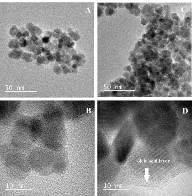
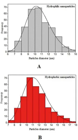
The morphology and size distribution of the nanoparticles were obtained via transmission electron microscopy (TEM). The images shown in Figure 1revealed that the synthesized nanoparticles have a nearly spherical shape, with homogeneous distribution. Hydrophilic nanoparticles are shown in Figure 1 - A and B, while hydrophobic nanoparticles are shown in Figure 1 - C and D. The layer of oleic acid adsorbed on the surface of the magnetic hydrophobic nanoparticles is shown to surround the particles. The hydrophilic magnetic nanoparticles have sizes ranging between 6 and 16 nm, with a mean diameter of 10.2 nm, while the hydrophobic magnetic nanoparticles, functionalized with oleic acid, have sizes ranging between 7 and 18 nm, with a mean diameter of 11.0 nm. The values of the diameters were calculated using the software Image J, with more than 300 values measured for each sample. Size histograms of both nanoparticles are shown in Figure 2. The distribution proile of the values of diameters follows the log-normal function, as had been observed in a previous study 25. Based on such results, it
can be concluded that the functionalization of the magnetic nanoparticles with oleic acid did not modify them regarding their morphology and size.
The surface area was determined by the BET method. The values of the hydrophilic and hydrophobic nanoparticles were 71.6 and 55.3 m2/g, respectively. Both materials present
large surface areas. Such character is desirable in order to enhance a higher mass transfer and chemical reaction rates on adsorption and ion exchange processes, in the case of the hydrophilic nanoparticles; and it is favorable to improve the distribution of the nanoparticles in the bulk of the organic phase on the solvent extraction process, in the case of the hydrophobic nanoparticles.
Figure 1. Transmission electronic microscopy images of the hydrophilic nanoparticles (A and B) and the hydrophobic nanoparticles functionalized with oleic acid (C and D).
spinel structure. This structure is characteristic of magnetite (Fe3O4) or maghemite (γ-Fe2O3), not being possible to identify which of the phases is present or if both phases are present in the material. The difractograms of hydrophilic and hydrophobic nanoparticles are similar, indicating that the functionalization process with oleic acid did not modify the crystallography character of the magnetic nanoparticles. The crystallite size of particles can be estimated by the Scherrer’s equation:
half-width of the maximum difraction peak expressed in radians and θ is the difraction angle of the peak position 23,26 The application of the Scherrer’s equation to the peak (311) with 2θ ~ 35.7º of the magnetic nanoparticles indicated that the crystalline size of hydrophilic and hydrophobic nanoparticles were of 11.2 and 12.1 nm, respectively. The calculated sizes are according to the average sizes found via transmission electron microscopy (TEM). Moreover, the results found by Scherrer’s equation are statistically similar, because the crystalline cores of both samples come from the exact same synthesis. In addition, the oleic acid present in the hydrophobic particles is not detected by the X-ray difraction method (it has no crystallographic organization), therefore it can not inluence on the crystallite size calculated by Scherrer’s equation.
( )
cos
D
K
1
$
b
i
m
=
Figure 2. Size distribution histogram of the hydrophilic nanoparticles (A) and the hydrophobic nanoparticles (B).
Figure 3. X-ray difractograms of the magnetic nanoparticles.
Raman spectroscopy analysis complements X-ray difractograms, sinceit diferentiates distinct phases of iron oxide present in the sample, such as magnetite (Fe3O4) and maghemite (γ-Fe2O3). Under the microscope linked to the
equipment, it was observed that the samples of hydrophilic
and hydrophobic nanoparticles presented areas with distinct colors, one brown and another black. For this reason, the analyses were performed on each distinct area of both samples, the results of which are shown in Figure 4. The vibrational modes of magnetite and maghemite structures in Raman spectroscopy are summarized in Table 1. A itting of the peaks of magnetite and maghemite spectra was performed via PeakFit function of the Origin software, which it can be seen in Figure 4. Analyzing the characteristic peaks of each phase, it can be concluded that magnetite and maghemite phases occur in both magnetic nanoparticles, and that the black area corresponds to the predominance of the magnetite phase, while the brown area is related to the predominance of the maghemite phase.
The occurrence of magnetite and maghemite was also identiied from the Mössbauer spectroscopy data for the samples at 150 K (Table 2 and Figure 5). Both 298 K spectra show broad line patterns with some small diferences in their six line shapes. The broad six lines of the 298 K spectra of both samples can be attributed to the inite-size efect and/ or spin relaxation of the analyzed nanoparticles. However, no superparamagnetic state (absence of doublet component) has been observed probably due to the presence of particles with a larger size indicated by XRD and TEM data and/or due to the intra-particle magnetic interactions. Therefore, in order to better quantify the phases, measurements at 150 K were performed. This temperature level is above the Verwey temperature, TV ~ 121 K, which is the temperature that the magnetic conductivity is signiicantly reduced and the magnetic behavior changes completely 30. Each spectra obtained at 150 K was itted with three sextets with line-width ~ 0.50 mm/s. The sextet with the largest hyperine magnetic ield (Table 2) was assigned to Fe3+ in maghemite. The other two sextets were assigned to Fe3+ in the tetrahedral sites and to the mixed valence Fe2+/Fe3+ in the octahedral sites of the magnetite. The numerical analysis of these Mössbauer spectra was carried out using the NORMOSTM software. Similar Debye temperatures were also assumed for both Fe-oxide phases, in order to have their relative areas. Results revealed that the hydrophilic nanoparticles have a composition of 60% maghemite and 40% magnetite, whereas the hydrophobic nanoparticles is composed by 53% maghemite and 47% magnetite. The lower amount of maghemite in the functionalized nanoparticles with oleic acid is likely due to a protective character of the organic layer, which prevents the magnetite nanoparticle from being oxidized in air.
The adsorption of oleic acid at the external surface of the nanoparticles occurs due to the high ainity for iron minerals. In fact, the negative charge of the carboxyl group is highly attracted to the positive surface of these minerals20,31, as schematically shown in Figure 6.
Figure 4. Raman spectrum of hydrophilic nanoparticles (A) and the hydrophobic nanoparticles (B), including analysis of the peaks using the Origin software.
Table 1: Vibrational modes of the magnetite and maghemite phases in Raman spectroscopy 27–29.
Phase Vibrational modes Wave number
(cm-1)
Magnetite (Fe3O4)
T2g 190 - 193
Eg 306 - 310
T2g 450 - 490
T2g 538 - 554
A1g 668 - 672
Maghemite (γ-Fe2O3)
T2g 350 - 365
Eg 500 - 511
A1g 700
isoelectric point of the hydrophilic magnetic nanoparticles found in this work is in fair agreement with the results available in the literature for similar particles, i.e., 5.6 32, 6.3 33, and 6.5
22. Based on this value, the synthesized nanoparticles have
a positive charge at pH < 6, thus favoring the adsorption of oleic acid molecules. In this study, the adsorption of oleic acid was carried out near pH 6, a value indicated as suitable to obtain the maximum chemisorption of the adsorbent. Moreover, the adsorption process occurred at 60 ºC, aimed at favoring the chemical linkage 22,34.
The Fourier transform infrared spectroscopy analyses conirmed the surface functionalization of magnetite nanoparticles (Figure 8). In relation to the peaks present in both spectra, the 578 cm-1 corresponded to the vibration of the Fe-O, while the bands 1630 and 3400 cm-1 are characteristic of hydroxyl groups related to the water presence on the surface of the particles 19,21. In the
spectrum of the nanoparticles functionalized with oleic acid (hydrophobic nanoparticles), bands 1425 and 1520 cm-1 are related to the vibration of the carboxyl group (COO-) of oleic acid, while bands 2851 and 2921 cm-1 correspond to the stretching of the C-H bonds 5,24.
Table 2: Mössbauer parameters for hydrophilic and hydrophobic magnetic nanoparticles at room temperature and 150K.
Sample Phases δ (mm/s) (± 0,05) Δ/2ξq (mm/s)(± 0,05) BHF (T) (± 0,2) Area (%) (± 1)
Hydrophilic nanoparticles (RT)
γ-Fe2O3 0.22 -0.06 47.0 38
(γ-Fe2O3/Fe3O4) 0.37 -0.06 41.6 62
Hydrophilic nanoparticles (150K)
γ-Fe2O3 0.40 -0.05 50.3 60
Fe3O4 (Fe3)
(tetrahedral site) 0.31 -0.08 48.1 14
Fe3O4 (Fe3+Fe2)
(octahedral site) 0.60 -0.02 46.4 26
Hydrophobic nanoparticles (RT)
γ-Fe2O3 0.29 -0.02 46.1 31
(γ-Fe2O3/Fe3O4) 0.38 -0.06 41.4 61
Hydrophobic nanoparticles (150 K)
γ-Fe2O3 0.41 -0.08 50.6 53
Fe3O4 (Fe3)
(tetrahedral site) 0.31 -0.02 48.1 16
Fe3O4 (Fe3+Fe2)
(octahedral site) 0.64 -0.02 45.4 31
Figure 5. Mössbauer spectra of the magnetic nanoparticles at room temperature (A) and 150 K (B).
and chemically adsorbed (5.6%) oleic acid, respectively, on the surface of the nanoparticles 5,19. These layers
represent 0.045 and 0.115 mL of oleic acid adsorbed physically and chemically, respectively, for each gram of magnetic nanoparticles. The third stage of weight loss is above 450 ºC – 500 ºC. According to Ayyappan at al. 35, the degradation of oleic acid produces reduction gases like CO and CO2, which are responsible for reducing
the magnetic nanoparticles. During heating, the possible reduction reactions may occur 35:
Fe O
3 4+
CO
3
FeO
+
CO
2(
CO
O
CO
2
1
2
)
2+
(
FeO
Fe O
Fe
3
)
2 3+
a
Figure 6. Adsorption of oleic acid on the surface of an iron oxide nanoparticle.
Figure 7. The isoelectric point (pHpzc) of the hydrophilic nanoparticles.
Figure 8. FTIR spectra of the magnetic nanoparticles.
Figure 9. Thermograms of the magnetic nanoparticles.
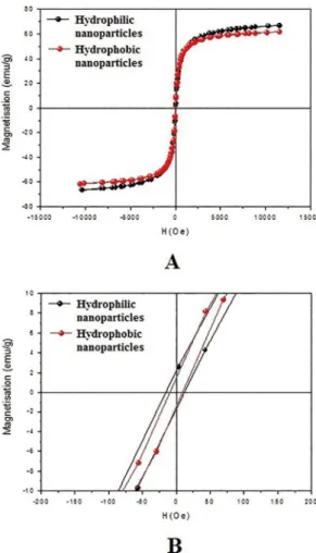
The hysteresis curves of the magnetic particles obtained using a magnetometer are show in Figure 10 - A and the coercivity ield values (HC) are show in Figure 10 - B. It can be observed that the samples have a ferro/ferrimagnetic behavior, because the coercive ields at room temperature are non-zero, which is in agreement with the Mössbauer data (six broad lines) above discussed. However, as the coercivity ield value is very small, it can be concluded that large part of the
particles are in the superparamagnetic state. This observation is in agreement with Mössbauer spectroscopy. The values of saturation magnetization and coercivity of the nanoparticles are shown in Table 3. The functionalization with oleic acid did not signiicantly change the magnetization values. In fact, the small decrease in the magnetization value on the hydrophobic nanoparticle is due to the presence of the organic compound 5,23 It is noted that the value of the percentage of nonmagnetic material adhered to the magnetic material (9%) is almost the same as the magnetization loss between the two samples (8%), thus indicating the reason why the nanoparticles functionalized with oleic acid (hydrophobic nanoparticles) lost magnetization.
Considering the fractions of each component (calculated by Mössbauer) and the saturation magnetization values of magnetite bulk phase (92 emu.g−1) 36 and maghemite bulk phase (74 emu.g−1) 26 the nominal magnetization values of the
Figure 10. Hysteresis curves (A), and coercivity ield values (B) of magnetic nanoparticles.
Table 3: Saturation magnetization and coercivity values of the magnetic nanoparticles.
Sample Coercivity (Gauss) Magnetization (emu/g)
Hydrophilic
nanoparticles 13.6 66.7
Hydrophobic
nanoparticles 9.3 61.5
The results of the chemical stability of the magnetic nanoparticles are shown in Table 4. Regarding the hydrophilic nanoparticles, no iron was determined in the aqueous solution at pH ≥ 4, while the hydrophobic nanoparticles are chemically stable to acid media at pH ≥ 3. Furthermore, the loss of iron of the hydrophobic particles is comparatively smaller than that of the hydrophilic nanoparticles when in contact with a medium with the same concentration of H+, which is attributed to the protective layer of oleic acid. The same result of chemical stability is shown in Figure 11 but in relation to the percentage of total iron that contaminated the water. It was observed that while the maximum iron loss of the hydrophobic nanoparticles (functionalized with oleic acid) is 31%, the iron loss in the hydrophilic nanoparticles
Table 4: Iron contamination of the water at changing acidities.
n.d. not detected
Acidity [H+] Hydrophilic
nanoparticles
Hydrophobic nanoparticles
10-7 n.d. n.d.
10-6 n.d. n.d.
10-5 n.d. n.d.
10-4 n.d. n.d.
10-3 8.2 ± 0.5 n.d.
10-2 83 ± 1 35.6 ± 0.4
10-1 883 ± 23 732 ± 44
0.5 4874 ± 75 1702 ± 22
1.0 5578 ± 2 1807 ± 36
2.0 5968 ± 248 2265 ± 32
Figure 11. Percentage of iron loss from the magnetic nanoparticles.
reaches 82%. Hence, under appropriate conditions, the magnetic nanoparticles can be employed with no chemical alteration of the environment in which they are applied and with no chemical or morphological alteration of the nanoparticles themselves. Moreover, in suitable conditions, the magnetic nanoparticles have a long service life and can be reused several times.
4. Conclusions
The magnetic nanoparticles synthesized through the co-precipitation method (hydrophilic nanoparticles) and subsequently functionalized with oleic acid (hydrophobic nanoparticles) have a spherical shape, with a average nanosize of approximately 10 nm, a large surface area (55 - 72 m2/g),
magnetization value. Regarding chemical stability, the hydrophilic nanoparticles are stable at pH ≥ 4, while the hydrophobic nanoparticles are stable at pH ≥ 3. It was observed that hydrophobic nanoparticles are relatively more resistant to acid environments than are hydrophilic nanoparticles due to the protection of the organic layer. Thus, under suitable conditions of pH and in processes involving aqueous and/or organic solutions, the nanoparticles developed in this work can be used for several cycles of the process with no chemical alteration of them or of the environment in which they are.
5. Acknowledgement
The authors acknowledge CNPq (nº 304050/2016-4), CAPES-PROEX, and FAPEMIG for their inancial support, the Center of Microscopy at Universidade Federal de Minas Gerais (UFMG) (http://www.microscopia.ufmg.br) for providing the equipment and technical support for experiments involving electron microscopy, and Centro de Desenvolvimento da Tecnologia Nuclear (CDTN) (http://www.cdtn.br/) for their support with Mössbauer spectroscopy and magnetic analysis.
6. References
1. Wang Q, Guan Y, Ren X, Cha G, Yang M. Rapid extraction of low concentration heavy metal ions by magnetic luids in high gradient magnetic separator. Separation and Puriication
Technology. 2011;82:185-189.
2. Franzreb M, Siemann-Herzberg M, Hobley TJ, Thomas ORT. Protein puriication using magnetic adsorbent particles. Applied Microbiology and Biotechnology. 2006;70(5):505-516.
3. Toma HE. Developing nanotechnological strategies for green industrial processes. Pure and Applied Chemistry. 2013;85(8):1655-1669.
4. Vatta LL, Sanderson RD, Koch KR. Magnetic nanoparticles: Properties and potential applications. Pure and Applied Chemistry.
2006;78(9):1793-1801.
5. Petcharoen K, Sirivat A. Synthesis and characterization of magnetite nanoparticles via the chemical co-precipitation method.
Materials Science and Engineering: B. 2012;177(5):421-427.
6. Im SH, Herricks T, Lee YT, Xia Y. Synthesis and characterization of monodisperse silica colloids loaded with superparamagnetic iron oxide nanoparticles. Chemical Physics Letters.
2005;401(1-3):19-23.
7. Hwang JY, inventor; Board of Control of Michigan Technological University, assignee. Magnetic solvent extraction. United States
patent US 5043070 A, 1991 Ago 27.
8. Palyska W, Chmielewski AG. Solvent Extraction and Emulsion Separation in Magnetic Fields. Separation Science and Technology.
1993;28(1-3):127-138.
9. Vatta LL, Koch KR, Sole KC. The potential use of hydrocarbon magnetic liquids in solvent extraction. In: Proceedings of 18th International Solvent Extraction Conference (ISEC 2008); 2008 Sep 15-19; Tucson, AZ, USA. Montreal: Canadian Institute of Mining, Metallurgy and Petroleum; 2008. p. 1513-1518.
10. Lobato NCC, Ferreira AM, Mansur MB. Evaluation of magnetic nanoparticles coated by oleic acid applied to solvent extraction processes. Separation and Puriication
Technology. 2016;168:93-100.
11. Ge F, Li MM, Ye H, Zhao BX. Efective removal of heavy metal ions Cd2+, Zn2+, Pb2+, Cu2+ from aqueous solution
by polymer-modiied magnetic nanoparticles. Journal of Hazardous Materials. 2012;211-212:366-372.
12. Silva GC, Almeida FS, Ferreira AM, Ciminelli VST. Preparation and application of a magnetic composite (Mn3O4/Fe3O4)
for removal of As(III) from aqueous solutions. Materials Research. 2012;15(3):403-408.
13. Yoon SY, Lee CG, Park JA, Kim SB, Kim JH, Lee SH, et al. Kinetic, equilibrium and thermodynamic studies for phosphate adsorption to magnetic iron oxide nanoparticles.
Chemical Engineering Journal. 2014;236:341-347.
14. Tang SC, Lo IMC. Magnetic nanoparticles: essential factors for sustainable environmental applications. Water Research.
2013;47(8):2613-2632.
15. Shan C, Ma Z, Tong M. Eicient removal of trace antimony(III) through adsorption by hematite modiied magnetic nanoparticles.
Journal of Hazardous Materials. 2014;268:229-236.
16. Drenkova-Tuhtan A, Mandel K, Paulus A, Meyer C, Hutter F, Gellermann C, et al. Phosphate recovery from wastewater using engineered superparamagnetic particles modiied with layered double hydroxide ion exchangers. Water Research.
2013;47(15):5670-5677.
17. Lee Y, Rho J, Jung B. Preparation of magnetic ion-exchange resins by the suspension polymerization of styrene with magnetite.
Journal of Applied Polymer Science. 2003;89(8):2058-2067.
18. Wikström P, Flygare S, Gröndalen A, Larsson PO. Magnetic aqueous two-phase separation: A new technique to increase rate of phase-separation, using dextran-ferroluid or larger iron oxide particles. Analytical Biochemistry.
1987;167(2):331-339.
19. Yang K, Peng H, Wen Y, Li N. Re-examination of characteristic FTIR spectrum of secondary layer in bilayer oleic acid-coated Fe3O4 nanoparticles. Applied Surface Science.
2010;256(10):3093-3097.
20. Mălăescu I, Gabor L, Claici F, Ştefu N. Study of some magnetic properties of ferroluids iltered in magnetic ield gradient. Journal of Magnetism and Magnetic Materials.
2000;222(1-2):8-12.
21. Cornell RM, Schwertmann U. The Iron Oxides: Structure, Properties, Reactions, Occurrences and Uses. Weinheim:
Wiley-VCH Verlag; 2003. 683 p.
22. López-López MT, Durán JDG, Delgado AV, González-Caballero F. Stability and magnetic characterization of oleate-covered magnetite ferroluids in diferent nonpolar carriers. Journal of Colloid and Interface Science. 2005;291(1):144-151.
24. Mauricio MR, de Barros HR, Guilherme MR, Radovanovic E, Rubira AF, de Carvalho GM. Synthesis of highly hydrophilic magnetic nanoparticles of Fe3O4 for potential use in biologic systems. Colloids and Surfaces A: Physicochemical and Engineering Aspects. 2013;417:224-229.
25. Kiss LB, Söderlund J, Niklasson GA, Granqvist CG. New approach to the origin of lognormal size distributions of nanoparticles. Nanotechnology. 1999;10(1):25.
26. Lemine OM, Omri K, Iglesias M, Velasco V, Crespo P, de la Presa P, et al. γ-Fe2O3 by sol–gel with large nanoparticles size
for magnetic hyperthermia application. Journal of Alloys and Compounds. 2014;607:125-131.
27. Jubb AM, Allen HC. Vibrational Spectroscopic Characterization of Hematite, Maghemite, and Magnetite Thin Films Produced by Vapor Deposition. ACS Applied Materials & Interfaces.
2010;2(10):2804-2812.
28. Shebanova ON, Lazor P. Raman spectroscopic study of magnetite (FeFe2O4): a new assignment for the vibrational spectrum.
Journal of Solid State Chemistry. 2003;174(2):424-430.
29. Slavov L, Abrashev MV, Merodiiska T, Gelev C, Vandenberghe RE, Markova-Deneva I, et al. Raman spectroscopy investigation of magnetite nanoparticles in ferroluids. Journal of Magnetism and Magnetic Materials. 2010;322(14):1904-1911.
30. Gorski CA, Scherer MM. Determination of nanoparticulate magnetite stoichiometry by Mössbauer spectroscopy, acidic dissolution, and powder X-ray difraction: A critical review.
American Mineralogist. 2010;95(7):1017-1026.
31. Soares PIP, Alves AMR, Pereira LCJ, Coutinho JT, Ferreira IMM, Novo CMM, et al. Efects of surfactants on the magnetic properties of iron oxide colloids. Journal of Colloid and Interface Science. 2014;419:46-51.
32. Viota JL, Arroyo FJ, Delgado AV, Horno J. Electrokinetic characterization of magnetite nanoparticles functionalized with amino acids. Journal of Colloid and Interface Science. 2010;344(1):144-149.
33. Vidojkovic S, Rodriguez-Santiago V, Fedkin MV, Wesolowski DJ, Lvov SN. Electrophoretic mobility of magnetite particles in high temperature water. Chemical Engineering Science.
2011;66(18):4029-4035.
34. Peck AS, Raby LH, Wadsworth ME. An Infrared Study of the Flotation of Hematite with Oleic Acid and Sodium Oleate. Transactions of The American Institute of Mining and Metallurgical Engineers. 1967;238:301-307.
35. Ayyappan S, Gnanaprakash G, Panneerselvam G, Antony MP, Philip J. Efect of Surfactant Monolayer on Reduction of Fe3O4 Nanoparticles under Vacuum. The Journal of Physical Chemistry C. 2008;112(47):18376-18383.
36. Araújo-Neto RP, Silva-Freitas EL, Carvalho JF, Pontes TRF, Silva KL, Damasceno IHM, et al. Monodisperse sodium oleate coated magnetite high susceptibility nanoparticles for hyperthermia applications. Journal of Magnetism and Magnetic Materials. 2014;364:72-79.
37. Iida H, Takayanagi K, Nakanishi T, Osaka T. Synthesis of Fe3O4
nanoparticles with various sizes and magnetic properties by controlled hydrolysis. Journal of Colloid and Interface Science.
2007;314(1):274-280.
38. Sato T, Iijima T, Seki M, Inagaki N. Magnetic properties of ultraine ferrite particles. Journal of Magnetism and Magnetic Materials. 1987;65(2-3):252-256.


