Maternal Filaggrin Mutations Increase the
Risk of Atopic Dermatitis in Children: An
Effect Independent of Mutation Inheritance
Jorge Esparza-Gordillo1,2, Anja Matanovic1,2, Ingo Marenholz1,2, Anja Bauerfeind1, Klaus Rohde1, Katja Nemat3, Min-Ae Lee-Kirsch3, Magnus Nordenskjöld4, Marten C. G. Winge4, Thomas Keil5, Renate Krüger6, Susanne Lau6, Kirsten Beyer6, Birgit Kalb6, Bodo Niggemann6, Norbert Hübner1, Heather J. Cordell7, Maria Bradley4,8, Young-Ae Lee1,2*
1Max-Delbrück-Centrum (MDC) for Molecular Medicine, Berlin, Germany,2Clinic for Pediatric Allergy, Experimental and Clinical Research Center, Charité Universitätsmedizin Berlin, Berlin, Germany,3Klinik fur Kinder- und Jugendmedizin, Technical University Dresden, Dresden, Germany,4Department of Molecular Medicine and Surgery, Karolinska Institutet, Stockholm, Sweden,5Institute for Social Medicine,
Epidemiology and Health Economics, Charité Universitätsmedizin Berlin, Berlin, Germany,6Pediatric Pneumology and Immunology, Charité Universitätsmedizin Berlin, Berlin, Germany,7Institute of Genetic Medicine, Newcastle University, Newcastle upon Tyne, United Kingdom,8Dermatology Unit, Department of Medicine, Solna Karolinska University Hospital, Stockholm, Solna, Sweden
Abstract
Epidemiological studies suggest that allergy risk is preferentially transmitted through moth-ers. This can be due to genomic imprinting, where the phenotype effect of an allele depends on its parental origin, or due to maternal effects reflecting the maternal genome's influence on the child during prenatal development. Loss-of-function mutations in the filaggrin gene (FLG) cause skin barrier deficiency and strongly predispose to atopic dermatitis (AD). We in-vestigated the 4 most prevalent EuropeanFLGmutations (c.2282del4, p.R501X, p.R2447X, and p.S3247X) in two samples including 759 and 450 AD families. We used the multinomial and maximum-likelihood approach implemented in the PREMIM/EMIM tool to model par-ent-of-origin effects. Beyond the known role of FLG inheritance in AD (R1meta-analysis= 2.4,
P = 1.0 x 10−36), we observed a strong maternalFLGgenotype effect that was consistent in
both independent family sets and for all 4 mutations analysed. Overall, children ofFLG -car-rier mothers had a 1.5-fold increased AD risk (S1 = 1.50, Pmeta-analysis= 8.4 x 10⁻⁸). Our
data point to two independent and additive effects ofFLGmutations: i) carrying a mutation and ii) having a mutation carrier mother. The maternal genotype effect was independent of mutation inheritance and can be seen as a non-genetic transmission of a genetic effect. TheFLGmaternal effect was observed only when mothers had allergic sensitization (ele-vated allergen-specific IgE antibody plasma levels), suggesting thatFLGmutation-induced systemic immune responses in the mother may influence AD risk in the child. Notably, the maternal effect reported here was stronger than most common genetic risk factors for AD recently identified through genome-wide association studies (GWAS). Our study highlights the power of family-based studies in the identification of new etiological mechanisms and OPEN ACCESS
Citation:Esparza-Gordillo J, Matanovic A, Marenholz I, Bauerfeind A, Rohde K, Nemat K, et al. (2015) Maternal Filaggrin Mutations Increase the Risk of Atopic Dermatitis in Children: An Effect
Independent of Mutation Inheritance. PLoS Genet 11 (3): e1005076. doi:10.1371/journal.pgen.1005076
Editor:Chris Cotsapas, Yale School of Medicine, UNITED STATES
Received:October 15, 2014
Accepted:February 16, 2015
Published:March 10, 2015
Copyright:© 2015 Esparza-Gordillo et al. This is an open access article distributed under the terms of the Creative Commons Attribution License, which permits unrestricted use, distribution, and reproduction in any medium, provided the original author and source are credited.
Data Availability Statement:All relevant data are within the paper and its Supporting Information files.
reveals, for the first time, a direct influence of the maternal genotype on the offspring’s sus-ceptibility to a common human disease.
Author Summary
Most human diseases are caused by a combination of multiple environmental and genetic influences. The widely used case/control approach aims to identify disease risk genes by comparing the genetic constitution of affected and healthy individuals. Although success-ful, this approach ignores additional mechanisms influencing disease risk. Here, we stud-ied mutations in the filaggrin gene (FLG), which are strong risk factors for atopic dermatitis (AD) and allergies, in a large number of families with AD. We found thatFLG
mutations in the mother, not the father, increased the AD risk of the children, even if the child did not inherit the mutation. Thus, our study revealed, for the first time, a direct in-fluence of a maternal mutation on the child’s risk for a common disease. The maternal
FLGeffect was only found when the mothers were allergic, and was absent in families of non-allergic mothers. This finding suggests thatFLG-induced changes in the maternal im-mune response shape the child’s imim-mune system during pregnancy and increase the child’s risk for AD. Our study indicates that maternalFLGmutations act as strong envi-ronmental risk factors for the child and highlights the potential of family-based studies in uncovering novel disease mechanisms in medical genetics.
Introduction
Atopic dermatitis (AD, eczema) is a chronic inflammatory skin disease with 10–20% preva-lence in industrialized countries. The etiology of AD is complex, with multiple genetic and en-vironmental factors influencing disease risk. Genome-wide association studies (GWAS) have successfully identified common genetic variants predisposing to AD, but the effect of these risk loci is small and altogether only account for a fraction of the disease heritability.
The filaggrin gene (FLG) encodes a structural protein playing a critical role in the terminal differentiation of the epidermis and in skin barrier function [1]. Loss-of-function mutations in
FLGwere identified as the cause of ichthyosis vulgaris, a common Mendelian trait character-ized by dry, scaly skin and frequent AD [2]. Subsequent studies revealed thatFLGmutations also strongly predispose to AD [3,4]. This observation has been widely replicated, rendering
FLGthe strongest and best characterized AD risk locus to date [1]. Overall, evidence from human and animal studies demonstrated that filaggrin deficiency results in altered skin struc-ture, impaired barrier function and enhanced antigen penetration through the skin, leading to the production of allergen-specific IgE antibodies (specific sensitization) and AD [5–7].
Epidemiological studies on allergic diseases have shown that maternal allergy is a stronger risk factor for the child than paternal allergy [8,9], although some conflicting results have been reported for AD [10,11]. The molecular basis of this preferential maternal transmission of al-lergy risk is currently unknown but it can potentially occur through two different biological mechanisms, genomic imprinting or direct maternal genotype effects. In genomic imprinting, either the maternally or the paternally inherited allele is expressed while the alternate allele is silenced. Thus, the effect of an allele depends on its parental origin resulting in phenotypic dif-ferences between reciprocal heterozygotes (parent-of-origin effects) [12,13]. Recent studies
variation in imprinted genes [12,13]. Alternatively, maternal genotype effects occur when the maternal genotype directly influences the child’s phenotype. This effect is independent of the child’s own genotype and occurs through the maternally provided environment during prena-tal development. Maternal genotype effects can lead to phenotypic differences between recipro-cal heterozygotes and are thus considered parent-of-origin effects [13,14].
We hypothesized that loss-of-function mutations inFLGmay show parent-of-origin effects. Analysis of 2 large family-based cohorts strongly supports that maternalFLGmutations direct-ly increase AD risk in the children.
Results
Allelic heterogeneity and population-specific mutations in
FLG
To systematically identify loss-of-function variants at theFLGlocus, we used data of the Exome Aggregation Consortium (ExAC [15]), which includes whole exome sequencing results of 61,486 individuals. Filtering by frameshift or non-sense mutations revealed 254 loss-of-func-tion mutaloss-of-func-tions in the gene (S1 Table). The majority ofFLGmutations were very rare, 227 of 254FLGmutations (89,4%) had an allele frequency (AF)<0.0001. Further analysis revealedthe presence of population-specific mutations. For example, the p.L4022X mutation was com-mon in East Asia (AF = 0.02) but absent from all other populations studied. This data confirms and extends previous reports of allelic heterogeneity and population-specific mutations inFLG
[1].
In the European (non-Finnish) population ExAC reported 146 loss-of-function mutations with a combined AF of 0.052. Of these, the 4 most prevalent mutations, accounting for 86% of all mutant alleles in this population, were selected for genotyping in the present study: p.761fsX35 (c.2282_2285delCAGT; rs558269137, referred to as c.2282del4), p.R501X
(c.1501C>T; rs61816761), p.R2447X (c.7339C>T; rs138726443), and p.S3247X (c.9740C>A;
rs150597413).
FLG
mutations are strong risk factors for AD
The 4 selected mutations were genotyped in 759 complete nuclear families from Central Eu-rope recruited through one or more children with AD (methods andTable 1). The allele fre-quencies in the founders were in good agreement with previous studies (0.059, 0.034, 0.01 and 0.002 for c.2282del4, p.R501X, p.R2447X and p.S3247X, respectively) [16,17].
As previously reported,FLGmutations showed a strong over-transmission from heterozy-gote parents to AD-affected children in a Transmission Disequilibrium Test (TDT;Table 2 [16,17]). We observed no linkage disequilibrium amongFLGmutations, since each mutation was on a different haplotype and 2 different mutations never occurred together in the same haplotype (S2 Table). Since previous studies reported that these 4 loss-of-functionFLG muta-tions have the same effect on AD risk, we decided to merge all variants into a combined geno-type [4]. This enabled us to work with one common instead of 4 low-frequency variants. Unless stated otherwise, the results presented below refer to the combined genotype.
Direct maternal genotype effect of
FLG
mutations
to detect them by improving the estimation of genotype frequencies in the general population (seemethods) [18,19].
We performed a step-by-step analysis starting with a basic genetic model and successively including additional risk parameters modelling parent-of-origin effects. The basic scenario ig-nored the available parental genotypes and tested the effect of the child´s genotype on his own phenotype. As expected, we observed large effects with relative risks of 3.1 for heterozygous (R1 parameter) and 10.5 for homozygousFLGmutation carriers (R2 parameter) (Child Geno-type or CG model; R1 = 3.1, R2 = 10.5;PCG= 5.9 x 10−74;Table 3).
Next maternal genotype effects were modelled by including an additional parameter, S1, to estimate the relative AD risk of children whose mother carried aFLGmutation. Children of
FLGmutation-carrier mothers had a striking 1.55 fold increase in AD risk independently of their own genotype (Mother-Child Genotype model or MCG; R1 = 2.57, R2 = 7.97, S1 = 1.55;
PMCG= 2.7 x 10−77;Table 3). A comparison of the Child Genotype and the Mother-Child
Ge-notype models by a likelihood-ratio test, strongly supported the existence of a maternal geno-type effect (PMCG vs CG= 5.0 x 10⁻⁶). In addition, we observed no evidence of interaction
between the child and maternal genotypes, indicating that carrying a mutation and having a
Table 1. Study populations.
Central European Northern European
Families Total number of families 759 450
Number of complete nuclear families 759 325
- families with 1 affected child (Trio) 323 35
- families with 2 affected siblings (ASP) 399 235
- families with>2 affected siblings 37 55
Number of incomplete families 0 125
Total number of affected children 1209 680
Unrelated individuals Number of cases 1147 0
Number of population-based controlsa 3339 1854
aAll population-based controls were included in the study irrespective of AD status. ASP refers to affected sib pairs.
doi:10.1371/journal.pgen.1005076.t001
Table 2. Results of the transmission disequilibrium test.
Central European Northern European
Mutation A1a A2b Freqc T / Ud ORe Pf Freqc T / Ud ORe Pf
c.2282del4 del Wild type 0.059 189 / 91 2.08 <10−6 0.047 83 / 40 2.07 7.26 x 10⁻⁵
p.R501X T C 0.034 97 / 47 2.06 5.70 x 10⁻⁵ 0.012 27 / 13 2.08 0.058
p.R2447X T C 0.010 29 / 17 1.71 0.27 0.014 8 / 22 0.36 0.01
p.S3247X A C 0.002 6 / 1 6 0.31 0.004 8 / 6 1.33 1
Combined Mutation Wild type 0.105 316 / 150 2.11 <10−6 0.085 125 / 78 1.60 0.001 aA1 is the mutant allele.
bA2 is the wild type allele.
cAllele frequency of A1 in the family founders.
dNumber of transmitted (T) and un-transmitted (U) A1 alleles.
eOdds ratio calculated as the ratio of transmitted versus un-transmitted alleles. fEmpirical p value calculated by
mutation carrier mother are independent risk factors with additive effect on disease risk (S3 Tableand methods). Thus, children with both risk factors, i.e. carrying aFLGmutation and having a mutation carrier mother, have a nearly 4-fold increased disease risk (R1 x S1 = 3.6).
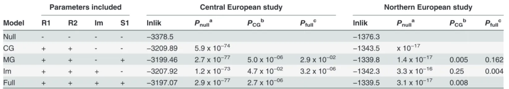
Importantly, both genomic imprinting and maternal genotype effects can lead to similar patterns of parent-of-origin effects and specific tests need to performed to distinguish them [13,14]. Finally, we tested an imprinting model by including the imprinting parameter, Im, which represents the relative risk of the child when inheriting a mutant allele from the mother as opposed to the father. A comparison with the Child Genotype model provided marginal support for the presence of imprinting (PIm vs CG= 0.047;Table 3). In order to test which
par-ent-of-origin scenario better fits our data we performed comparisons versus a full model con-taining all risk parameter (R1, R2, S1 and Im). Interestingly, adding the maternal genotype parameter S1 to a model already containing Im resulted in a significantly better model
(Table 4; p = 3.2 x 10⁻⁶). On the contrary, adding Im to a model already containing S1 provided
only a marginal improvement (p = 0.03). These results favour the existence of a direct maternal genotype effect ofFLG.
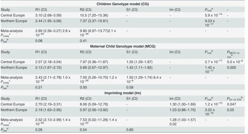
Table 3. Parent-of-origin analysis of the combinedFLGmutations.
Children Genotype model (CG)
Study R1 (CI) R2 (CI) S1 (CI) Im (CI) Pnulla
-Central Europe 3.10 (2.68–3.59) 10.5 (7.20–15.36) - - 5.9 x 10−74
-Northern Europe 2.44 (1.95–3.06) 7.37 (3.37–15.81) - - 9.23 x
10−17
-Meta-analysis Pmetac
2.89 (2.56–3.27) 2.8 x
10−65 9.80 (6.9710−39 –13.77)2.1 x - - -
-Phetd 0.08 0.41
Maternal Child Genotype model (MCG)
Study R1 (CI) R2 (CI) S1 (CI) Im (CI) Pnulla PMCG vs
CGb
Central Europe 2.57 (2.18–3.04) 7.97 (5.36–11.87) 1.55 (1.29–1.87) - 2.7 x 10−77 5.0 x 10⁻⁶
Northern Europe 2.13 (1.67–2.72) 5.89 (2.67–12.97) 1.42 (1.11–1.82) - 1.42 x
10−17 0.005
Meta-analysis Pmetac
2.43 (2.11–2.78) 1.0 x
10−36 7.50 (5.2610−28 –10.70) 1.2 x 1.50 (1.2910−8 –1.74) 8.4 x - -
-Phetd 0.21 0.50 0.58
Imprinting model (Im)
Study R1 (CI) R2 (CI) S1 (CI) Im (CI) Pnulla PIm vs CGb
Central Europe 2.70 (2.19–3.31) 8.06 (5.09–12.78) - 1.30 (1.00–1.69) 1.2 x 10−73 0.047
Northern Europe 2.19 (1.63–2.95) 5.97 (2.56–13.92) - 1.23 (0.86–1.75) 3.22 x
10−16 0.25
Meta-analysis Pmetac
2.52 (2.13–2.99) 1.4 x
10−26 7.53 (5.0210−22 –11.28) 1.4 x - 1.28 (1.030.02 –1.57) -
-Phetd 0.26 0.54 0.80
aPvalue for the comparison of each model versus the null model with no effects. bPvalue for the comparison of each model versus the Child Genotype model.
cPvalue for the meta-analysis of each estimated parameter (seemethods). CI indicates 95% con
fidence interval.
dP value for a test of heterogeneity. All results correspond to the combinedFLGmutations.
Replication in an independent sample and meta-analysis
We aimed to replicate our findings by examining the same 4FLGmutations in an independent Northern European population including 450 AD families and 1854 population-based control individuals (methods andTable 1) [17,20–22]. Step-by-step analysis with PREMIM/EMIM again supported a maternal genotype effect. The genotypes of both children and mothers had an independent effect on AD risk, and children ofFLG-carrier mothers showed a 1.4 fold in-creased risk (R1 = 2.13, R2 = 5.89, S1 = 1.42;PMCG= 1.4 x 10⁻¹⁷;PMCG vs CG= 0.005;Table 3
andS3 Table). Importantly, the results obtained were consistent in both populations studied providing strong support to the existence of maternal genotype effects on FLG.
A meta-analysis was performed using the inverse variance method as implemented in METAL [23], which uses the effects estimates and standard errors from each risk parameter. This revealed a highly significant 1.5 fold increased AD risk in children ofFLG-carrier mothers (S1meta-analysis= 1.50;P =8.4 x 10⁻⁸;Table 3).
FLG
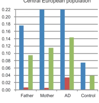
mutations are more frequent in mothers than in fathers
Analysis of parental genotypes revealed a higher prevalence ofFLGmutations in mothers than in fathers in both study populations (Fig. 1). This is consistent with the maternal genotype ef-fect observed. Additionally the frequency ofFLGmutations in the parental population (moth-ers and fath(moth-ers together) was remarkably higher than in population-based controls of
unknown phenotype. This is likely due to the recruitment of families with multiple affected children leading to a parental population enriched in strong genetic risk factors.
Robustness of the
FLG
maternal effect
At this stage we considered the potential weaknesses of our study in order to discard false posi-tives due to methodological issues and to gain further support to the existence of aFLG mater-nal genotype effect.
We analysed theFLGc.2282del4, p.R501X and p.R2247X mutations independently (this was not possible for p.S3247X since it was too rare). The maternal effect and the increased fre-quency of mutations in mothers were found for all 3 mutations in both study populations (S4 TableandS5 Table). This suggests that the maternal effect is not specific to a given variant but a general characteristic ofFLGloss-of-function mutations.
Table 4. Comparison of the MCG and Imprinting models with the full model.
Parameters included Central European study Northern European study
Model R1 R2 Im S1 lnlik Pnulla P
CGb Pfullc lnlik Pnulla PCGb Pfullc
Null - - - - −3378.5 −1376.3
CG + + - - −3209.89 5.9 x 10–74 −1343.5 x 10−17
MG + + - + −3199.46 2.7 x 10−77 5.0 x 10−06 2.9 x 10−02 −1339.8 1.4 x 10⁻¹⁷ 0.005 0.162
Im + + + - −3207.92 1.2 x 10−73 4.7 x 10−02 3.2 x 10−06 −1342.3 3.3 x 10⁻¹⁶ 0.25 0.004
Full + + + + −3197.07 2.9 x 10−77 2.7 x 10−06 −1339.5 3.1 x 10⁻¹⁷ 0.008
The“+”and“-”symbols indicate whether a given risk parameter was included or excluded in the corresponding model. lnlik is the maximized ln likelihood for each model.
aP value for the comparison of each model versus the null model with no effects.
In the populations studied, mothers typically have a more prominent role than fathers in children’s health care [24]. We hypothesized that preferential ascertainment of AD-affected mothers carryingFLGmutations may be the cause of the observed maternal effect. Indeed, we observed a higher frequency of AD-affected mothers than fathers, which could be due to a gen-uine maternal effect or to ascertainment bias (AD prevalence in Central European mothers = 0.23 and fathers = 0.12; Northern European mothers = 0.35 and fathers = 0.19). In order to
Fig 1. Frequency of FLG mutations in fathers, mothers, individuals with atopic dermatitis and controls.Allele and genotype frequencies of the combinedFLG-mutations in fathers and mothers were calculated using all available parents. AD refers to the frequency in the AD-affected children including the families and the unrelated AD-cases (available only in the Central European study). Frequency in controls corresponds to population-based individuals with unknown disease status. Results of the Central and Northern European populations are shown in panels A and B, respectively.
avoid this potential bias we repeated our analysis including only families in which both parents had a negative history of AD. Importantly, the maternal genotype effect remained strong and significant in the remaining population (Meta-S1 = 1.38;P =0.003;S6 Table).
We also tested the potential effect of the paternalFLGgenotype on the children. Since this option is not available in PREMIM/EMIM, we performed the analysis after exchanging the pa-ternal and mapa-ternal genotypes on our genotype files. This analysis revealed no significant effect of the paternalFLGstatus (S7 Table).
A large proportion of the families included in the present study (60%) were recruited through an affected sib pair. Aiming to maximize power, all previous analyses were performed considering all affected siblings as independent individuals which may lead to biased risk pa-rameter estimates. We therefore repeated the analysis including only one affected child per family and found that the magnitude of the maternal effect remained constant in this set of in-dependent trios (S1meta-analysis= 1.45;P =1.1 x 10⁻⁴;S8 Table).
Potential influence of maternal immunity
Filaggrin has a major role in cutaneous barrier function [1]. According to publicly available datasets [25–27]FLGexpression is highest in skin and absent in tissues relevant for mother-child interactions such as uterus, placenta, or mammary gland (S1 Fig.andS2 Fig.). However, recent studies demonstrated thatFLGmutations result in increased antigen penetration through the skin and the production of allergen-specific antibodies (IgE, specific sensitization) [6,7,28]. Thus, we hypothesized that systemic inflammatory responses inFLGcarrier mothers may influence AD risk in children via feto-maternal immune crosstalk.
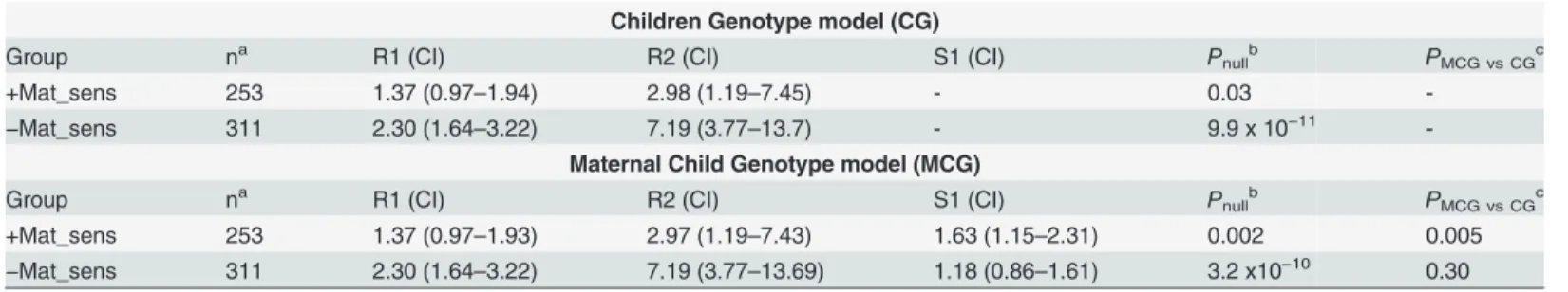
All available maternal plasma samples from the Central European study were therefore test-ed for the presence of allergen-specific IgE, which is a well-characteriztest-ed biochemical marker of allergy [29]. We performed a stratified analysis in 253 families with and 311 families without maternal specific sensitization. In the families with maternal specific sensitization (+Mat_sens) we observed a strong maternal genotype (S1 = 1.63;PMCG vs CG= 0.005) and a weak child
geno-type effect (R1 = 1.37; Pnull= 0.03;Table 5). This was consistent with a marginal
over-trans-mission ofFLGmutations from parents to affected offspring in a transmission disequilibrium test (TDT, transmitted (T): untransmitted (U) = 97:65,P =0.04;Table 6). In contrast, the op-posite pattern was observed in the group of families with non-allergic mothers (−Mat_sens).
Table 5. Parent-of-origin-analysis analysis stratified by maternal specific sensitization. Children Genotype model (CG)
Group na R1 (CI) R2 (CI) S1 (CI) P
nullb PMCG vs CGc
+Mat_sens 253 1.37 (0.97–1.94) 2.98 (1.19–7.45) - 0.03
-−Mat_sens 311 2.30 (1.64–3.22) 7.19 (3.77–13.7) - 9.9 x 10⁻¹¹
-Maternal Child Genotype model (MCG)
Group na R1 (CI) R2 (CI) S1 (CI) P
nullb PMCG vs CGc
+Mat_sens 253 1.37 (0.97–1.93) 2.97 (1.19–7.43) 1.63 (1.15–2.31) 0.002 0.005
−Mat_sens 311 2.30 (1.64–3.22) 7.19 (3.77–13.69) 1.18 (0.86–1.61) 3.2 x10⁻¹⁰ 0.30 aNumber of families.
bPvalue for the comparison with the null model with no effects. cPvalue for the comparison with the Child Genotype model.
Here, the maternal genotype effect was not significant (S1 = 1.18;PMCG vs CG= 0.3) while the
child genotype effect was strong (R1 = 2.3; Pnull= 9.9 x 10⁻¹¹). This observation was confirmed
by a striking over-transmission ofFLGmutations in the TDT (T:U = 169:68,P =5.3 x 10⁻⁸). In
concordance with the different rates of mutation transmission observed in both groups, the fre-quency ofFLGmutations was significantly higher in AD-children of non-allergic mothers (mutation frequency 0.16 and 0.11 in affected children of−Mat_sens and +Mat_sens families, respectively; OR = 1.48;P =0.005;Table 6).
Discussion
We report here that maternal loss-of-function mutations inFLGdirectly influence AD risk in the offspring independently of the child’s own genotype. Importantly, this maternal effect was observed consistently for all differentFLGmutations tested and in 2 independent populations. Given that genomic imprinting and maternal genotype effects can lead to similar patterns of parent-of-origin effects [14], we specifically modelled both scenarios. Although we cannot completely exclude an imprinting effect, our data supports a direct maternal genotype effect of
FLGmutations. This is consistent with the lack of known imprinted genes in the 1q21.3 geno-mic region containingFLG[25].
It is not obvious how maternal mutations in a skin-barrier gene can influence the child’s phenotype. However, given that filaggrin deficiency promotes specific sensitization, we hy-pothesized that systemic immune responses may play a role in theFLGmaternal effect. Our observation that theFLGmaternal effect is significant only in the group of sensitized mothers supports this hypothesis and indicates thatFLGmutations in allergic mothers act as a strong environmental risk factor for AD in the child.
The importance of prenatal mother-child interaction in shaping the child’s immune pheno-type is highlighted by studies in pregnant mice: while the induction of Th2 immune responses increased susceptibility to allergic asthma in the offspring, a protective effect was observed upon induction of Th1 responses, emphasizing the importance of maternal immune status during pregnancy [30–32]. Supporting evidence in humans arise from epidemiological studies showing that exposure to a microbial-rich farm environment during pregnancy protects chil-dren from the development of allergic diseases [33]. It is unknown how this“immunological imprinting”may be transmitted from mother to child, but animal and human studies suggest the induction of epigenetic modifications in relevant immune cells in the offspring [31,34]. Fu-ture large population-based studies with parental DNA, data on maternal allergic sensitization and biological material available for epigenetic analysis will be required to further explore this interesting hypothesis.
Table 6. TDT analysis stratified by maternal specific sensitization.
Transmission Disequilibrium Test (TDT) Allelic test in childrend
Group na T / Ub ORc P Mut freq OR (P)
+Mat_sens 253 97 / 65 1.49 0.04 0.11 1
−Mat_sens 311 169 / 68 2.49 5.3 x10⁻⁸ 0.16 1.48 (0.005)
aNumber of families.
bNumber of transmitted (T) and un-transmitted (U) alleles. Allele counts are referred to the combinedFLGmutations. cOdds ratio calculated as the ratio of transmitted versus un-transmitted alleles.
dComparison of allelic frequency between AD-affected children of the
A recent mouse study found that parent-of-origin effects are widespread and account for an unexpectedly large proportion of complex trait heritability [35]. This is supported by human studies demonstrating that the parental origin of an allele inherited by the offspring can affect disease susceptibility to complex diseases [12,13,36–38]. However, evidence for the existence of maternal genotype effects, which occur without transmission of the risk allele to the offspring, comes mainly from animal studies analysing the effect of maternal gene knockouts in wild-type offspring [39]. Examples of such effects in humans are scarce and refer to rare congenital malformations [40,41]. The present work is, to our knowledge, the first report of a large mater-nal genotype effect in a common human disease. Notably, the magnitude of the matermater-nal effect (RR = 1.5) was consistent in both data sets and exceeded that reported for most AD genetic risk factors identified to date.
Interestingly, AD is commonly the first manifestation of allergic disease and filaggrin defi-ciency is a risk factor for the transition from AD to other atopic diseases such as food allergy, hay fever, and asthma [5,16]. Thus, it is tempting to speculate that maternalFLGmutations may influence the risk of a much wider range of allergic disorders.
This and other studies provide proof-of-principle that associations originally discovered by case-control analysis can arise as a consequence of parent-of-origin effects, although with an underestimation of the effect size due to inaccurate genetic modelling [12]. Family-based stud-ies re-evaluating previously identified susceptibility loci will enable the identification of parent-of-origin effects and help characterize part of the missing heritability in complex traits.
Methods
Ethics statement
The study was carried out in accordance to the approval of the ethics commission of the Char-ité—Universitätsmedizin Berlin (ref EA2/054/10) and following the guidelines of the declara-tion of Helsinki. Informed consent was obtained from all probands or their legal guardians.
Subjects
We investigated samples originating from European family-based and population-based stud-ies. All samples were divided, according to the country of origin, into a Central and a Northern European study population (Table 1).
The GENUFAD study (Genetic Analysis of Nuclear Families with Atopic Dermatitis) re-cruited complete nuclear families with at least two children affected with early-onset (<2 years
of age) and moderate to severe AD as previously described [20]. A doctor’s diagnosis of AD was made according to standard criteria [42]. The GENUFAD study contributed 522 complete German nuclear families to the Central European study and 32 Swedish families to the North-ern European data set. A large proportion of these families have been reported in previous studies [16,43].
The MAS (Multicenter Allergy Study) is a previously described population-based birth co-hort in which 1314 German infants were followed since 1990 to investigate the epidemiology of allergic diseases [44]. The diagnosis of AD was made as previously described [16]. 112 Ger-man MAS trios, consisting of a child with AD and both parents, were included in the Central European study.
Additionally, 125 ETAC trios from different European countries were included in the Central European study (48 from the Netherlands, 23 from Italy, 20 from the UK, 15 from France, 13 from the Czech Republic, and 6 from Germany).
A previous study from Sweden contributed 397 families to the Northern European study group [17]. This included 272 complete affected sib pair families with AD diagnosed according to the U.K. Working Party’s Diagnostic Criteria [46]. The remaining 125 families were incom-plete nuclear families including mother-child or father-child pairs.
In all family-based studies, information regarding the parental history of AD was obtained by a questionnaire at the time of family recruitment. The analysis shown inS6 Tablewas per-formed in families in which both parents had a negative history of AD. Families in which one or both parents had a positive or unknown disease history were excluded.
As described below, the analytical methods used allowed the incorporation of unrelated in-dividuals to increase power. Thus, the Central European study also included previously pub-lishedFLGgenotypes of 772 unrelated German AD cases and 373 German controls from a previous GWAS [43]. In addition, we genotypedFLGmutations in 375 German children with AD diagnosed at a tertiary care center for pediatric allergy at Charité Universitätsmedizin Ber-lin. Also, previously publishedFLGgenotype counts of 2,963 population-based German indi-viduals from the International Study of Asthma and Allergies in Childhood II Study [47] were included. Likewise, in our Northern European study population we included genotypes of the 3 most prevalentFLGmutations (c.2282del4, p.R501X and p.R2447X) previously reported in the Swedish population-based BAMSE cohort [22]. The rarestFLGmutation (p.S3247X) was not available in the BAMSE dataset.
Data on specific allergic sensitization was available in a large proportion of mothers from the GENUFAD and MAS studies. Plasma levels of specific IgE against grass and birch pollen, ribwort, cat and dog dander, mold (Cladosporidium herbarum, Alternaria tenuis), hen’s egg, cow’s milk, fish, peanut, and house dust mite were determined using CAP-RAST-FEIA (Phar-macia). A mother was defined as sensitized if specific IgE0.7 kU/l (CAP2) to at least one al-lergen was detected.
Genotyping of
FLG
mutations
Genomic DNA was prepared from whole blood by standard methods. TheFLGc.2282del4 var-iant was analyzed with fluorescence-based semiautomated genotyping [16] and theFLGp. R501X, p.R2447X and p.S3247X mutations with Taqman allelic discrimination (Applied Bio-systems, Foster City, California, USA) as previously described [4]. Genotyping of p.R2447X in the Northern European families was performed using a fluorescent Kaspar assay (KASP-By-Design genotyping assays, LGC group, Teddington, UK). Genotyping with Taqman and Kas-par was performed using a ViiA 7 Real-Time PCR System (Applied Biosystems, Foster City, California, USA). When analyzing the rare p.R2447X and p.S3247X mutations, a sample known to be a mutation carrier was included on each genotyping plate as a positive control.
TheFLGmutations are named according to the nomenclature recommendations by den Dunnen and Antonarakis [48]. The positions of the mutations in the cDNA refer to the A of the ATG-translation initiation codon of NM_002016.1.
Statistical analysis
the analysis to increase the power to detect parent-of-origin effects [18,19]. Since the EMIM analysis is based on the assumption that the genotype frequencies in controls correspond to those in the general population, all population-based controls were included in the analysis irrespectively of disease status. Starting values for allele frequencies of theFLGmutations in the study population (including controls) were estimated with PREMIM (–a option).
The trios were analyzed using the EMIM tool, which uses a multinomial modelling ap-proach to estimate genotype relative risk parameters on the basis of observed counts of geno-type combinations in case-parent trios. The following parameters influencing the disease risk in the child were modelled with EMIM:
• R1 (R2), the factor by which an individual’s disease risk is multiplied if they carry one (two) risk alleles at a given locus.
• S1 (S2), the factor by which an individual’s disease risk is multiplied if the mother carries one (two) risk alleles at that locus.
• Im (Ip) the factor by which an individual’s disease risk is multiplied if inheriting a risk allele from the mother (or father).
• γ11 (interaction term), the factor by which an individual’s disease risk is multiplied if both
mother and child have 1 copy of the risk allele.
Previous data indicated thatFLGmutations do not fit an additive genetic model, since the risk of AD in the homozygous carriers is too high. Thus, instead of using the default EMIM set-tings assuming an additive model we choose to independently estimate the R1 and R2 parame-ters. Modelling of maternal genotype effects was done using the default additive model. All analyses were performed under the“conditional on exchangeable parental genotype”(CEPG) assumption. This assumption should protect from potential biases in parameter estimation due to the inclusion of families recruited through multiple affected individuals, at the cost of re-duced power to detect parent-of-origin effects compared to assuming only Hardy-Weinberg equilibrium [18,19]. A step-by-step analysis was performed by including additional risk param-eters in the model as indicated inS9 Table.
Maximum likelihood estimates were obtained from each model and a likelihood ratio test was performed to assess the significance among nested models. Note that it is not possible to directly compare the MCG and Im models in a likelihood ratio test, since they are not nested. However, they can be compared indirectly by comparison to the full model (Table 4).
In order to test for genetic interaction between the child and the maternal genotypes we in-cluded and interaction term in the model (γ11). This parameter estimates the factor by which
an individual’s disease risk is multiplied if both mother and child have one copy of the risk al-lele. A likelihood ratio test comparing the Maternal Child Genotype (MCG) and the MCG-In-teraction model was then performed (seeS10 Table).
A meta-analysis of the results from the Central and Northern European populations was performed using METAL [23]. The inverse variance method was used and the corresponding betas and standard errors were obtained from the EMIM summary file. The meta-analysis was performed on the single risk parameters estimates (R1, R2, S1 or Im) from each model. An analysis of heterogeneity was also performed with METAL in order to evaluate if the observed effect sizes were homogeneous across datasets.
exclude this potential bias we repeated the analysis including one affected child per family (omitting the−xa option in PREMIM;S8 Table).
The Transmission Disequilibrium Test (TDT) was performed with PLINK [49]. In order to account for multiple affected offspring within families, empirical p-values were calculated with the—tdt—perm option, which flips the allele transmitted from parent to offspring with 50:50 probability. Allelic effects were calculated with PLINK—assoc using the offspring of the +Mat_Sens as controls and those from the−Mat_Sens as cases.
Haplotype frequencies on the central European Study were calculated with FAMHAP [50], which computes maximum-likelihood estimates obtained with the expectation-maximization algorithm.
Supporting Information
S1 Fig. Tissue expression pattern ofFLGfrom BioGPS.Data from the BioGPS Portal where
mRNA levels were quantified with expression arrays in human tissues. [25,26]. (TIF)
S2 Fig. Tissue expression pattern ofFLGfrom GTEx.Data from the GTEx Consortium
where mRNA was quantified by Next generation Sequencing. [27] (TIF)
S1 Table.FLGloss-of-function mutations in the ExAC project.Only high quality mutations
were considered (filter = pass).aHuman genome build GRCh37.p13 (chromosome 1).bAlias used in this manuscript.cAllele frequency in the combined set of 61,486 unrelated individuals.
dAlternative allele count of genotypes in the combined populationeTotal number of called
ge-notypes in the combined population. Data was accessed on December 12, 2014. (XLSB)
S2 Table. Lack of linkage disequilibrium amongFLGmutations.
(DOCX)
S3 Table. Analysis of interaction between child and maternalFLGmutations.
(DOCX)
S4 Table. Analysis of individualFLGmutations.
(DOCX)
S5 Table. Frequencies of each of the 4FLGmutations analyzed independently.
(DOCX)
S6 Table. Parent-of-origin analysis after exclusion of families with parental history of AD. (DOCX)
S7 Table. Analysis for paternal genotype effects. (DOCX)
S8 Table. Parent-of-origin analysis with one single AD-affected child per family. (DOC)
S9 Table. Step-by-step analysis with PREMIM/EMIM. (DOCX)
Acknowledgments
We thank all individuals and families for their participation in this study. We thank all physi-cians and nurses involved in patient recruitment for their valuable contribution to the study. We are grateful to C. Flachmeier and S. Kolberg for excellent technical support.
The authors would like to thank the Exome Aggregation Consortium and the groups that provided exome variant data for comparison. A full list of contributing groups can be found at http://exac.broadinstitute.org/about.
Author Contributions
Conceived and designed the experiments: JEG YAL. Performed the experiments: JEG AM IM. Analyzed the data: JEG KR AB HJC. Contributed reagents/materials/analysis tools: KN MALK MN MCGW TK RK SL KB BK BN MB NH YAL. Wrote the paper: JEG IM HJC NH YAL.
References
1. Brown SJ, McLean WH (2012) One remarkable molecule: filaggrin. J Invest Dermatol 132: 751–762. doi:10.1038/jid.2011.393PMID:22158554
2. Smith FJ, Irvine AD, Terron-Kwiatkowski A, Sandilands A, Campbell LE et al. (2006) Loss-of-function mutations in the gene encoding filaggrin cause ichthyosis vulgaris. Nat Genet 38: 337–342. PMID: 16444271
3. Palmer CN, Irvine AD, Terron-Kwiatkowski A, Zhao Y, Liao H et al. (2006) Common loss-of-function variants of the epidermal barrier protein filaggrin are a major predisposing factor for atopic dermatitis. Nat Genet 38: 441–446. PMID:16550169
4. Sandilands A, Terron-Kwiatkowski A, Hull PR, O'Regan GM, Clayton TH et al. (2007) Comprehensive analysis of the gene encoding filaggrin uncovers prevalent and rare mutations in ichthyosis vulgaris and atopic eczema. Nat Genet 39: 650–654. PMID:17417636
5. Brown SJ, Asai Y, Cordell HJ, Campbell LE, Zhao Y et al. (2011) Loss-of-function variants in the filag-grin gene are a significant risk factor for peanut allergy. J Allergy Clin Immunol 127: 661–667. doi:10. 1016/j.jaci.2011.01.031PMID:21377035
6. Fallon PG, Sasaki T, Sandilands A, Campbell LE, Saunders SP et al. (2009) A homozygous frameshift mutation in the mouse Flg gene facilitates enhanced percutaneous allergen priming. Nat Genet 41: 602–608. doi:10.1038/ng.358PMID:19349982
7. Kawasaki H, Nagao K, Kubo A, Hata T, Shimizu A et al. (2012) Altered stratum corneum barrier and en-hanced percutaneous immune responses in filaggrin-null mice. J Allergy Clin Immunol 129: 1538–
1546. doi:10.1016/j.jaci.2012.01.068PMID:22409988
8. Lim RH, Kobzik L, Dahl M (2010) Risk for asthma in offspring of asthmatic mothers versus fathers: a meta-analysis. PLoS One 5: e10134. doi:10.1371/journal.pone.0010134PMID:20405032
9. Goldberg M, Eisenberg E, Elizur A, Rajuan N, Rachmiel M et al. (2013) Role of parental atopy in cow's milk allergy: a population-based study. Ann Allergy Asthma Immunol 110: 279–283. doi:10.1016/j. anai.2013.01.017PMID:23535093
10. Bisgaard H, Halkjaer LB, Hinge R, Giwercman C, Palmer C et al. (2009) Risk analysis of early child-hood eczema. J Allergy Clin Immunol 123: 1355–1360. doi:10.1016/j.jaci.2009.03.046PMID: 19501236
11. Wadonda-Kabondo N, Sterne JA, Golding J, Kennedy CT, Archer CB et al. (2004) Association of pa-rental eczema, hayfever, and asthma with atopic dermatitis in infancy: birth cohort study. Arch Dis Child 89: 917–921. PMID:15383434
12. Kong A, Steinthorsdottir V, Masson G, Thorleifsson G, Sulem P et al. (2009) Parental origin of se-quence variants associated with complex diseases. Nature 462: 868–874. doi:10.1038/nature08625 PMID:20016592
13. Lawson HA, Cheverud JM, Wolf JB (2013) Genomic imprinting and parent-of-origin effects on complex traits. Nat Rev Genet 14: 609–617. doi:10.1038/nrg3543PMID:23917626
14. Hager R, Cheverud JM, Wolf JB (2008) Maternal effects as the cause of parent-of-origin effects that mimic genomic imprinting. Genetics 178: 1755–1762. doi:10.1534/genetics.107.080697PMID: 18245362
16. Marenholz I, Nickel R, Ruschendorf F, Schulz F, Esparza-Gordillo J et al. (2006) Filaggrin loss-of-func-tion mutaloss-of-func-tions predispose to phenotypes involved in the atopic march. J Allergy Clin Immunol 118: 866–871. PMID:17030239
17. Ekelund E, Lieden A, Link J, Lee SP, d'Amato M et al. (2008) Loss-of-function variants of the filaggrin gene are associated with atopic eczema and associated phenotypes in Swedish families. Acta Derm Venereol 88: 15–19. doi:10.2340/00015555-0383PMID:18176743
18. Howey R, Cordell HJ (2012) PREMIM and EMIM: tools for estimation of maternal, imprinting and inter-action effects using multinomial modelling. BMC Bioinformatics 13: 149. doi: 10.1186/1471-2105-13-149PMID:22738121
19. Ainsworth HF, Unwin J, Jamison DL, Cordell HJ (2011) Investigation of maternal effects, maternal-fetal interactions and parent-of-origin effects (imprinting), using mothers and their offspring. Genet Epidemiol 35: 19–45. doi:10.1002/gepi.20547PMID:21181895
20. Lee YA, Wahn U, Kehrt R, Tarani L, Businco L et al. (2000) A major susceptibility locus for atopic der-matitis maps to chromosome 3q21. Nat Genet 26: 470–473. PMID:11101848
21. Muller S, Marenholz I, Lee YA, Sengler C, Zitnik SE et al. (2009) Association of Filaggrin loss-of-func-tion-mutations with atopic dermatitis and asthma in the Early Treatment of the Atopic Child (ETAC) pop-ulation. Pediatr Allergy Immunol 20: 358–361. doi:10.1111/j.1399-3038.2008.00808.xPMID: 19538357
22. Ballardini N, Kull I, Soderhall C, Lilja G, Wickman M et al. (2013) Eczema severity in preadolescent chil-dren and its relation to sex, filaggrin mutations, asthma, rhinitis, aggravating factors and topical treat-ment: a report from the BAMSE birth cohort. Br J Dermatol 168: 588–594. doi:10.1111/bjd.12196 PMID:23445315
23. Willer CJ, Li Y, Abecasis GR (2010) METAL: fast and efficient meta-analysis of genomewide associa-tion scans. Bioinformatics 26: 2190–2191. doi:10.1093/bioinformatics/btq340PMID:20616382
24. Case A, Paxson C (2001) Mothers and others: who invests in children's health? J Health Econ 20: 301–328. PMID:11373833
25. Su AI, Wiltshire T, Batalov S, Lapp H, Ching KA et al. (2004) A gene atlas of the mouse and human pro-tein-encoding transcriptomes. Proc Natl Acad Sci U S A 101: 6062–6067. PMID:15075390
26. Wu C, Orozco C, Boyer J, Leglise M, Goodale J et al. (2009) BioGPS: an extensible and customizable portal for querying and organizing gene annotation resources. Genome Biol 10: R130. doi:10.1186/ gb-2009-10-11-r130PMID:19919682
27. GTEx Consortium. (2013) The Genotype-Tissue Expression (GTEx) project. Nat Genet 45: 580–585. doi:10.1038/ng.2653PMID:23715323
28. van den Oord RA, Sheikh A (2009) Filaggrin gene defects and risk of developing allergic sensitisation and allergic disorders: systematic review and meta-analysis. BMJ 339: b2433. doi:10.1136/bmj.b2433 PMID:19589816
29. Liu FT, Goodarzi H, Chen HY (2011) IgE, mast cells, and eosinophils in atopic dermatitis. Clin Rev Al-lergy Immunol 41: 298–310. doi:10.1007/s12016-011-8252-4PMID:21249468
30. Hamada K, Suzaki Y, Goldman A, Ning YY, Goldsmith C et al. (2003) Allergen-independent maternal transmission of asthma susceptibility. J Immunol 170: 1683–1689. PMID:12574331
31. Fedulov AV, Kobzik L (2011) Allergy risk is mediated by dendritic cells with congenital epigenetic changes. Am J Respir Cell Mol Biol 44: 285–292. doi:10.1165/rcmb.2009-0400OCPMID:20118218
32. Straubinger K, Paul S, Prazeres da CO, Ritter M, Buch T et al. (2014) Maternal immune response to helminth infection during pregnancy determines offspring susceptibility to allergic airway inflammation. J Allergy Clin Immunol.
33. von ME, Vercelli D (2010) Farm living: effects on childhood asthma and allergy. Nat Rev Immunol 10: 861–868. doi:10.1038/nri2871PMID:21060319
34. Brand S, Teich R, Dicke T, Harb H, Yildirim AO et al. (2011) Epigenetic regulation in murine offspring as a novel mechanism for transmaternal asthma protection induced by microbes. J Allergy Clin Immunol 128: 618–625. doi:10.1016/j.jaci.2011.04.035PMID:21680015
35. Mott R, Yuan W, Kaisaki P, Gan X, Cleak J et al. (2014) The architecture of parent-of-origin effects in mice. Cell 156: 332–342. doi:10.1016/j.cell.2013.11.043PMID:24439386
36. Shirakawa T, Li A, Dubowitz M, Dekker JW, Shaw AE et al. (1994) Association between atopy and vari-ants of the beta subunit of the high- affinity immunoglobulin E receptor. Nat Genet 7: 125–129. PMID: 7920628
38. Soderhall C, Marenholz I, Kerscher T, Ruschendorf F, Esparza-Gordillo J et al. (2007) Variants in a novel epidermal collagen gene (COL29A1) are associated with atopic dermatitis. PLoS Biol 5: e242. PMID:17850181
39. Gleason G, Liu B, Bruening S, Zupan B, Auerbach A et al. (2010) The serotonin1A receptor gene as a genetic and prenatal maternal environmental factor in anxiety. Proc Natl Acad Sci U S A 107: 7592–
7597. doi:10.1073/pnas.0914805107PMID:20368423
40. Yan L, Zhao L, Long Y, Zou P, Ji G et al. (2012) Association of the maternal MTHFR C677T polymor-phism with susceptibility to neural tube defects in offsprings: evidence from 25 case-control studies. PLoS One 7: e41689. doi:10.1371/journal.pone.0041689PMID:23056169
41. Gordeeva LA, Voronina EN, Sokolova EA, Ermolenko NA, Gareeva JV et al. (2013) Association GSTT1, GSTM1 and GSTP1 (Ile105Val) genetic polymorphisms in mothers with risk of congenital mal-formations in their children in Western Siberia: a case-control study. Prenat Diagn 1–7.
42. Hanifin JM, Rajka G (1980) Diagnostic Features of Atopic Dermatitis. Acta Derm (Stockholm) 92 (Suppl.): 44–47.
43. Esparza-Gordillo J, Weidinger S, Folster-Holst R, Bauerfeind A, Ruschendorf F et al. (2009) A common variant on chromosome 11q13 is associated with atopic dermatitis. Nat Genet 41: 596–601. doi:10. 1038/ng.347PMID:19349984
44. Lau S, Illi S, Sommerfeld C, Niggemann B, Bergmann R et al. (2000) Early exposure to house-dust mite and cat allergens and development of childhood asthma: a cohort study. Multicentre Allergy Study Group. Lancet 356: 1392–1397. PMID:11052581
45. Warner JO (2001) A double-blinded, randomized, placebo-controlled trial of cetirizine in preventing the onset of asthma in children with atopic dermatitis: 18 months' treatment and 18 months' posttreatment follow-up. J Allergy Clin Immunol 108: 929–937. PMID:11742270
46. Williams HC, Burney PG, Hay RJ, Archer CB, Shipley MJ et al. (1994) The U.K. Working Party's Diag-nostic Criteria for Atopic Dermatitis. I. Derivation of a minimum set of discriminators for atopic dermati-tis. Br J Dermatol 131: 383–396. PMID:7918015
47. Weidinger S, O'Sullivan M, Illig T, Baurecht H, Depner M et al. (2008) Filaggrin mutations, atopic ecze-ma, hay fever, and asthma in children. J Allergy Clin Immunol 121: 1203–1209. doi:10.1016/j.jaci. 2008.02.014PMID:18396323
48. Den Dunnen JT, Antonarakis SE (2001) Nomenclature for the description of human sequence varia-tions. Hum Genet 109: 121–124. PMID:11479744
49. Purcell S, Neale B, Todd-Brown K, Thomas L, Ferreira MA et al. (2007) PLINK: a tool set for whole-ge-nome association and population-based linkage analyses. Am J Hum Genet 81: 559–575. PMID: 17701901