Structure and Function of the Su(H)-Hairless
Repressor Complex, the Major Antagonist of
Notch Signaling in
Drosophila melanogaster
Zhenyu Yuan1☯, Heiko Praxenthaler2☯, Nassif Tabaja1, Rubben Torella3, Anette Preiss2, Dieter Maier2, Rhett A. Kovall1*
1Department of Molecular Genetics, Biochemistry and Microbiology, University of Cincinnati College of Medicine, Cincinnati, Ohio, United States of America,2Universität Hohenheim, Institut für Genetik, Stuttgart, Germany,3Centre for Molecular Informatics, Department of Chemistry, University of Cambridge,
Cambridge, United Kingdom
☯These authors contributed equally to this work. *[email protected]
Abstract
Notch is a conserved signaling pathway that specifies cell fates in metazoans. Receptor-ligand interactions induce changes in gene expression, which is regulated by the transcrip-tion factor CBF1/Su(H)/Lag-1 (CSL). CSL interacts with coregulators to repress and activate transcription from Notch target genes. While the molecular details of the activator complex are relatively well understood, the structure-function of CSL-mediated repressor complexes is poorly defined. InDrosophila, the antagonist Hairless directly binds Su(H) (the fly CSL ortholog) to repress transcription from Notch targets. Here, we determine the X-ray structure of the Su(H)-Hairless complex bound to DNA. Hairless binding produces a large conforma-tional change in Su(H) by interacting with residues in the hydrophobic core of Su(H), illus-trating the structural plasticity of CSL molecules to interact with different binding partners. Based on the structure, we designed mutants in Hairless and Su(H) that affect binding, but do not affect formation of the activator complex. These mutants were validated in vitro by isothermal titration calorimetry and yeast two- and three-hybrid assays. Moreover, these mutants allowed us to solely characterize the repressor function of Su(H) in vivo.
Author Summary
Notch signaling is a form of cell-to-cell communication, in which extracellular receptor-ligand interactions ultimately result in changes in gene expression. The Notch pathway is highly conserved from the model organismDrosophila melanogasterto humans. When mutations occur within Notch pathway components, this often leads to human disease, such as certain types of cancers and birth defects. Transcription of Notch target genes is regulated by the transcription factor CSL (for CBF1/RBP-J in mammals, Su(H) in Dro-sophila, and Lag-1 inCaenorhabditis elegans). CSL functions as both a transcriptional acti-vator and repressor by forming complexes with coactiacti-vator and corepressor proteins,
a11111
OPEN ACCESS
Citation:Yuan Z, Praxenthaler H, Tabaja N, Torella R, Preiss A, Maier D, et al. (2016) Structure and Function of the Su(H)-Hairless Repressor Complex, the Major Antagonist of Notch Signaling inDrosophila melanogaster. PLoS Biol 14(7): e1002509. doi:10.1371/journal.pbio.1002509
Academic Editor:Hugo J. Bellen, Baylor College of Medicine, UNITED STATES
Received:February 1, 2016
Accepted:June 10, 2016
Published:July 12, 2016
Copyright:© 2016 Yuan et al. This is an open access article distributed under the terms of the
Creative Commons Attribution License, which permits unrestricted use, distribution, and reproduction in any medium, provided the original author and source are credited.
Data Availability Statement:The PDB file has been deposited in the RCSB Protein Data Bank (www.rcsb. org) with the accession code 5E24.http://www.rcsb. org/pdb/explore/explore.do?structureId=5e24
respectively. Here we determine the high-resolution X-ray structure of Su(H) (the fly CSL ortholog) in complex with the corepressor Hairless, which is the major antagonist of Notch signaling in Drosophila. The structure unexpectedly reveals that Hairless binding results in a dramatic conformational change in Su(H). In parallel, we designed mutations in Su(H) and Hairless based on our structure and showed that these mutants are defective in complex formation in vitro and display functional deficiencies in in vivo assays. Taken together, our work provides significant molecular insights into how CSL functions as a transcriptional repressor in the Notch pathway.
Introduction
The Notch pathway is a highly conserved cell-to-cell signaling mechanism that is essential for cell fate decisions during embryogenesis and postnatal tissue homeostasis [1]. In humans, aber-rant Notch signaling underlies the pathogenesis of many diseases, including certain types of cancer [2], congenital syndromes [3], and cardiovascular defects [3]. The involvement of Notch in human disease has led to considerable efforts to identify pharmaceuticals that modu-late Notch signaling for therapeutic purposes [2].
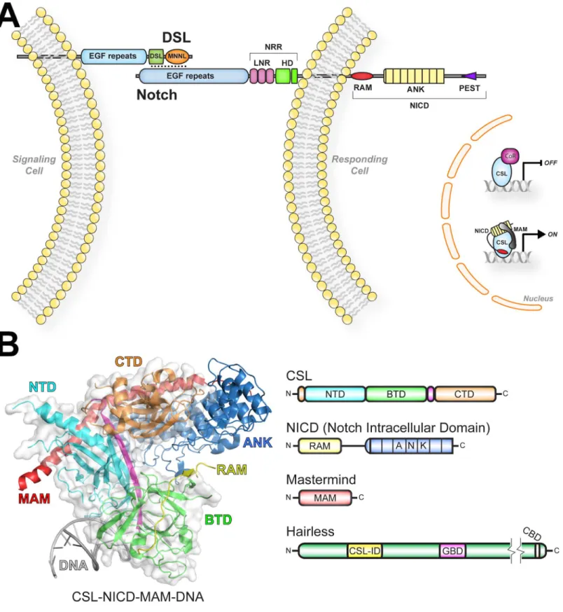
The central components consist of the receptor Notch, the ligand DSL (Delta, Serrate, Lag-2), and the nuclear effector CSL (CBF1/RBP-J, Su(H), Lag-1) (Fig 1A) [4]. DSL and CSL are initialisms for the mammalian, fly, and worm orthologous proteins, respectively. Notch and DSL are multidomain transmembrane proteins with a single transmembrane spanning region, and CSL is a DNA binding transcription factor [4]. As shown inFig 1A, signaling occurs when Notch and DSL molecules on neighboring cells interact, which results in cleavage of Notch and release of the Notch intracellular domain (NICD) from the cell membrane [1]. Subsequently, NICD localizes to the nucleus and directly binds CSL and the coactivator Mastermind (MAM), forming the transcriptionally active CSL-NICD-MAM ternary complex [4]. CSL-NICD-MAM binds at the promoter and enhancer regions of Notch target genes, recruits coactivators, such as p300/CBP and Mediator [5,6], and up-regulates transcription at these sites [7]. Signaling is terminated when NICD is phosphorylated within its PEST domain, resulting in its ubiquitin-mediated degradation [5].
At the structural level, much is known about CSL and the activator complex it forms with NICD and MAM [8–12]. As shown inFig 1B, the structure of CSL consists of three domains: NTD (N-terminal domain), BTD (β-trefoil domain), and CTD (C-terminal domain) [11]. The NTD and CTD are immunoglobulin (Ig) domains structurally related to the Rel family of tran-scription factors (e.g., NF-κB and NFAT). CSL binds DNA as a monomer (e.g., CGTGGGAA), in which its NTD and BTD specifically bind bases in the major and minor grooves of DNA, respectively. In addition, CSL-NICD-MAM complexes can also bind as cooperative dimers on DNA at SPS (Su(H) paired site) elements [13,14]. The RAM (RBP-J associated molecule) and ANK (ankyrin repeats) domains of NICD bind the BTD and CTD of CSL, respectively. MAM adopts an extendedα-helix that binds the CTD-ANK interface and the NTD of CSL [8,10,12].
CSL can also function as a repressor by interacting with transcriptional corepressor proteins [7], such as Hairless in flies [15], and SMRT/HDAC1 associated repressor protein (SHARP) [16,17] (also known as MSX-2 interacting nuclear target [MINT]) and KyoT2 [18] in mammals (Fig 1). Corepressors are components of large multiprotein complexes that contain histone modification activity, which convert the local chromatin into a repressive environment. While biochemical/cellular studies in mammals demonstrate that CSL interacts with corepressors and functions as a repressor [17,19–21], there is limited genetic data on CSL repressor function. On
Competing Interests:The authors have declared that no competing interests exist.
the other hand, there is extensive biochemical, cellular, and genetic data from the model organ-ismDrosophila melanogasterdemonstrating that Su(H) (Suppressor of Hairless—the fly ortho-log of CSL) functions as a transcriptional repressor at Notch target genes [22]. In this case, Su(H) binds the antagonist Hairless (Fig 1B) [15], which in turn interacts with the corepressors Groucho and CtBP (C-terminal binding protein) to repress transcription [23–25]. Previously, we defined the region of Hairless that interacts with Su(H) and showed that it binds with low nanomolar affinity to Su(H) [26]. Nonetheless, the structural details of Su(H)-Hairless interac-tions are unknown.
Here, we determine the 2.14 Å X-ray structure of the Su(H)-Hairless repressor complex bound to DNA. As predicted from our previous studies [26], Hairless binds exclusively to the CTD of Su(H), but does so in a strikingly unusual manner. Hairless wedges itself between the twoβ-sheets that compose the Ig fold of the CTD, significantly distorting the overall fold of this domain. This results in Hairless largely interacting with residues that form the hydropho-bic core of the CTD. We designed site-directed mutations to validate our structure and identify the residues critical for Su(H)-Hairless complex formation. Moreover, we were able to design Su(H) mutants that largely affect Hairless binding, but not NICD or MAM, which allowed us to solely characterize its repressor function in cellular assays and in flies. Taken together, our studies provide significant molecular insights into how the antagonist Hairless interacts with the transcription factor Su(H), reveal the remarkable structural plasticity of CSL molecules, and identify a novel binding cleft on the CTD of CSL that could potentially be exploited for modulating Notch signaling.
Results
High Resolution Structure of the Su(H)-Hairless Repressor Complex
To determine the Su(H)-Hairless-DNA crystal structure, we purified recombinant Su(H) (98–
523) and Hairless (232–269) proteins from bacteria and stoichiometrically formed a complex with a 15-mer duplex DNA, containing a single Su(H) binding site. Su(H) (98–523) corre-sponds to the structural core of CSL proteins (NTD, BTD, CTD) [11] and Hairless (232–269) comprises the conserved CSL-ID previously shown to be sufficient for Su(H) binding [26]. While crystals were obtained of the Su(H)-H-DNA complex, the crystals diffracted weakly, precluding structure determination. We took two approaches to improve the diffraction prop-erties of our complex crystals: (1) we introduced surface entropy reduction (SER) mutations [27] into our Su(H) construct (R155T and N281G); and (2), we employed a fixed-arm carrier approach [28], in which Hairless (232–269) was purified as a maltose binding protein (MBP) fusion protein (MBP-H). Subsequently, we were able to isolate Su(H)/MBP-H/DNA crystals that diffract to 2.14 Å at a synchrotron source and belong to the space group C2. The Su(H)/ MBP-H/DNA complex structure (PDB ID: 5E24) was solved by molecular replacement and refined to a final R factor and free R factor of 17.5% and 19.6%, respectively (Table 1). The con-tents of the asymmetric unit and representative electron density from the complex structure are shown inS1 Fig. In subsequent figures, which illustrate the details of the Su(H)-Hairless complex, the MBP moiety is not shown for clarity.
Ligands), NRR (Negative Regulatory Region), LNR (LIN12-Notch Repeats), HD (heterodimerization domain), RAM (RBP-J associated molecule), and ANK (ankyrin repeats). (B) Structure of the CSL-NICD-MAM ternary complex bound to DNA (PDB ID: 2FO1) [8]. The NTD, BTD, and CTD domains of CSL are colored cyan, green, and orange, respectively. Aβ-strand that makes hydrogen-bonding interactions with all three domains is colored magenta. The RAM and ANK domains of NICD are colored yellow and blue, respectively. MAM and DNA are colored red and gray, respectively. Domain schematics are colored similarly to the structure. NTD (N-terminal domain), BTD (β-trefoil domain), CTD (C-terminal domain), CSL-ID (CSL interaction domain), GBD (Groucho binding domain), and CBD (C-terminal binding protein binding domain).
As shown in Figs2and3, Hairless binds the CTD of Su(H), severely perturbing the overall fold of CTD when compared to anapostructure of mouse CSL (also known as RBP-J) [9]. It should be mentioned that there are noapostructures of Su(H) solved, necessitating the com-parison with theapoRBP-J structure. However, given the high degree of sequence similarity between fly and mouse CSL proteins (S2 Fig), we reason that theapoRBP-J structure is a good approximation of theapoSu(H) structure. For example, within the structural core of CSL, the primary sequence of Su(H) and RBP-J are 82% identical (90% similar); and within the CTD, fly and mouse orthologs are 75% identical (88% similar). Moreover, of the 33 residues in the CTD that are different between Su(H) and RBP-J, 27 of these are surface exposed, likely having mini-mal effects on folding; and of the remaining 6 residues that are either partially or entirely bur-ied in the CTD, there are only conservative differences between fly and mouse (e.g., Leu!Met, Thr!Val, Ile!Val, and Ser!Thr) (S2 Fig). Nonetheless, in the absence of anapoSu(H) struc-ture we cannot rule out minor differences in strucstruc-ture between mouse and fly CSL orthologs.
The Ig-fold of CTD is a seven-strandedβ-sandwich, in which fourβ-strands (a, b, e, d) form half of the sandwich, and the remaining three strands (c, f, g) form the other half (Fig 3A and 3B). Strikingly, Hairless wedges itself between the first and last strands of theβ-sandwich (Fig 2A and 2B), making extensive interactions with the hydrophobic core of the CTD and burying ~900 Å2in surface area. For example, Hairless interacts with 10 residues in the CTD that have less than 20 Å2in solvent accessible surface area in a correspondingapostructure of CSL (Fig
2C). The site of Hairless binding is remarkably well conserved in mammalian CSL orthologs (S2 Fig). Hairless binding produces relatively modest changes in the first fiveβ-strands of the
CTD, with the exception of the firstβ-hairpin formed by residues L436-M444, which is trans-lated outward by as much as 6 Å (Fig 3C). However, the region betweenβ-strands e and f
Table 1. Data collection and refinement statistics.
Data Collection Statistics
Resolution (Å) 31.3–2.14 (2.15–2.14)
Space Group C2
Wavelength (Å) 0.97872
Unit Cell a, b, c (Å) 177.7, 93.9, 154.4
Unit Cellα,β,γ(°) 90.00, 109.8, 90.00
Rmerge 0.11 (0.65)
I/σI 12.5 (3.1)
Completeness (%) 99.9 (99.8)
Redundancy 7.6 (7.7)
Refinement Statistics
Rwork/Rfree(%) 0.175 / 0.196
Number of reflections 131,091
Number of atoms: Protein/DNA 13,766
Number of atoms: Ligand/Ion 117 (115 EDO, 2MTT)
Number of atoms: Water 681
B factors: Protein/DNA 53.18
B factors: Ligand/Ion 62.74
B factors: Water 47.81
RMSD Bond Lengths (Å) 0.01
RMSD Bond Angles (°) 1.04
Ramachandran (favored/outliers) 97.44% / 0.30%
Highest resolution shell shown in parentheses.
undergoes the largest structural change upon Hairless binding (Fig 3B), resulting in a
completely new conformation of this loop, as well as displacement of the terminalβ-strand by as much as 4.5 Å (Fig 3C). We queried the Dali server [29] to identify similar protein com-plexes. Given the multitude of Ig structures in the database, it was surprising that the search did not uncover any other structurally related complexes. These data suggest that the Su(H)-Hairless complex represent a heretofore novel interaction mode between an Ig domain and its cognate binding partner.
Previously, we showed that the region of Hairless that binds Su(H) is a random coil in solu-tion [26]; however, Hairless assumes an extendedβ-hairpin conformation when bound to the
CTD of Su(H) (Fig 2A and 2B). Hairless residues G232-R249 form a classicβ-hairpin, which is
followed by an extended loop structure and a thirdβ-strand that makes an anti-parallel
interac-tion with theβ-hairpin (Fig 2B). The Hairlessβ-hairpin is amphipathic, in which its hydropho-bic surface, created by residues L235, F237, L245, L247, and W258, buries itself within the core of the CTD (Fig 2B and 2C). In particular, F237 is buried the deepest, anchoring the interaction between Hairless and Su(H).
To analyze the structural changes that occur within CTD when bound to Hairless, we per-formed molecular dynamics (MD) simulations of the Su(H)-Hairless-DNA complex structure and compared these results with MD simulations of theapoRBPJ-DNA structure (PDB ID: 3BRG) (Fig 3DandS3 Fig) [30]. Two simulations were performed, in which Hairless was removed from the model and the resulting Su(H)-DNA structure was allowed to sample differ-ent conformations over the time course of the experimdiffer-ent. Both simulations converged to a root mean square deviation (RMSD) value of ~0.35 nm for the cαatoms (S3 Fig), which was similar to theapoCSL-DNA structure simulation. Root mean square fluctuation (RMSF) anal-ysis revealed that the largest changes occurred within the CTD of Su(H) (residues 473–498, the region betweenβ-strands e and f) (S3 Fig), which was expected, given that this is the region in
Su(H) that incurs the largest structural change upon Hairless binding. This structural rear-rangement results in the CTD assuming a more compact conformation that is similar to the
apoCSL-DNA structure (S3 Fig). Closer inspection revealed that Su(H) residue F516 under-goes a dramatic change in its side chain dihedral angle (~100°) when comparing bound and unbound structures (S3 Fig). Interestingly, the shift in residue F516 of Su(H) is the result of Hairless residue F237 occupying a similar position deep within the core of the CTD. Moreover, when Hairless is removed from the structure, within the first 2–3 ns of the simulation F516 flips its dihedral angle to a conformation similar to the unbound CSL-DNA conformational state and remains in this position for the rest of the simulation (S3 Fig). We also performed principal component analysis of the simulations (Fig 3D), which revealed Su(H) samples two distinct conformational regions (Fig 3D): (1) when bound to Hairless (colored cyan) or (2) in an unbound state (colored yellow). Interestingly, when Hairless is removed from the complex Fig 2. Structure of the Su(H)-Hairless repressor complex bound to DNA.(A) Ribbon diagram of the Su(H)-Hairless structure (PDB ID: 5E24). The NTD, BTD, and CTD are colored similar toFig 1. Hairless is colored yellow, and the DNA is colored purple and pink. (B) Magnified view of the Su(H)-Hairless interaction. Su(H) is shown as a molecular surface with residues that contact Hairless colored orange. Hairless is shown as cαtrace with side chains that directly contact the CTD shown in stick representation. Hairless residues that were mutated in this study are labeled. (C) Open book representation of the Su(H)-Hairless complex.Left, Su(H) is shown as a molecular surface with its Hairless binding site colored orange and yellow. Su(H) residues that were mutated here are colored yellow and labeled.Right, molecular surface representation of Hairless colored yellow and orange. Residues mutated here are colored orange and labeled. (D) Side-by-side comparison of Su(H)-Hairless repression complex (left) and the CSL-NICD-MAM activation complex (right, PDB ID: 3V79) [12], which illustrates the partially overlapping binding sites of Hairless, and NICD and MAM on CSL. CSL is represented as a gray surface with the residues that contact Hairless colored orange. Hairless is colored yellow; the RAM and ANK domains of NICD are colored yellow and blue; MAM is colored red; and the DNA is colored purple and pink.
(colored magenta), very early in the simulations Su(H) moves from the bound to unbound con-formational region and doesn’t sample the bound conformational region for the remainder of the simulation. Taken together, these data suggest that Hairless binds and stabilizes a strained conformation of Su(H), which is likely a rare high-energy conformer within the ensemble of Su(H) molecules in solution.
Su(H) interacts with the antagonist Hairless and the coactivators NICD and MAM to repress and activate, respectively, transcription from Notch target genes [15,22]. Previous stud-ies suggest that Hairless and NICD/MAM binding to Su(H) are mutually exclusive [26].Fig 2D
shows a side-by-side comparison of the Hairless binding site mapped onto the surface of CSL for the Su(H)-Hairless repression complex and the CSL-NICD-MAM transcriptional activa-tion complex. Clearly, the binding sites for Hairless and NICD partially overlap, and the con-formational changes induced in CTD by Hairless binding would sterically block interactions with ANK and MAM. Thus, the Su(H)-Hairless structure supports a model in which Hairless and NICD/MAM binding to Su(H) are mutually exclusive. However, in spite of their partially overlapping binding sites, we speculated that due to their different modes of binding we could design mutations in Su(H) that primarily affect Hairless binding, but largely leave interactions with NICD unaffected. Characterization of these and other mutants by ITC, cellular assays, and in vivo assays in the fly are described below.
Binding Studies of Su(H) and Hairless Mutants
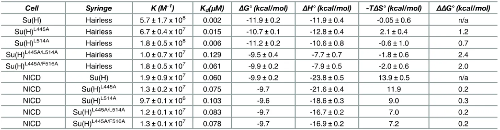
To analyze the contributions individual residues make to the Su(H)-Hairless complex, we designed mutations in Su(H) and Hairless, based on the structure, and tested the effect these mutations had on binding using ITC. The thermodynamic binding parameters (ΔG°,ΔH°, TΔS
°) of these interactions are contained within Tables2and3. As shownFig 4A(Table 2), Hair-less strongly binds Su(H) with 2 nM affinity. Consistent with HairHair-less being a random coil in solution prior to interacting with Su(H), complex formation is enthalpically driven at 25°C (Table 2). We made alanine substitutions at Hairless residues L235, F237, L245, L247, and W258, which are buried at the interface with Su(H) (Fig 2B and 2C), and determined their affinity for Su(H). HL235Aand HF237Aaffect binding ~30-fold and 140-fold, respectively (Fig 4B
and 4CandTable 2), consistent with these residues burying the most amount of surface area at the Su(H)-Hairless interface. HL245A, HL247A, and HW258A, whose side chains are more surface
exposed, reduce binding to a lesser extent, 5–12-fold (Fig 4D–4FandTable 2).
Next, we performed alanine-scanning mutagenesis of the residues in Su(H) that contact Hairless (Fig 4G–4I,Table 3andS1 Table). Some of these residues lie within the hydrophobic Fig 3. Conformational changes in Su(H) as a result of Hairless binding.(A) Figure shows Ig domain fold of the CTD from theapostructure of mouse CSL (RBP-J) (PDB ID: 3BRG) [9], which is composed of sevenβ-strands (a through g), wherein strands a, b, d, and e form one-half of theβ-sandwich, and strands f, g, and c form the other half. (B) Figure shows conformation of the CTD when Hairless is bound. Hairless binds between strands a and g, distorting the overall fold. The largest conformational change occurs in the loop region between strands e and f (colored green). (C) Structural overlay of the cαtraces from theapoRBP-J structure (3BRG) with the structure of Su(H) from the Su(H)-Hairless complex.Left, overall alignment of RBP-J (yellow) with Su(H) (orange). Note the good structural correspondence between the NTD and BTD, but poor structural similarity between the CTD domains.Right, zoomed view of the overlay of the CTDs fromapoRBP-J (yellow) and Su(H) (orange), highlighting the different conformations. Theβ-hairpin loop between strands a and b (L436-M444) and the C-terminus of the CTD from the Su(H)-Hairless complex undergo a translation of 6Åand 4.5Å, respectively, when compared to the
structure ofapoCSL. (D) Principal component analysis of the MD simulations for the Su(H)-Hairless structure (+H, cyan), the structure of Su(H) when Hairless is removed (-H,magenta), and anapoCSL structure(yellow). The covariance matrix of the trajectories were constructed, based on the 3-D fluctuations of cαatoms from their average position, and diagonalized, creating eigenvectors and eigenvalues that represent the direction and the amplitude of the motion, respectively. All the trajectories were projected on the first two eigenvectors and eigenvalues of these trajectories.
core of the CTD, e.g., F460 and I500, and when these were mutated to alanine resulted in insol-uble protein, precluding analysis. Alanine substitutions at other sites within the CTD resulted in binding comparable to wild type (S1 Table). However, we were able to identify two mutants, Su(H)L445Aand Su(H)L514A, which individually only affected binding 8-fold and 3-fold,
respec-tively, but in combination [Su(H)L445A/L514A] reduced binding 65-fold (Fig 4G and 4Hand
Table 3), suggesting that these residues are coupled. We reasoned that side chains near L445 and L514, when substituted for alanine, might also display nonadditive effects on binding. This led us to identify the double mutant Su(H)L445A/F516A, which reduced binding to Hairless
31-fold (Fig 4IandTable 3). We also screened numerous combinations of mutants by yeast two-hybrid (Y2H) assays that led to the triple mutants Su(H)L434A/L445A/L514Aand Su(H)L445A/ L514A/F516A, which nearly completely abrogated binding to Hairless (Fig 5A). The Y2H assay
also confirmed that the double mutant Su(H)L445A/L514Ahas reduced binding to Hairless. To
support that the Hairless binding site is conserved in mammals, we performed Y2H assays with wild-type and triple-leucine mutant RBP-J molecules. Similar to Su(H), wild-type RBP-J, but not the triple-leucine mutant, binds Hairless (Fig 5A).
Table 2. ITC data for the binding of Hairless mutants to Su(H).
Cell Syringe BSA (Å2) K (M-1) Kd(
μM) ΔG° (kcal/mol) ΔH° (kcal/mol) -TΔS° (kcal/mol) ΔΔG° (kcal/mol)
Su(H) Hairless (H) n/a 5.7±1.7 x 10⁸ 0.002 -11.9±0.2 -11.9±0.4 -0.05±0.6 n/a
Su(H) HL235A 71 1.7±0.4 x 10⁷ 0.063 -9.8±0.2 -9.6±1.1 -0.2±1.3 2.1
Su(H) HF237A 83 4.3±1.9 x 10⁶ 0.279 -9.0±0.3 -4.4±1.4 -4.6±1.7 2.9
Su(H) HL245A 56 1.1±0.4 x 10⁸ 0.010 -10.9±0.2 -9.3±0.4 -1.6±0.3 1.0
Su(H) HL247A 34 5.1±0.6 x 10⁷ 0.020 -10.5±0.1 -11.1±0.4 0.6±0.4 1.4
Su(H) HW258A 78 4.3±0.6 x 10⁷ 0.024 -10.4±0.1 -8.5±0.2 -1.9±0.3 1.5
ITC data for the binding of Hairless mutants to Su(H). All experiments were performed at 25°C. Table values are the mean of at least three independent experiments and errors represent the standard deviation of multiple experiments. BSA refers to the amount of surface area buried by the side chain in the native complex structure.
doi:10.1371/journal.pbio.1002509.t002
Table 3. ITC data for the binding of Su(H) mutants to Hairless and NICD.
Cell Syringe K (M-1) Kd(
μM) ΔG° (kcal/mol) ΔH° (kcal/mol) -TΔS° (kcal/mol) ΔΔG° (kcal/mol)
Su(H) Hairless 5.7±1.7 x 10⁸ 0.002 -11.9±0.2 -11.9±0.4 -0.05±0.6 n/a
Su(H)L445A Hairless 6.7±0.4 x 10⁷ 0.015 -10.7±0.1 -12.8±0.4 2.1±0.4 1.2
Su(H)L514A Hairless 1.8±0.5 x 10⁸ 0.006 -11.2±0.2 -10.6±0.8 -0.6±1.0 0.7
Su(H)L445A/L514A Hairless 1.0±0.7 x 10⁷
0.129 -9.5±0.4 -7.7±0.7 -1.8±0.6 2.4
Su(H)L445A/F516A Hairless 1.8±0.5 x 10⁷ 0.061 -9.9±0.2 -7.9±0.5 -2.0±0.6 2.0
NICD Su(H) 1.9±0.9 x 10⁷ 0.060 -9.9±0.2 -23.8±0.5 13.9±0.5 n/a
NICD Su(H)L445A 1.3±0.2 x 10⁷ 0.075 -9.7 -21.6±0.4 11.9 0.2
NICD Su(H)L514A 9.7±0.1 x 10⁶
0.103 -9.6 -18.6±0.3 9.0 0.3
NICD Su(H)L445A/L514A 1.2±0.1 x 10⁷ 0.083 -9.7 -16.7±0.2 7.0 0.2
NICD Su(H)L445A/F516A 1.3±0.1 x 10⁷ 0.078 -9.7 -16.9±0.2 7.2 0.2
ITC data for the binding of Su(H) mutants to Hairless and NICD. All Su(H)-Hairless binding experiments were performed at 25°C. Table values are the mean of at least three independent experiments and errors represent the standard deviation of multiple experiments. For native Su(H)-NICD binding studies, the table values are from our publication Contreras et al. 2015, and are the mean of at least three independent experiments and errors represent the standard deviation of multiple experiments. For the NICD binding experiments with Su(H) mutants, the errors represent the standard deviation of the nonlinear least squaresfit of the data to the titration curves and the table values representΔGobs,ΔHobs, and -TΔSobs. n/a = not applicable.
We performed several stability and functional assays on the single, double, and triple mutants described above to ensure that these mutant constructs were folded correctly and functionally active for binding to NICD. For the single and double Su(H) mutants, far UV cir-cular dichroism (CD) experiments showed some differences in the spectra between wild-type and mutant proteins; however, analysis of the CD data with Contin-LL [31] revealed similar amounts of secondary structure between native and mutant Su(H) proteins (S4 Fig). Similarly, thermal shift assays showed some destabilization of the mutants compared to wild type, but not substantial misfolding, and EMSA confirmed that the mutants bind DNA comparable to wild-type Su(H) (S4 Fig). As NICD also binds the CTD of Su(H) [26,32], we used ITC to test whether the mutants affect NICD binding. As shown inFig 5B–5F(Table 3), NICD binds Su(H) with 60 nM affinity, and importantly, the single and double mutants of Su(H) have little to no effect on NICD binding, suggesting that the mutations do not significantly affect the fold or function of the CTD. Unfortunately, we were unable to purify enough protein of the Su(H) triple mutants to analyze these constructs in vitro. However, we do demonstrate that the triple mutants can interact with NICD and MAM in a yeast three-hybrid assay (Fig 5A), again sug-gesting generally correct folding and function. Taken together, despite their partially overlap-ping binding sites on Su(H), we have identified mutations in Su(H) that affect complexes with Hairless, but leave interactions with NICD and MAM largely intact; albeit, with the caveat that the mutations do effect the stability of Su(H) to some degree. Henceforth, we refer to these mutants as Su(H)LL/AA, Su(H)LLL/AAA, and Su(H)LLF/AAA, respectively.
In Vivo Characterization of Su(H) Mutants
To test our Su(H) mutants within cells, we transfectedDrosophilaSchneider S2 cells with the NRE (Notch Response Element) reporter, which contains Su(H) binding sites coupled to the luciferase gene [33]. S2 cells express Su(H) and Hairless, but require the cotransfection of NICD to activate the reporter (Fig 6A) [23,33]. Providing additional Su(H) in the cells raises the activity about 3-fold, as observed previously [23,26,34]. Cotransfection of the Su(H) mutant constructs (Su(H)LL/AA, Su(H)LLF/AAA, Su(H)LLL/AAA) result in a very similar increase in reporter activity, in accordance with activator complex (Su(H)-NICD-MAM) assembly on DNA (Fig 6A). To address whether the repressor activity of Su(H) is compromised by the mutations, S2 cells were cotransfected with Hairless. Cotransfection of NICD and Hairless results in a 2-fold loss in reporter activity, which is restored by transfection of Su(H) (Fig 6A) [23,26,34]. Apparently, the competition of NICD and Hairless for Su(H) determines the overall reporter activity level. How-ever, cotransfection of the Su(H) mutants, which are deficient for Hairless binding, result in acti-vation of the reporter similar to cotransfection of Su(H) (native or mutant) and NICD (Fig 6A). We attribute the slight differences in reporter activity to repression mediated by endogenous Su(H) with the addition of exogenous Hairless and/or the mutations having a minor effect on NICD binding and/or residual binding of the mutants to Hairless. Nonetheless, these data sug-gest that in S2 cells our Su(H) mutants are competent to form an activator complex with NICD and MAM, but are defective for interacting with Hairless.
Fig 4. Thermodynamic binding analysis of structure based Hairless and Su(H) mutants.Figure shows representative thermograms (raw heat signal and nonlinear least squares fit to the integrated data) for Su(H) binding to Hairless molecules (wild type and mutants). Each experiment was performed at 25°C, with 40 titrations of 7μl injections spaced 120 s apart. The dissociation constants (Kd) shown for each experiment are from Tables2and3. (A) Wild-type Su(H) and Hairless interact with 2
nM affinity. The Hairless point mutants L235A (B) and F237A (C), which have the largest effect on binding, interact with Su(H) with 63 nM and 279 nM affinity, respectively. The Hairless point mutants L245A (D), L247A (E), and W258A (F) have more modest effects on interactions with Su(H), with 10 nM, 20 nM, and 24 nM dissociation constants, respectively. The Su(H) point mutants L445A (G) and L514A have minor effects on binding to Hairless, whereas the Su(H) double mutants L445A/L514A (H) and L445A/F516A (I) have considerable reduced binding to Hairless with 129 nM and 61 nM affinity, respectively.
To address the in vivo activity of our Su(H) mutants, we established transgenic fly lines that allow for tissue-specific overexpression [26]. Insertion at the identical site (96E) avoided unwanted position effects [26,35]. In addition, combined overexpression of Hairless and the Su(H) constructs was made possible by recombination [26]. The capacity of our Su(H) mutants to activate transcription was analyzed with thevgBE-lacZreporter gene, a readout for the Notch target genevestigial[36]. Ectopic expression of the constructs (wild type and mutants) was induced in the central domain of the wing anlagen. In response to Notch activation, thevgBE -lacZreporter is expressed along the presumptive wing margin [36] (red stripe,Fig 6Bpanel 1), which can be easily scored for differences in Notch signaling. Similar to ectopic expression of wild-type Su(H), the three mutant Su(H) constructs (Su(H)LL/AA, Su(H)LLF/AAA, Su(H)LLL/ AAA) result in a weak expansion of
vgBE-lacZexpression (Fig 6Bpanels 2–5). In addition, acti-vation of Notch, due to ectopic expression of Su(H), causes an overgrowth of the wing disc [23,26,34]. Similar overgrowth is observed for the three Su(H) mutants when compared to wild-type Su(H) (asterisks inFig 6Bpanels 2–5), suggesting that the mutations have little to no effect on assembly of the activator complex and activation of a Notch target gene in vivo.
To address whether the Su(H) mutants affect repressor complex formation in vivo, they were coexpressed with Hairless andvgBE-lacZreporter expression was monitored (Fig 6B). As a control, we ectopically expressed Hairless alone and in combination with wild-type Su(H), obtaining the expected results from a down-regulation of Notch signaling. Ectopic Hairless expression strongly repressed thevgBE-lacZreporter, which was accompanied by a loss of tissue in the overexpression domain (Fig 6Bpanel 6). Combined overexpression of Su(H) and Hair-less resulted in widespread silencing of thevgBE-lacZreporter and a nearly complete loss of tis-sue within the overexpression domain (Fig 6Bpanel 7), consistent with a hyper-repression of Notch signaling in this tissue [26,34]. This phenotype has been interpreted as a result of ectopic repressor complex formation, reflecting Su(H) and Hairless protein binding in a highly sensi-tized manner [24,37].
In the case of a complete loss of binding between the Su(H) mutants and Hairless, i.e., inde-pendent activity of the two components, the combined overexpression should result in an addi-tive phenotype: (1) activation of Notch signaling by the Su(H) mutant defecaddi-tive in Hairless binding and (2) repression by Hairless. The two should level out each other, and hence, wing discs resembling wild type would be expected. Any residual binding between Su(H) mutants and Hairless, however, would be uncovered by the strong super-repression effect resulting from their combined overexpression. As shown inFig 6B(panel 10), ectopic expression of the triple mutant Su(H)LLL/AAAwith Hairless resulted in normal expression of thevgBE-lacZ
reporter and the wing disc was similar to the control, suggesting an almost complete loss of Hairless binding by the mutant. Ectopic expression of Su(H)LLF/AAAwith Hairless was less
effective (Fig 6Bpanel 9), perhaps reflecting residual binding to Hairless and/or potentially subtle defects in activator complex formation, which our other assays were not sensitive enough to detect. The appreciable binding of the double mutant Su(H)LL/AAto Hairless Fig 5. Su(H) double and triple mutants are altered for Hairless binding, but unaffected for binding to NICD. (A) Figure shows yeast two-hybrid results for Hairless (H, 171–357) binding to wild-type and mutant Su(H) and RBP-J constructs. Reduced binding to Hairless is observed for the double mutant Su(H)L445A/L514A, whereas the
triple mutants (Su(H)L434A/L445A/L514Aand Su(H)L445A/L514A/F516A) display little to no binding to Hairless. Similar results are observed for RBP-J and RBP-JL386A/L397A/L466A. A yeast three-hybrid assay withDrosophilaMAM (118
–
194), NICD (1762–2176), and Su(H) shows that the double and triple mutants do not affect formation of the Su(H)-NICD-MAM activator complex. (B–F) Figure shows representative thermograms (raw heat signal and nonlinear least squares fit to the integrated data) for native and mutant Su(H) constructs binding to wild-type NICD (RAMANK, 1762–2142). Each experiment was performed at 25°C, with 40 titrations of 7μl injections spaced 120 s apart. The dissociation constants (Kd) shown for each experiment are fromTable 3. Su(H) single and double mutants (C–F)
have no effect on NICD binding.
observed in vitro was uncovered in our in vivo experiment with a repression of Notch activity similar to wild type Su(H) (Fig 6Bpanel 8).
Finally, these results were confirmed by a phenotypic analysis of adult flies, in which the Su(H) mutant constructs and Hairless were coexpressed during eye development. As shown inS5 Fig, and consistent with our previous results, ectopic induction of all three Su(H) mutants in the eye imaginal discs effects eye morphology indistinguishable from wild-type Su(H), but the triple mutants Su(H)LLL/AAAand Su(H)LLF/AAAretain little to no binding of
Hairless in vivo. However, in this in vivo context Su(H)LL/AAretained considerably less
Hair-less-mediated repressor activity, reflected by a milder phenotype compared to the Su(H)/H co-overexpression.
Discussion
In the Notch pathway, extracellular interactions are transduced into changes in gene expres-sion via the transcription factor CSL [1]. To activate transcription, CSL forms a ternary complex with the intracellular domain of the Notch receptor (NICD) and the coactivator Mas-termind (MAM), which binds at the promoter and enhancer regions of Notch target genes. A number of crystal structures that correspond to the CSL-NICD-MAM complex have been determined, and accompanying functional studies have scrutinized its role as an activator [4]. To repress transcription, CSL binds corepressor proteins, such as Hairless fromDrosophila, and SHARP or KyoT2 from mammals, which recruit other factors involved in transcriptional repression, such as Groucho, CtBP, NCoR, and Polycomb-group proteins, thus localizing the transcriptional repression machinery to Notch target genes [7]. While CSL is absolutely required to activate transcription at all target genes, its role as a repressor in different organ-isms is not as clear-cut. In mammals, RBP-J (CSL ortholog in mammals) has been shown to directly bind SHARP [16,17,20] and KyoT2 [18,19], and cellular studies have shown that core-pressors and their associated complexes are recruited to Notch target genes [21,38]; for certain cases, loss of RBP-J or its associated corepressor results in the de-repression of some, but not all target genes [21,38,39]. These in vitro data, however, have not yet been fully replicated in vivo, as there a only a few examples in mouse knockout studies that suggest RBP-J functions as a transcriptional repressor [40–42].
On the contrary, inDrosophilathere is substantial evidence both in vitro and in vivo that Su(H) (CSL ortholog in flies) binds the antagonist Hairless and functions as a transcriptional repressor at Notch target genes [15,22]. To provide a detailed structural basis for this interac-tion and gain addiinterac-tional insights into its funcinterac-tion, here we determined the high-resoluinterac-tion structure of the Su(H)-Hairless repressor complex bound to DNA (Fig 2), and presented con-vincing in vitro and in vivo mechanistic studies that complement and support our structure (Figs4–6). The most striking feature of the Su(H)-Hairless complex is the substantial confor-mational change that occurs in the CTD of Su(H) upon Hairless binding (Fig 3). This results in experiments were performed as indicated. Statistical significance was assessed using Dunnett’s test with***p0.001. The underlying data can be found inS1 Data. (B) Figure shows in vivo assays for formation of the activator and repressor complex in the wing imaginal discs of stage-matched larvae. Expression from thevgBE–lacZreporter is colored red and was
determined in the presence of ectopically expressed UAS-Su(H) constructs (green) as indicated, either singly (panels 1–5)
or in combination with ectopically expressed Hairless (UAS-H,panels 6–10). All constructs were overexpressed in the
central domain of the wing anlagen using the omb-Gal4 driver (encircled with a dashed line, Su(H) is colored green). UAS-GFP served as control (panel 1). Hairless is colored blue and in overlays with Su(H) appears turquoise (third row). Overexpression of Su(H) isoforms causes a subtle induction of thevgBE–lacZreporter, denoted by arrows, and an
overproliferation of the affected tissue (asterisks,panels 2–5). Concomitant overexpression of Su(H) isoforms and Hairless
(panels 7–10) results in repression of thevgBE–lacZreporter, denoted by bars, and tissue loss, as observed by loss of green
and blue staining (panels 6–8). Discs were framed to visualize the morphology in lower panels. Size bars = 100μm.
the twoβ-sheets that compose the Ig-fold of the CTD to be splayed apart, exposing residues in
the hydrophobic core of the CTD that form the basis for the Su(H)-Hairless interaction. To our knowledge, this conformational change and binding mode observed in our Su(H)-Hairless complex structure is without precedence in other Ig domain-containing structures. On the other hand, this illustrates the remarkable structural plasticity of this Ig domain in CSL. It will be interesting to see whether this occurs in other Ig-fold proteins, i.e., whether this binding mode is a basic principle of Ig domains or is particular to CSL proteins.
The structural change in Su(H) upon Hairless binding represents the largest and most strik-ing conformational change observed in all CSL-mediated complex structures determined to date. This raises the question as to what role the structural change plays in Su(H) function. One possibility is that the conformation Su(H) assumes when bound to Hairless serves to recruit specific binding partners or target the Su(H)-Hairless complex to specific sites in the genome. While the identities of these potentially new binding partners are unknown, it would be interesting in future studies to use screening approaches in order to identify factors that only bind the Su(H)-Hairless complex. A similar approach was used previously to identify Mastermind inCaenorhabditis elegans[43], a factor that only binds CSL when NICD is pres-ent. Moreover, our Su(H) mutants that are defective in Hairless binding may be useful in genome-wide studies to identify DNA sites bound by the Su(H)-Hairless complex.
Our MD simulations of the Su(H)-Hairless complex structure demonstrate that the confor-mation Su(H) must assume to bind Hairless is energetically unfavorable, and therefore, likely to be a rare conformer in the ensemble of Su(H) molecules in solution. The fact that the region of Hairless that interacts with Su(H) is unstructured prior to complex formation [26] suggests that the kinetics of Su(H)-Hairless association (kon) is a relatively slow process. If this is indeed
the case, then this would require the off rate (koff) to also be relatively slow in order to achieve
the 2 nM affinity (Kd= koff/kon) determined for Su(H)-Hairless complexes, which is the
stron-gest CSL binding partner measured to date. While the functional consequences of these slow on/off rates for Su(H)-Hairless complexes are not immediately evident, intriguingly these maybe important for its role as a transcriptional repressor, its subcellular localization, and/or replacement of activation complexes at Notch target genes. In the latter case, given that dimeric Su(H)-NICD-MAM complexes are stabilized at SPS sites by cooperative interactions between the ANK domain of NICD [13,14], it is conceivable that the displacement dynamics between monomeric versus dimeric Notch activation complexes and Su(H)-Hairless repressor com-plexes at Notch target genes are different. However, to our knowledge, there are no data avail-able to support this hypothesis, but it would be interesting to investigate this in the future.
Regarding the subcellular localization of Su(H), Hairless shuttles Su(H) into the nucleus [44,45], similar to NICD [46,47]. Moreover, there are two examples that demonstrate the amount of nuclear Su(H) is dependent on Hairless: (1) when Hairless is overexpressed, then Su(H) accumulates in the nucleus [44,45]; and (2) when cell clones are lacking Hairless, then Su(H) protein displays a conspicuously lower abundance [45]. Interestingly, an inter-depen-dence of Notch and Su(H) protein levels has been reported in theDrosophilaembryo [48]. Per-haps, Hairless and/or NICD binding to Su(H), and the nuclear import of these complexes may protect Su(H) from degradation in the cytosol. In mammals, it has been shown that RBP-J requires interactions with either corepressors or NICD for nuclear localization [49], and RBP-J is subjected to degradation in response to phosphorylation by p38 MAP kinase [50]. Interest-ingly, these data raise the possibility that a similar mechanism in flies and mammals exists to regulate CSL subcellular localization and turnover.
provides a potential mechanism whereby corepressors and NICD could compete for binding to RBP-J. For example, both the corepressor KyoT2 and RAM interact with the BTD of CSL in very similar conformations [19], whereas Hairless exclusively binds the CTD to a newly identi-fied binding cleft on Su(H). Moreover, there are no known corepressors inDrosophilathat interact with the BTD of Su(H). Interestingly, the corepressor SHARP, which has been sug-gested to be a Hairless analogue in mammals, interacts with both the BTD and CTD of RBP-J [17,20]. Given the high degree of sequence similarity between fly and mammalian CSL proteins (S2 Fig), perhaps SHARP binds the CTD of RBP-J in a manner similar to Hairless. Nonetheless, future studies that focus on the structure and function of the RBPJ-SHARP corepressor com-plex will be important for elucidating the enigmatic repressor role RBP-J plays in mammals.
Finally, there have been extensive efforts in the pharmaceutical industry and academic labs to identify reagents that modulate Notch signaling for therapeutic endpoints [2]. For example,
γ-secretase inhibitors and monoclonal antibodies that target the extracellular domains of
Notch receptors and ligands have been developed to treat certain types of cancer. However, in comparison, there has been very little progress in identifying and developing small molecules that directly target RBP-J and the transcription complexes it forms with coregulators. Our Su(H)-Hairless structure provides a new binding cleft that could be targeted. Consistent with this notion, previous studies from our groups demonstrated that Hairless binds RBP-J and when only the CSL-ID of Hairless is expressed in mammalian cells, it acts as a potent antago-nist of Notch signaling in transcriptional reporter assays [26].
Materials and Methods
Protein Overexpression and Purification
The codons that correspond to Su(H) residues 98–523, which represents the structural core of CSL (NTD, BTD, CTD), were cloned into pGEX-6P1. Site directed mutagenesis was used to introduce surface entropy reduction (SER) mutations (R155T and N281G) into pGEX-6P-1-Su(H) for purification and crystallization purposes. Wild-type and R155T/N281G Su(H) proteins were demonstrated to have nearly identical binding to Hairless [26]. Similarly, site directed mutagenesis was used to make the Su(H) mutant constructs described herein. As described previously [26], GST-Su(H) was overexpressed in BL21 Tuner cells (Novagen) and purified to homogeneity using a combination of affinity (glutathione resin), ion exchange (SP), and size exclusion chromatography. The purified protein was concentrated, flash frozen in liq-uid nitrogen, and stored at -80°C in a buffer containing 20 mM MES pH 6.0, 0.5 M NaCl, 1% ethylene glycol, and 0.1 mM TCEP.
Crystallization, Data Collection, and Structure Determination
In order to crystallize and determine the X-ray structure of the Su(H)-Hairless-DNA complex, recombinant Su(H) (98–523) and MBP-Hairless (232–269) proteins were purified to homoge-neity from bacteria and stoichiometrically bound to an oligomeric 15-mer duplex DNA, con-taining a single Su(H) binding site (TTACTGTGGGAAAGA, AATCTTTCCCACAGT). Crystals were grown out of a mother liquor containing 0.1 M Tris pH 8.0, 19% PEG3350, 0.2M (NH4)2SO4, were cryoprotected with 20% ethylene glycol, and were flash frozen in liquid
nitro-gen for data collection. X-ray diffraction data were collected on frozen crystals (200 K) at the Advanced Photon Source on the LS-CAT beamline 21-ID-F (λ= 0.97872 Å) and processed
with XDS [51]. Su(H)/MBP-H/DNA complex crystals belong to space group C2 with unit cell dimensions (a = 177.7, b = 93.9, c = 154.4,β= 109.8°) (Table 1). The Su(H)/MBP-H/DNA
structure was solved using molecular replacement (Phaser) [52] with the search models 3BRG [9] and 3H4Z [28], which contain RBP-J bound to DNA and MBP, respectively. The asymmet-ric unit of the crystals contains two Su(H), two MBP-H, and one DNA duplex (S1 Fig). The Hairless structure was built manually with COOT [53], and the structure was refined with Phe-nix [54] and Buster [55] to a final R factor and free R factor of 17.0% and 19.4%, respectively (Table 1). The model was evaluated with Molprobity [56] and the Ramachandran statistics for the final structure are 97.2% of the residues in the favored region with 0.24% as outliers.
The structure indicates that Hairless residues G232-R263 form the core structural motif that interacts with Su(H), whereas residues K264-P269 neither contact Su(H) nor theβ-hairpin motif of Hairless, and their extended structure is stabilized by interactions with the crystal lat-tice. Representative electron density of the CTD-Hairless interaction is shown inS1 Fig. The asymmetric unit (AU) of the crystals contains two Su(H)/MBP-H complexes, but surprisingly, only one of the complexes is bound to DNA (S1 Fig). While the explanation for the crystalliza-tion of these asymmetric complexes is unclear, the overall conformacrystalliza-tion of Su(H)-Hairless in the two complexes is similar (RMSD 0.92 for 442 cαatoms). The primary difference lies in the temperature factors (B factors) between the two complexes in the AU, in which the regions in the NTD and BTD that bind DNA have much higher B factors for the complex that is missing DNA (S1 Fig). All structure figures were created with PyMOL [57].
Isothermal Titration Calorimetry
ITC experiments were carried out using a MicroCal VP-ITC microcalorimeter. All experiments were performed at 25°C in a buffer composed of 50 mM sodium phosphate pH 6.5 and 150 mM NaCl. Su(H) and SMT3-H (Hairless) proteins were degassed and buffer-matched using dialysis and size exclusion chromatography. A typical experiment contained 5μM Su(H)
in the cell and 50μM SMT3-H in the syringe. The data were analyzed using ORIGIN software
and fit to a one-site binding model.
Molecular Dynamics Simulations Analysis
calculations were performed with the CUDA-enabled version of PMEMD [60], using TESLA GPUs at the High Performance Computing (HPC) cluster of the University of Cambridge. Analysis of the trajectories was performed with the AMBERTOOLS 1.5, GROMACS [61] and RStudio packages. Cavity analysis of the most representative structures of the principal clusters of the Su(H) trajectory was performed using trj_cavity [62].
Yeast Two- and Three-Hybrid Assay
The yeast two- and three-hybrid experiments were performed in triplicate as described previ-ously [26,34]. pEG-MAM corresponds toD.melanogasterMastermind, comprising codons 118 to 194. pESC-NICD corresponds to the RAM and ANK domains ofDrosophilaNotch, comprising codons 1762 to 2176. pEG-Hairless corresponds toDrosophilaHairless, compris-ing codons 171 to 357. Amino acid substitutions in full-length Su(H) and RBP-J were intro-duced using QuickChange II XL Site directed Mutagenesis Kit (Agilent) and ultimately cloned into pJG. The three promising mutants (L445A/L514A, L445A/L514A/F516A, L434A/L445A/ L514A) were shuttled into pUAST-attB and pMT vectors. All constructs were sequence verified.
Cellular Reporter Assays
Reporter assays were performed in triplicate as described in Maier et al. [26]. Schneider S2 cells (obtained from the DGRC) were transiently transfected with 1μg of the NRE-luciferase
reporter [33] and 0.2μg of control Renilla plasmid (Promega) to normalize transfection.
pMT-NICD was cotransfected with 0.5μg of the relevant pMT-Su(H) construct and/or 0.5μg
pMT-Hairless. The total amount of transfected DNA was kept constant at 3μg by using the
pMT-A vector (Promega). Constructs were induced 6 h after transfection by adding 0.5 mM CuSO4; 18 h later, cells were harvested and luciferase activity was measured in duplicate with a
luminometer (Lumat LB9507), using the dual-luciferase reporter assay system according to the manufacturer’s protocol (Promega). The effects of the addition of respective Su(H) variants rel-ative to the controls were assessed statistically using ANOVA and Dunnett’s test
(
p0.001).
In Vivo Analysis of Hairless and Su(H) Transgenes
Circular Dichroism
CD measurements were taken in triplicate using an Aviv Circular Dichroism Spectrometer model 215 at 25°C in a 0.02 cm cuvette. Wavelength scans were performed between 190 and 240 nm using 1.0 nm increments. Su(H) proteins were in a buffer containing 10 mM Tris-phosphoric acid pH 7.4 and 50 mM NaF. Protein concentrations were between 2–4 mg/ml. CD data were analyzed using Contin-LL (Provencher and Glockner Method) [31] with the
SMP180 reference set.
Thermal Shift Assays
An Applied Biosystems StepOne Real Time PCR system was used to collect the thermal shift data and the data were processed with their proprietary Protein Thermal Shift Software v1.2. Su(H) proteins were used at a concentration of 7μM in a buffer containing 25 mM MES pH
6.0, 0.5 M NaCl, 1 mM EDTA, and 1 mM TCEP.
EMSA
EMSAs were performed as described previously [9,26]. Wild-type or mutant Su(H) constructs were bound to an oligomeric 19-mer duplex that contains a single CSL-binding site and sepa-rated on a 7% polyacrylamide gel containing 0.5x Tris-borate buffer, pH 7, for several hours at 4°C. Complexes were visualized on the gel using SYBR-GOLD stain (Invitrogen).
Supporting Information
S1 Data. Excel file containing in separate sheets the numerical data for Figs6AandS4.
(XLSX)
S1 Fig. Additional details of the Su(H)-Hairless-DNA X-ray structure determination.(A) Figure shows orthogonal views of the molecules contained within the asymmetric unit of the crystal. The asymmetric unit contains two Su(H) molecules (colored yellow and magenta), two MBP-Hairless fusion molecules (colored cyan and green), and one DNA duplex (colored gray). MBP/H-A, MBP/H-B, Su(H)-C, and, Su(H)-D refer to chains A, B, C, and D, respectively, con-tained within the PDB file. (B) Figure shows a temperature factor (B-factor) comparison of the two Su(H) molecules contained within the asymmetric unit of the crystals. The color gradient from blue to red represents low to high B-factors, respectively. Cαworm thickness also
corre-sponds to B-factor magnitude, with low and high B-factors corresponding to thinner and thicker worms, respectively. (C) Figure shows representative electron density for the two less molecules within the asymmetric unit. Su(H) is represented as a gray surface and the Hair-less molecules (MBP/H-A and MBP/H-B) are shown in a stick representation with carbon, oxygen, and nitrogen atoms colored yellow, red, and blue, respectively. Electron density (slate blue mesh) corresponds to a simulated annealing composite omit map contoured at 1σ.
(PNG)
representing only main chain contacts. Overall, within the structural core of CSL, the primary sequence of Su(H) is 82% identical (90% similarity) to RBP-J; within the CTD, there is 75% identicalness (88% similarity) between fly and mouse CSL proteins. Of the 33 residues that are different between fly and mouse CSL, 27 of these residues (82%) have side chains that are sur-face exposed. Only 6 of the 33 residues that are different are buried or partially buried in the apo structure of CSL (3BRG), and in these cases the amino acid changes are very conservative, e.g., leucine to methionine, threonine to valine, isoleucine to valine, and serine to threonine. (PNG)
S3 Fig. Computational analysis of the Su(H)-Hairless structure.(A) Figure shows two inde-pendent MD simulations of the Su(H) structure after Hairless is removed from the complex, using the starting structure as the point of reference. Thex- andy-axes correspond to the time (picoseconds) and the cαRMSD (root mean square deviation), respectively. (B) Figure shows the cαRMSF (root mean square fluctuation), as a function of residue number, for the two
Su(H) simulations (red and black), and for comparative purposes, to a simulation for anapo
structure of CSL (green, PDB ID: 3BRG) [9]. The black circle denotes large fluctuations in the CTD of Su(H), compared toapoCSL, which are indicative of large rearrangements in the CTD of Su(H) when Hairless is removed from the complex. (C) Figure shows cavity calculations (denoted at gray spheres with accompanying volume in Å3) of the CTD of Su(H) (colored orange, ribbon diagram) for the most representative structures during the time course of the MD simulations:“Start”refers to the first frame of the MD simulations;“Cluster 1, 2, and 3”
describes the three most representative structures of the MD simulations during the early, mid-dle, and late frames, respectively; and“Final”represents the final frame of the MD simulations. (D) Figure shows color-coded, time-based projection of the first eigenvector/eigenvalue for the PCA (principal component analysis) of the Su(H) structure from the MD simulations. The blue structure represents the first frame of the MD simulations and the red structure indicates the final frame. (E) Figure shows time-based F516 dihedral angle calculation during the time course of the two Su(H) simulations (red and green), as compared to the simulation ofapoCSL (blue). As shown in the inset, F516 occupies two distinct conformations: (1) when bound to Hairless (yellow structure, corresponding to ~-50° dihedral angle) or (2) unbound (green struc-ture, corresponding to ~50° dihedral angle). Very early in the Su(H) simulations (red and green), when Hairless is removed from the complex F516 immediately assumes a dihedral angle similar to theapostructure of CSL (blue), consistent with F516 assuming a high energy conformer in the presence of Hairless, which is rarely sampled.
(PNG)
L445A/L514A, and lanes 8–10 Su(H) mutant L445A/F516A. (PNG)
S5 Fig. Effects on eye morphology by the overexpression of Su(H) variants.Wild-type or mutant Su(H) protein variants as indicated were overexpressed singly or in combination with full-length Hairless in the eye imaginal disc using thegmr-Gal4driver line. UAS-GFP served as control. Crosses were kept at 25°C and resultant adult eyes are shown. Ectopic expression of wild-type Su(H), as well as the mutant isoforms Su(H)LL/AA, Su(H)LLF/AAA, and Su(H)LLL/AAA,
affects eye morphology: eyes appear more bulgy and have slightly irregular ommatidia. Ani-mals that only overexpress Hairless have smaller eyes with a rough appearance [63,64]. Co-overexpression of both Su(H) and Hairless leads to a strong reduction or a complete loss of the eye, and the flies die as pharate adults in their pupal case. Lethality is also observed when Hair-less is combined with Su(H)LL/AA; however, the flies are able to eclose if incubated at 18°C. In contrast, the combined overexpression of the triple mutants Su(H)LLF/AAAor Su(H)LLL/AAA
with Hairless is viable, and the eye phenotype is normalized. Notably, the combination of Hair-less with Su(H)LLL/AAAresults in an almost normal fly, indicative of a complete lack of Hairless
binding by this mutant. (PDF)
S1 Table. ITC data for the binding of Su(H) mutants to Hairless.All experiments were per-formed at 25°C. For native Su(H)-Hairless binding studies, table values are fromTable 2and are the mean of at least three independent experiments, and errors represent the standard devi-ation of multiple experiments. For all other table entries, the values representΔGobs,ΔHobs,
and -TΔSobsand the errors represent the standard deviation of the nonlinear least squares fit of
the data to the titration curves. N/A = not applicable; for Su(H) mutants F460A and I500A we were unable to purify the recombinant protein for ITC binding studies.
(DOC)
Acknowledgments
The authors thank members of the Kovall lab for their technical support and helpful com-ments. We acknowledge S. Artavanis-Tsakonas, S. Bray, and F. Karch for fly lines and DNA constructs, and Robert Glen for access to the computing facilities of the Centre for Molecular Informatics, University of Cambridge. We are grateful to A. Schulz for superb technical assis-tance, T. K. Smylla for assistance with statistical evaluation, and A. C. Nagel for her constant help during the work.
Author Contributions
Conceived and designed the experiments: RAK DM AP. Performed the experiments: ZY NT HP RT. Analyzed the data: RAK ZY HP RT DM AP. Wrote the paper: RAK DM AP.
References
1. Hori K, Sen A, Artavanis-Tsakonas S (2013) Notch signaling at a glance. J Cell Sci 126: 2135–2140. doi:10.1242/jcs.127308PMID:23729744
2. Ntziachristos P, Lim JS, Sage J, Aifantis I (2014) From fly wings to targeted cancer therapies: a centen-nial for Notch signaling. Cancer Cell 25: 318–334. doi:10.1016/j.ccr.2014.02.018PMID:24651013
3. Louvi A, Artavanis-Tsakonas S (2012) Notch and disease: a growing field. Semin Cell Dev Biol 23: 473–480. doi:10.1016/j.semcdb.2012.02.005PMID:22373641
4. Kovall RA, Blacklow SC (2010) Mechanistic insights into Notch receptor signaling from structural and biochemical studies. Curr Top Dev Biol 92: 31–71. doi:10.1016/S0070-2153(10)92002-4PMID:
5. Fryer CJ, White JB, Jones KA (2004) Mastermind recruits CycC:CDK8 to phosphorylate the Notch ICD and coordinate activation with turnover. Mol Cell 16: 509–520. PMID:15546612
6. Wallberg AE, Pedersen K, Lendahl U, Roeder RG (2002) p300 and PCAF act cooperatively to mediate transcriptional activation from chromatin templates by notch intracellular domains in vitro. Mol Cell Biol 22: 7812–7819. PMID:12391150
7. Borggrefe T, Oswald F (2009) The Notch signaling pathway: transcriptional regulation at Notch target genes. Cell Mol Life Sci 66: 1631–1646. doi:10.1007/s00018-009-8668-7PMID:19165418
8. Wilson JJ, Kovall RA (2006) Crystal structure of the CSL-Notch-Mastermind ternary complex bound to DNA. Cell 124: 985–996. PMID:16530045
9. Friedmann DR, Wilson JJ, Kovall RA (2008) RAM-induced allostery facilitates assembly of a Notch pathway active transcription complex. J Biol Chem 283: 14781–14791. doi:10.1074/jbc.M709501200
PMID:18381292
10. Nam Y, Sliz P, Song L, Aster JC, Blacklow SC (2006) Structural basis for cooperativity in recruitment of MAML coactivators to Notch transcription complexes. Cell 124: 973–983. PMID:16530044
11. Kovall RA, Hendrickson WA (2004) Crystal structure of the nuclear effector of Notch signaling, CSL, bound to DNA. Embo J 23: 3441–3451. PMID:15297877
12. Choi SH, Wales TE, Nam Y, O'Donovan DJ, Sliz P, et al. (2012) Conformational locking upon coopera-tive assembly of Notch transcription complexes. Structure 20: 340–349. doi:10.1016/j.str.2011.12.011
PMID:22325781
13. Arnett KL, Hass M, McArthur DG, Ilagan MX, Aster JC, et al. (2010) Structural and mechanistic insights into cooperative assembly of dimeric Notch transcription complexes. Nat Struct Mol Biol 17: 1312–
1317. doi:10.1038/nsmb.1938PMID:20972443
14. Nam Y, Sliz P, Pear WS, Aster JC, Blacklow SC (2007) Cooperative assembly of higher-order Notch complexes functions as a switch to induce transcription. Proc Natl Acad Sci U S A 104: 2103–2108. PMID:17284587
15. Maier D (2006) Hairless: the ignored antagonist of the Notch signalling pathway. Hereditas 143: 212–
221. PMID:17362357
16. Kuroda K, Han H, Tani S, Tanigaki K, Tun T, et al. (2003) Regulation of marginal zone B cell develop-ment by MINT, a suppressor of Notch/RBP-J signaling pathway. Immunity 18: 301–312. PMID:
12594956
17. Oswald F, Kostezka U, Astrahantseff K, Bourteele S, Dillinger K, et al. (2002) SHARP is a novel compo-nent of the Notch/RBP-Jkappa signalling pathway. Embo J 21: 5417–5426. PMID:12374742
18. Taniguchi Y, Furukawa T, Tun T, Han H, Honjo T (1998) LIM protein KyoT2 negatively regulates tran-scription by association with the RBP-J DNA-binding protein. Mol Cell Biol 18: 644–654. PMID:
9418910
19. Collins KJ, Yuan Z, Kovall RA (2014) Structure and function of the CSL-KyoT2 corepressor complex: a negative regulator of Notch signaling. Structure 22: 70–81. doi:10.1016/j.str.2013.10.010PMID:
24290140
20. VanderWielen BD, Yuan Z, Friedmann DR, Kovall RA (2011) Transcriptional repression in the Notch pathway: thermodynamic characterization of CSL-MINT (Msx2-interacting nuclear target protein) com-plexes. J Biol Chem 286: 14892–14902. doi:10.1074/jbc.M110.181156PMID:21372128
21. Liefke R, Oswald F, Alvarado C, Ferres-Marco D, Mittler G, et al. (2010) Histone demethylase KDM5A is an integral part of the core Notch-RBP-J repressor complex. Genes Dev 24: 590–601. doi:10.1101/ gad.563210PMID:20231316
22. Bray S, Furriols M (2001) Notch pathway: making sense of Suppressor of Hairless. Curr Biol 11: R217–221. PMID:11301266
23. Nagel AC, Krejci A, Tenin G, Bravo-Patino A, Bray S, et al. (2005) Hairless-mediated repression of notch target genes requires the combined activity of Groucho and CtBP corepressors. Mol Cell Biol 25: 10433–10441. PMID:16287856
24. Morel V, Lecourtois M, Massiani O, Maier D, Preiss A, et al. (2001) Transcriptional repression by Sup-pressor of Hairless involves the binding of a Hairless-dCtBP complex in Drosophila. Curr Biol 11: 789–
792. PMID:11378391
25. Barolo S, Stone T, Bang AG, Posakony JW (2002) Default repression and Notch signaling: Hairless acts as an adaptor to recruit the corepressors Groucho and dCtBP to Suppressor of Hairless. Genes Dev 16: 1964–1976. PMID:12154126
26. Maier D, Kurth P, Schulz A, Russell A, Yuan Z, et al. (2011) Structural and functional analysis of the repressor complex in the Notch signaling pathway of Drosophila melanogaster. Mol Biol Cell 22: 3242–
27. Derewenda ZS (2004) Rational protein crystallization by mutational surface engineering. Structure 12: 529–535. PMID:15062076
28. Moon AF, Mueller GA, Zhong X, Pedersen LC (2010) A synergistic approach to protein crystallization: combination of a fixed-arm carrier with surface entropy reduction. Protein Sci 19: 901–913. doi:10. 1002/pro.368PMID:20196072
29. Holm L, Rosenstrom P (2010) Dali server: conservation mapping in 3D. Nucleic Acids Res 38: W545–
549. doi:10.1093/nar/gkq366PMID:20457744
30. Weinkam P, Pons J, Sali A (2012) Structure-based model of allostery predicts coupling between distant sites. Proc Natl Acad Sci U S A 109: 4875–4880. doi:10.1073/pnas.1116274109PMID:22403063
31. Provencher SW, Glockner J (1981) Estimation of globular protein secondary structure from circular dichroism. Biochemistry 20: 33–37. PMID:7470476
32. Contreras AN, Yuan Z, Kovall RA (2015) Thermodynamic binding analysis of Notch transcription com-plexes from Drosophila melanogaster. Protein Sci 24: 812–822. doi:10.1002/pro.2652PMID:
25650119
33. Bray S, Musisi H, Bienz M (2005) Bre1 is required for Notch signaling and histone modification. Dev Cell 8: 279–286. PMID:15691768
34. Kurth P, Preiss A, Kovall RA, Maier D (2011) Molecular analysis of the Notch repressor-complex in Dro-sophila: characterization of potential Hairless binding sites on Suppressor of Hairless. PLoS ONE 6: e27986. doi:10.1371/journal.pone.0027986PMID:22125648
35. Bischof J, Maeda RK, Hediger M, Karch F, Basler K (2007) An optimized transgenesis system for Dro-sophila using germ-line-specific phiC31 integrases. Proc Natl Acad Sci U S A 104: 3312–3317. PMID:
17360644
36. Kim J, Sebring A, Esch JJ, Kraus ME, Vorwerk K, et al. (1996) Integration of positional signals and regu-lation of wing formation and identity by Drosophila vestigial gene. Nature 382: 133–138. PMID:
8700202
37. Furriols M, Bray S (2000) Dissecting the mechanisms of Suppressor of Hairless function. Dev Biol 227: 520–532. PMID:11071771
38. Yatim A, Benne C, Sobhian B, Laurent-Chabalier S, Deas O, et al. (2012) NOTCH1 nuclear interactome reveals key regulators of its transcriptional activity and oncogenic function. Mol Cell 48: 445–458. doi:
10.1016/j.molcel.2012.08.022PMID:23022380
39. Castel D, Mourikis P, Bartels SJ, Brinkman AB, Tajbakhsh S, et al. (2013) Dynamic binding of RBPJ is determined by Notch signaling status. Genes Dev 27: 1059–1071. doi:10.1101/gad.211912.112
PMID:23651858
40. Hu B, Castillo E, Harewood L, Ostano P, Reymond A, et al. (2012) Multifocal epithelial tumors and field cancerization from loss of mesenchymal CSL signaling. Cell 149: 1207–1220. doi:10.1016/j.cell.2012. 03.048PMID:22682244
41. Surendran K, Boyle S, Barak H, Kim M, Stomberski C, et al. (2010) The contribution of Notch1 to neph-ron segmentation in the developing kidney is revealed in a sensitized Notch2 background and can be augmented by reducing Mint dosage. Developmental Biology 337: 386–395. doi:10.1016/j.ydbio. 2009.11.017PMID:19914235
42. Tsuji M, Shinkura R, Kuroda K, Yabe D, Honjo T (2007) Msx2-interacting nuclear target protein (Mint) deficiency reveals negative regulation of early thymocyte differentiation by Notch/RBP-J signaling. Proc Natl Acad Sci USA 104: 1610–1615. PMID:17242367
43. Petcherski AG, Kimble J (2000) LAG-3 is a putative transcriptional activator in the C. elegans Notch pathway. Nature 405: 364–368. PMID:10830967
44. Maier D, Praxenthaler H, Schulz A, Preiss A (2013) Gain of function Notch phenotypes associated with ectopic expression of the Su(H) C-terminal domain illustrate separability of Notch and Hairless-medi-ated activities. PLoS ONE 8: e81578. doi:10.1371/journal.pone.0081578PMID:24282610
45. Maier D, Nagel AC, Johannes B, Preiss A (1999) Subcellular localization of Hairless protein shows a major focus of activity within the nucleus. Mech Dev 89: 195–199. PMID:10559498
46. Gho M, Lecourtois M, Geraud G, Posakony JW, Schweisguth F (1996) Subcellular localization of Sup-pressor of Hairless in Drosophila sense organ cells during Notch signalling. Development 122: 1673–
1682. PMID:8674407
47. Fortini ME, Artavanis-Tsakonas S (1994) The Suppressor of Hairless protein participates in Notch receptor signaling. Cell 79: 273–282. PMID:7954795
48. Wesley CS, Mok LP (2003) Regulation of Notch signaling by a novel mechanism involving Suppressor of Hairless stability and carboxyl terminus-truncated Notch. Mol Cell Biol 23: 5581–5593. PMID: