J. Appl. Oral Sci. vol.26
Texto
Imagem

Documentos relacionados
Pindborg tumor, also known as calcifying odontogenic epithe- lial tumor, is a highly rare neoplasm, characterized by local invasiveness and presenting amyloid material.. This
The calcifying odontogenic cyst is an uncommon odontogenic lesion that can have intra- or extraosseous occurrence with both cystic or tumor behavior.. A report of an
The diagnostic and surgical management of a multifocal calciiyng epithelial odontogenic tumor in the mandible and maxilla associated with a squamous odontogenic tumor:
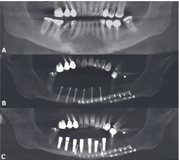
Key words: cone beam computed tomography, keratocystic odontogenic tumor, magnetic resonance imaging, odontogenic keratocyst, oral diagnosis.. T he keratocystic odontogenic tumor
- La simulación de la producción horaria y diaria de destilado mediante el modelo teórico de Dunkle proporciona valores simulados entre aproximadamente 40-50%
Ameloblastic fibrodentinosarcoma (AFDS) is a rare odonto- genic tumor histologically characterized by a sarcomatous ectomesenchymal component associated with variable amounts of
A case of ameloblastic carcinoma of the mandible diagnosed by FNAB is presented in this report, illustrating its effectiveness for preoperative diagnosis of odontogenic tumors..
Peripheral ameloblastoma is a rare variant of benign odontogenic tumor that exhibits the histologic characteristics of its intraosseous counterpart but occurs in the soft
