Supracostal Access: Does it affect Tubeless Percutaneous
Nephrolithotomy Eficacy and Safety?
J. Jun-Ou, Bannakij Lojanapiwat
Division of Urology, Department of Surgery, Chiangmai University, Chiangmai, Thailand
ABSTRACT
Introduction: Tubeless percutaneous nephrolithotomy (PCNL) in selected patients has been found to be safe and can reduce postoperative discomfort without increasing complications. The challenges of tubeless PCNL via supracostal access are inadequate drainage and postoperative bleeding, conditions that may increase pulmonary complications. We compare the eficacy and safety of the tubeless supracostal versus the standard supracostal PCNL.
Materials and Methods: Supracostal PCNL with one percutaneous renal access, no signiicant bleeding, extravasation and residual stone was performed in 95 patients. Of these, 43 were tubeless PCNL (Group-I) and 52 were PCNL with standard routine postoperative nephrostomy tube (Group-II). In group-I, PCNL was done by the standard supracostal technique with the placement of a postoperative external ureteral catheter for 48 hours. The operative time, success rate, hospital stay and ensuing complications were compared between group-I and group-II.
Results: Patients in the tubeless PCNL group (Group-I) were 90.7% stone -free while those with standard routine postop-erative nephrostomy tube(Group-II) were 84.6% stone -free. Additionally, stone fragments of less than 4 mm in diameter were found in 9.3% of patients in group-I and 25.4% in group-II. The success rate, hematocrit change and complication were not signiicantly different between both groups. The analgesic requirement, operative time and hospital stay were all signiicantly less in the tubeless supracostal group (Group-I). None of group I and only one patient of group II needed intercostal drainage.
Conclusion: Tubeless supracostal percutaneous nephrolithotomy in selected patients is effective with acceptable compli-cations. This technique offers the advantage of lower analgesic requirement, shorter operative time and hospital stay. The pulmonary complication is the same as the standard supracostal percutaneous nephrolithotomy.
Key words: percutaneous nephrolithotomy; eficacy; complications Int Braz J Urol. 2010; 36: 171-6
INTRODUCTION
Percutaneous nephrolithotomy (PCNL) is the accepted treatment for large renal and upper ureteral stones. The four stages of PCNL are: (1) renal ac-cess, (2) tract dilatation, (3) nephroscopy and stone disintegration, and (4) nephrostomy tube placement.
In uncomplicated PCNL where there is no signiicant
doi: 10.1590/S1677-55382010000200006
extravasation, signiicant bleeding, or any need for a
second nephroscopy, the placement of the nephros-tomy tube may not be necessary (tubeless PCNL)
(1-4). In speciic situations of PCNL, a supracostal renal approach is necessary (5-8). Pulmonary com -plication is more common with this approach due to
the anatomy of the kidney. Extravasation and bleeding
may increase the incidence of postoperative
pulmo-nary complications in supracostal access. The eficacy
and complication of tubeless PCNL via the supracostal route were compared to those of supracostal PCNL with routine nephrostomy tube placement.
MATERIALS AND METHODS
Patients
A total of 95 patients underwent PCNL
via supracostal. The patients were divided into two groups, 43 receiving tubeless supracostal PCNL (Group-I) and 52 receiving supracostal PCNL with routine nephrostomy tube placement (Group-II). Four criteria were established for PCNL of both groups in this study regardless of the stone burden, namely, (1) a single access site, (2) non obstructive renal unit,
(3) no signiicant perforation or bleeding, and (4) a
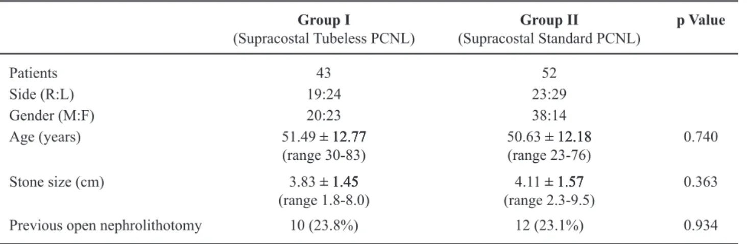
second look would not be required. The mean age of
group-I and II was 51.49 � 1�.�� years (range 3�-83� 1�.�� years (range 3�-83 1�.�� years (range 3�-83 years) and 5�.63 � 1�.18 years (range �3-�6 years),� 1�.18 years (range �3-�6 years), 1�.18 years (range �3-�6 years),
respectively. The mean stone size of group I and II
was 3.83 � 1.45 cm (range 1.8-8.� cm) and 4.11 � � 1.45 cm (range 1.8-8.� cm) and 4.11 � 1.45 cm (range 1.8-8.� cm) and 4.11 �� 1.57 cm (range 2.3-9.5 cm), respectively. Ten patients
(�3.8%) in group I and 1� patients (�3.1%) in group II had previous open nephrolithotomy. Patient proiles
are shown in Table-1. The positions of the calculi are
shown in Table-�. All patients were operated by a
single surgeon.
Methods
Single stage percutaneous nephrolithotomy was done in all patients. Intravenous antibiotic was
given before the operation in all cases. After the in
-duction of general anesthesia, an open-end 6F ureteral
catheter was placed via a transurethral approach into the ureter with the patient in a supine position. The tip of the ureteral catheter was placed at the uretero-pelvic junction or at the renal pelvis. The percutane-ous access was created by a single urologist (BL) in
all cases. Under luoroscopic guidance in the prone
position and after injection of contrast media via ureteral catheter, 95 sites were supracostal upper pole access. The needle was pushed through the diaphragm
and retroperitoneum in full expiration, whereas the
needle was passed through the kidney during deep inspiration. The working and safety guide-wires were inserted after the tip of the needle was in the col-lecting system. Tract dilatations were performed by
Amplatz fascial dilators (Cook Urological Spencer, Indiana, USA) or telescopic metal dilators sizes from 8F-3�F, with an inserted 3�F Amplatz sheath. Using a standard nephroscope (�6F), stone disintegration
was obtained with ultrasonic and/or pneumatic litho-tripsy. Fluoroscopy and contrast nephrostogram with systematic nephroscopy were performed to evaluate the stone-free status.
As regards tubeless PCNL, the ureteral cath
-eter (the same 6F ur-eteral cath-eter that was placed at
the beginning of the operation) was adjusted
neph-Table 1 – Proiles of patients.
Group I
(Supracostal Tubeless PCNL)
Group II
(Supracostal Standard PCNL)
p Value
Patients 43 52
Side (R:L) 19:24 23:29
Gender (M:F) 20:23 38:14
Age (years) 51.49 � 12.77� 12.77 12.77
(range 3�-83) 5�.63 � 1�.18(range �3-�6) � 1�.18 1�.18 0.740 Stone size (cm) 3.83 � 1.45� 1.45 1.45
(range 1.8-8.�) (range 2.3-9.5)4.11 � 1.57 � 1.57 1.57
�.363
roscopically, the tip being placed at the renal pelvis. The working sheath was removed with the safety guide wire still in placed. The nephrostomy site was
examined and, if there was no evidence of active
bleeding for 5 minutes, the wound was closed with sutures. The guide wire was then removed and the ureteral catheter was left attached to the Foley catheter
for 48 hours. The nephrostomy tube sized ��F was
routinely inserted in the remained cases (Group-II). The prolong placement of the ureteral catheter and nephrostomy tube depended on postoperative fever, bleeding or other complications.
Before and on the irst day of the surgery, all
patients were tested for complete blood count to deter-mine any change in the hematocrit level. Postoperative
chest x-ray (CXR) was routinely done in all cases. The patient’s symptoms and CXRs were used to evaluate
pulmonary complications. If the patients developed shortness of breath, chest pain and desaturation in the
recovery room and CXR revealed pleural effusion,
the intercostal drainages were done immediately.
Postoperative plain ilm KUB at day 1 was done
for evaluation of the stone free status. Meperidine injection was given when the patients complained of pain. Statistical analysis with Chi-square for qualita-tive variables and Student’s-t-tests for quantitaqualita-tive variables with p < 0.05 was considered statistically
signiicant.
RESULTS
The stone-free rate was 90.7% in group I and
84.6% in group II, and fragments � 4 mm occurred� 4 mm occurred 4 mm occurred 9.3% and 15.4% in groups I and II, respectively. The
operative time was 4�.38 � 16.93 min (range �5-9�� 16.93 min (range �5-9� 16.93 min (range �5-9� min) in group I and 58.85 � 18.46 (range 3�-1�5� 18.46 (range 3�-1�5 18.46 (range 3�-1�5 min) in group II, which was signiicantly different
(p = 0.03). The hospital stay was 3.45 � 1.01 (range� 1.01 (range 1.01 (range
�-� days) and 4.83 � 1.44 (range 3-1� days) in group� 1.44 (range 3-10 days) in group 1.44 (range 3-10 days) in group I and group II, respectively. Meperidine usage was
3� � 31(�-15�) mg in group I and �� � 36 (�-15�) � 31(�-15�) mg in group I and �� � 36 (�-15�) 31(�-15�) mg in group I and �� � 36 (�-15�)� 36 (�-15�) 36 (�-15�) in group II which was signiicantly different (p < �.��1). Decrease in the hematocrit level was �.65 � �
�.59 mg% in group I and �.31 � �.46 mg% which� �.46 mg% which �.46 mg% which
was not statistically different (p = 0.522). There were
4 patients (9.3� %) in group I and 5 patients (9.6� %) in group II who had hydrothorax as indicated by respiratory symptoms or postoperative CXRs Only
1 patient (1.92 %) of group II needed intercostal drainage (Table-3).
COMMENTS
Percutaneous nephrolithotomy has replaced open stone surgery for large renal or upper ureteral calculi because it is a less minimally invasive tech-nique. The last stage after completion of PCNL is the placement of the nephrostomy tube. The purpose of the nephrostomy tube is to provide hemostasis along
the tract, avoid urinary extravasation and maintain
adequate drainage of the kidney. In selected patients, tubeless percutaneous nephrolithotomy, with only
an externalized ureteral catheter or double J stent, is
safe, economical and provides reduced postoperative discomfort with the same outcome (1-4). Inclusion criteria of tubeless PCNL are the use of a single ac-cess site where the renal unit is not obstructive, no
signiicant perforation, bleeding and no need for a
Table 2 – Stone positions.
Group I
(Supracostal tubeless PCNL)
Group II
(Standard supracostal PCNL)
p Value
Staghorn stone (%) 14 (32.5) 15 (�8.8) 0.227
Calyceal stone (%) 12 (27.9 ) 9 (17.3)
Pelvic + Calyceal stone (%) 13 (30.2 ) �5 (48.1)
Upper ureteral stone (%) 2 (4.7) 3 (5.8)
second look (1-4). The stone burden may not neces-sarily to be taken into account.
Winield and associates reported signiicant
complications after premature removal of nephrostomy tube after PCNL (9). Bellman and colleagues reported
tubeless PCNL with only a double-J stent for one week without compromising eficacy and safety (1).
We previously reported tubeless PCNL in 37 patients
with only the placement of an externalized ureteral catheter for 48 hours to provide adequate drainage for
the upper tract without increasing complications and blood transfusion (2). Due to the selected patients in tubeless PCNL, the stone free status had to be assessed during the operation. The systematic nephroscopy,
intraoperative luoroscopy with contrast nephrosto -gram were used for evaluation of the stone free status.
Karami and Gholamrezaie (1�) and Aghamir et al.
(11) reported the technique of tubeless PCNL without
any externalized ureteral catheter or double J stent.
They found that the totally tubeless PCNL technique was safe and effective, requires less hospital stay and analgesics and led to a fast recovery time. No urinoma was found on postoperative ultrasound with an average
length of hospital stay of 1.6 days.
Under specific conditions, access to the kidney may require the upper pole approach. The
indications for the upper pole approach are staghorn calculi, large or multiple upper calyceal stones, renal calculi associated with ureteropelvic junction or upper ureteral pathology, large upper ureteral calculi and
calculi in speciic anatomy (8,1�,13). The upper pole approach provides a straight tract along the long axis of
the kidney and ensures reaching most of the collecting system, which provides easier manipulation of the rigid nephroscope and other rigid instruments. This approach can achieve a better stone clearance (12,13). Upper pole access can be achieved via the supracostal and infra-costal approaches. Due to the anatomy of the kidney, pulmonary complications are more common with the
supracostal approach (8,13). We previously reported
170 supracostal PCNL compared with 294 infracostal PCNL. We found that both approaches provide the same effective results, but pulmonary complications are higher when using the supracostal approach as compared with the subcostal approach. The pulmonary complications that needed intercostal drainage were 5% and 0.3% in supracostal and subcostal approach, respectively. There was a 17-fold greater possibility of pulmonary complication in the supracostal when compared to the subcostal approach (12).
Postoperative pulmonary complications after PCNL can be detected by postoperative symptoms and Table 3 – Results and complications.
Group I
(Supracostal tubeless PCNL)
Group II
(Standard supracostal PCNL)
p Value
Stone free (%) 39 (90.7) 44 (84.6) 0.131
Stone fragment � 4 mm (%)� 4 mm (%) 4 mm (%) 4 ( 9.30) 7(15.4) Operative time (min) 4�.38 � 16.93 � 16.93 16.93
(range 25-90)
58.85 � 18.46 � 18.46 18.46 (range 30-105)
0.030
Hospital stay (days) 3.45 � 1.01 � 1.01 1.01 (range 2-7)
4.83 � 1.44 � 1.44 1.44 (range 3-10)
< 0.001
Meperidine usage (mg) 37.00 � 31.00 � 31.00 31.00 (range 0-150)
��.�� � 36.�� � 36.�� 36.�� (range 0-150)
< 0.001
Hematocrit change (mg%) �.65 � �.59 � 2.59 2.59 �.3 � �.46 � �.46 �.46 0.522
Pleural complication (%) 4 (9.30) 5 (9.6�) 0.492
postoperative CXRs. The symptoms of pulmonary complications are poor oxygen saturation, dyspnea
and tachypnea postoperatively. The abnormality
of postoperative CXRs depends on the volume of pleural effusion (14). The treatment of hydrothorax depends on the amount of hydrothorax and the patients
symptoms. Conservative treatment is preferentially for those with no or mild symptoms and minimal
ef-fusion. Patients with signiicant symptoms and a large
amount of pleural effusion need intercostal drainage. The incidence of pulmonary complications after 12th supracostal approach that need surgical intervention
was from �% to �3% (range 5-8).
The techniques of supracostal approach require coordination with the anesthetist to control respiration. To avoid injuring the intercostal vessels, the intercostal puncture is made in the lower half of the
intercostal space. During full expiration, the needle
is passed through the retroperitoneum and diaphragm to prevent injury to the lung. The needle insertion, which is passed through the parenchyma to the col-lecting system, is done during deep inspiration for
downward displacement of the kidney. An Amplatz
sheath is used in all patients during the percutaneous supracostal approach to maintain low pressure irriga-tion that can reduce the risk of pleural effusion and
extravasation. After supracostal access, postoperative CXRs were routinely used in all cases for evaluation
of pulmonary complications (12).
One concern with the tubeless PCNL
tech-nique is to ensure adequate drainage and no signiicant bleeding postoperatively. Extravasation and bleeding can be signiicant problems of this technique after
supracostal approach and may lead to pulmonary complications. There is limited published data in the literature to date on tubeless PCNL in percutane-ous nephrolithotomy via supracostal access. Shah and colleagues (15) reported 72 patients of tubeless PCNL via supracostal access compared with 72 pa-tients with routine standard supracostal PCNL. Only
a single 6F double J stent was placed in the tubeless group, whereas both the 6F double J stent and a �8F
nephrostomy tube were placed in control group.
The double J stent was removed at 1-� weeks after
the surgery and nephrostomy tube was removed in 12-24 hours. Stone free at 1 to 2 weeks was 99.44%
and 91.66% in tubeless and in control group, respec
-tively. Blood transfusion was required in 3 patients of the tubeless group and in 4 of the control group
with only 1 patient in control group had hydrothorax
that needed intervention. They concluded that tube-less PCNL via supracostal is safe and effective with lower analgesic requirement and shorter hospital stay without increasing thoracic complication. Shah and
associates (16) reported 3� bilateral simultaneous
supracostal tubeless accesses in 51 urinary tracts of 45
renal units. As regards the supracostal access tracts,
no urine leakage or major chest complication were found, and patients were stone free or had residual stone fragment of less than 5 mm in 39 and 4 renal units, respectively. Gonen et al. reported 10 tubeless and stentless supracostal PCNL. These authors found that this technique is safe and offers advantages of a lower analgesia and shorter hospital stay without increasing of pulmonary complication (17).
In our series, all patients in the present study had the same criteria for the tubeless PCNL. We found that the incidence of pulmonary complications in tubeless supracostal PCNL was not different from the standard supracostal PCNL. The pleural compli-cation was 9.30% in tubeless supracostal PCNL and
9.6�% in standard PCNL. Almost all of these patients
were resolved after conservative treatment. Only one standard PCNL patient needed intercostal drainage.
The results of the treatment, as justiied by stone free and insigniicant residual fragment condition, were
the same in both groups. The analgesic requirement, operating time and hospital stay were less in tubeless group compared to the standard supracostal PCNL.
All patients in both groups, who had previous open
nephrolithotomy (10 of group I and 12 of group II) were found to be safe with the same outcome. The longer hospital stay in our series compared to the other studies was obviously related to our tubeless PCNL technique which was different from the other patients where we routinely left a ureteral catheter
indwelling for 48 hours after the surgery (�).
CONCLUSIONS
Tubeless supracostal percutaneous
nephroli-thotomy in selected patients with externalized ureteral
re-quirement, shorter operative time and shorter hospital stay. This technique does not increase hemorrhage or pulmonary complications when compared with the standard supracostal percutaneous nephrolithotomy.
CONFLICT OF INTEREST
None declared.REFERENCES
1. Bellman GC, Davidoff R, Candela J, Gerspach J, Kurtz S, Stout L: Tubeless percutaneous renal surgery. J Urol. 199�; 15�: 15�8-8�.
2. Lojanapiwat B, Soonthornphan S, Wudhikarn S: Tube-less percutaneous nephrolithotomy in selected patients. J Endourol. ���1; 15: �11-3.
3. Goh M, Wolf JS Jr: Almost totally tubeless percu -taneous nephrolithotomy: further evolution of the technique. J Endourol. 1999; 13: 1��-8�.
4. Shah HN, Kausik VB, Hegde SS, Shah JN, Bansal MB: Tubeless percutaneous nephrolithotomy: a prospective feasibility study and review of previous reports. BJU Int. ���5; 96: 8�9-83.
5. Stening SG, Bourne S: Supracostal percutaneous neph-rolithotomy for upper pole caliceal calculi. J Endourol. 1998; 1�: 359-6�.
6. Golijanin D, Katz R, Verstandig A, Sasson T, Landau EH, Meretyk S: The supracostal percutaneous neph-rostomy for treatment of staghorn and complex kidney stones. J Endourol. 1998; 1�: 4�3-5.
7. Munver R, Delvecchio FC, Newman GE, Preminger GM: Critical analysis of supracostal access for percu-taneous renal surgery. J Urol. ���1; 166: 1�4�-6.
8. Gupta R, Kumar A, Kapoor R, Srivastava A, Mandhani A: Prospective evaluation of safety and eficacy of the supracostal approach for percutaneous nephroli-thotomy. BJU Int. ����; 9�: 8�9-13.
9. Winield HN, Weyman P, Clayman RV: Percutane -ous nephrostolithotomy: complications of premature nephrostomy tube removal. J Urol. 1986; 136: ��-9. 1�. Karami H, Gholamrezaie HR: Totally tubeless per
-cutaneous nephrolithotomy in selected patients. J Endourol. ���4; 18: 4�5-6.
11. Aghamir SM, Hosseini SR, Gooran S: Totally tubeless percutaneous nephrolithotomy. J Endourol. ���4; 18: 64�-8.
12. Lojanapiwat B, Prasopsuk S: Upper-pole access for percutaneous nephrolithotomy: comparison of supra-costal and infrasupra-costal approaches. J Endourol. ���6; 20: 491-4.
13. Lang EK, Thomas R, Davis R, Colon I, Cheung W, Sethi E, et al.: Risks and beneits of the intercostal approach for percutaneous nephrolithotripsy. Int Braz J Urol. ���9; 35: ��1-81; discussion �81-3.
14. Woodring JH: Detection of pleural effusion on supine chest radiographs. AJR Am J Roentgenol. 198�; 149: 858-9.
15. Shah HN, Hegde SS, Shah JN, Bansal MB: Safety and eficacy of supracostal access in tubeless percutaneous nephrolithotomy. J Endourol. ���6; ��: 1�16-�1. 16. Shah HN, Kausik VB, Hegde SS, Shah JN, Bansal MB:
Safety and eficacy of bilateral simultaneous tubeless percutaneous nephrolithotomy. Urology. ���5; 66: 500-4.
17. Gonen M, Cicek T, Ozkardes H: Tubeless and stentless percutaneous nephrolithotomy in patients requiring supracostal access. Urol Int. ���9; 8�: 44�-3.
Accepted after revision: October 3, 2009
Correspondence address: Dr. Bannakij Lojanapiwat Division of Urology Department of Surgery Chiangmai University Chiangmai, 50200, Thailand
Fax: + 66 53 945-154