Features of the female prostate according to age:
an autopsy study
Características da próstata feminina de acordo com a idade: um estudo de autópsia
Thalita Cristina M. Costa; Patrícia M. Cury; Ana Maria G. Custódio
Faculdade de Medicina de São José do Rio Preto, São Paulo, Brazil.
First submission on 19/12/15; last submission on 23/04/16; accepted for publication on 18/05/16; published on 20/08/16
aBStraCt
Introduction: The prostate gland plays an important role in male and female reproductive system. Data on this organ have not been fully
explored in women since its irst description, probably because it is considered a vestigial gland. Objective: To correlate the morphology of the female prostate with age in autopsy. Material and methods: Thirty-two female cadavers, 31 adults and one newborn, underwent
dissection of the region corresponding to the prostate for histological analysis. The urethral region was divided into three portions: proximal, median, and distal. All the glands present in the samples were counted. Clinical data were collected, including age and previous diagnosis of menopause. Results: There were no macroscopically visible prostate. Morphological analyses showed glands surrounding the urethra with a stratiied epithelium, ranging from squamous to columnar types, with prevalence of basophilic cells and some presenting with secretion inside. A signiicant correlation with prostate tissue was found between the median and the proximal urethra, as well as between the median and distal urethra, suggesting that when the glandular structures increase in the median region, there is also an increase in the anterior and distal structures. Moreover, a prevalence of the glands in the median urethra was observed in post-menopausal women.
Conclusion: This study suggests that the number of female prostate glands increases after menopause, with proliferative spread and growth
of the median portion to the proximal and distal portions.
Key words: prostate; women; menopause.
introDuCtion
The prostate gland plays a role in both male and female reproductive systems. Data on this organ have not been fully explored in women since its irst description, probably because it is considered a vestigial gland and due to its limited study in autopsy material(1). Recent experimental studies with rodents have
increased the knowledge of the female prostate(2-8).
In 1672, Renier de Graaf identiied the irst mammal female prostate as a group of glands surrounding the urethra, similar to the male prostate. In 1880, Alexander J. C. Skene observed the presence of two paraurethral ducts opening around the urethra, with no obvious function, which were identiied as Skene’s paraurethral glands, considered a vestigial organ. In 1906, Barnett also identiied these deep urethral glands and described their location between the bladder neck and the external urethral meatus, near to the urethra, and above the pubocervical fascia. The
anatomy of the female prostate differs from that of the male organ. In men, the prostate surrounds the urethra and is encapsulated, while in women it is not well deined, does not have a capsule, and can not be macroscopically observed(1).
Histologically, the female prostate has more ducts than glands; the opposite occurs in men. The glands are presented either in groups or isolated; they are composed of basal and secreting cells; the latter may be cylindrical, cubic, or columnar. Intermediate cells may be observed by electron microscopy among these cell types(1).
The prostate location in the urethra may vary, and is more commonly seen in the distal portion. However, it may be observed in the proximal (meatal type), distal (posterior type), and in the entire urethra (rudimentary type)(1).
The female prostate plays an important role in reproduction, since it supports spermatozoa in the female body, providing nutrients through the prostate luid, which has the same features
as that the male(9), and affects the sexual behavior(1). Some studies
have associated the female prostate with the Gräfenberg point (G-point), reporting that ejaculation may be stimulated by the G-point(10), while other authors advocate the idea that they are
the same structure(11).
Few studies have investigated the prostate in healthy women; thus, compared to the extensive knowledge about male prostate, the female prostate features are still not fully understood. According to Dodson (1994)(12), prostate markers and prostate speciic antigen
(PSA) were found in urethral syndrome, urethral carcinoma, and breast cancer, but the normal female prostate pattern has not been fully described. Thus, the present study was performed in order to correlate prostate morphology with age in female cadavers.
MatEriaL anD MEthoDS
Female cadavers were submitted to autopsy at a Death Certiication Service, over a period of 10 months, the regions corresponding to the prostate were subjected to dissection and analysis. The research ethics committee of the institution approved the study. The exclusion criteria were: presence of body decomposition, loss of urethral region integrity, and stillborn child.
The bladder and urethra were dissected and abdominally removed in a single portion during autopsy, and the material was preserved in 10% buffered formalin. The entire urethral region was transversally divided into three segments after being separated from the bladder – proximal, median, and distal samples – and embedded in parafin.
These 3-5 μm blocks from each cadaver were sectioned at three different levels (proximal, median, and distal portions), with the distal portion corresponding to the urethral meatus. The slides were stained with hematoxylin-eosin (HE) and examined by light microscopy. To characterize the presence of prostate tissue and its prevalent site, the number of glands on each slice (anterior, median, posterior samples) was determined.
Clinical data including age, leading cause of death, and underlying causes of death were obtained from the death certiicate of each subject. Information regarding the diagnosis of menopause and urinary tract infection for the case studies were obtained from medical records.
Data on morphological features of prostate at different ages were statistically analyzed using Fisher’s exact test, analysis of variance and the Kruskal-Wallis test, and Spearman’s correlation coeficient was calculated.
rESuLtS
Clinical analysis
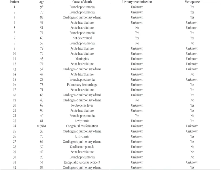
Thirty-two female cadavers were studied; there were 31 adults with an average age of 60.6 years (range: 25-96 years) and one newborn. The main causes of death reported in the death certiicate were acute heart failure and bronchopneumonia (nine and seven cases, respectively). Among 21 patients with available data on menopause, menopausal status was determined in 14. Moreover, only three patients had a history of urinary tract infection among the six patients with available data (Table 1).
Morphological analysis
Thirty-two urethras were dissected from the female cadavers studied over a period of 10 months, but seven could not be analyzed due to autolysis on the material. One case of a child was not included in the statistical analysis due to the lack of similar cases for comparison.
No macroscopic prostate was visualized in the cases. The prostate tissue was histologically identiied in all cadavers studied (Table 2). Glands were predominantly found in the
proximal, median, and distal regions of 10, seven, and six cases, respectively. In one case, the urethra showed no prevalent region for the gland.
Morphological analysis showed glands surrounding the urethra, which presented a stratiied epithelium ranging from squamous to columnar types; basophils were prevalent, and some glands showed secretion. A dense stromal tissue was observed around the glands (Figure 1).
Nine prostate tissues were considered as cystic, and most showed internal calculus (Figure 2). One case of paraurethral bleeding was found (Figure 3), 12 cases showed an increase in vascular structures (paraurethral ectasia), and chronic lymphocytic inlammation was observed in 62% of cases (Figure 4).
Statistical analysis
Data were analyzed aiming to establish a statistical correlation between the histological data from the autopsy material with individual’s age and clinical parameters. The material of a stillborn and seven prostate tissues could not be analyzed due to a lack of similar cases for comparison and because of poor preservation, respectively. Therefore, 24 prostate tissues were considered for statistical analysis.
taBLE 1 − Clinical data from the female cadavers studied
Patient Age Cause of death Urinary tract infection Menopause
1 96 Bronchopneumonia Unknown Yes
2 75 Bronchopneumonia Unknown Yes
3 85 Cardiogenic pulmonary edema Unknown Yes
4 54 Acute heart failure Unknown Unknown
5 51 Acute heart failure No Unknown
6 74 Bronchopneumonia Yes Yes
7 60 Not determined Yes Yes
8 58 Bronchopneumonia No No
9 72 Acute heart failure Unknown Unknown
10 76 Acute heart failure Unknown Unknown
11 43 Meningitis Unknown Unknown
12 74 Acute heart failure Unknown Unknown
13 54 Cardiogenic pulmonary edema Unknown Unknown
14 47 Acute heart failure Unknown No
15 25 Bronchopneumonia Unknown Unknown
16 75 Pulmonary hemorrhage Unknown Yes
17 71 Acute heart failure Unknown Yes
18 65 Cardiogenic pulmonary edema Unknown Yes
19 45 Cardiogenic pulmonary edema No No
20 68 Neutropenic fever Unknown Yes
21 96 Acute heart failure Unknown Yes
22 40 Bronchopneumonia Yes No
23 81 Arrhythmia Unknown Yes
24 0 (NB) Congenital malformation Unknown Unknown
25 38 Cardiogenic pulmonary edema Unknown Unknown
26 76 Arrhythmia Unknown Yes
27 64 Cardiogenic pulmonary edema Unknown Yes
28 39 Cardiac tamponade Unknown No
29 42 Acute heart failure Unknown No
30 25 Bronchopneumonia Unknown No
31 53 Encephalic vascular accident Unknown Unknown
32 85 Cardiogenic pulmonary edema Unknown Yes
NB: newborn.
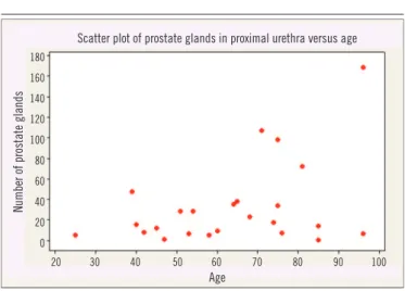
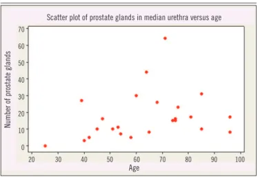
0.15, p = 1, p = 0.17, p = 0.40, respectively). No statistically signiicant correlation between age and prevalent regions for gland was found by analysis of variance (p = 0.53) (Figures 5
to 8).
Since the number of glands did not show a regular pattern according to age, Spearman’s test was used instead of the Pearson’s correlation test. Signiicant correlations were found between the median and proximal urethral portions (Spearman’s correlation: 0.430, p = 0.034), as well as between
the median and distal urethras (Spearman’s correlation: 0.438,
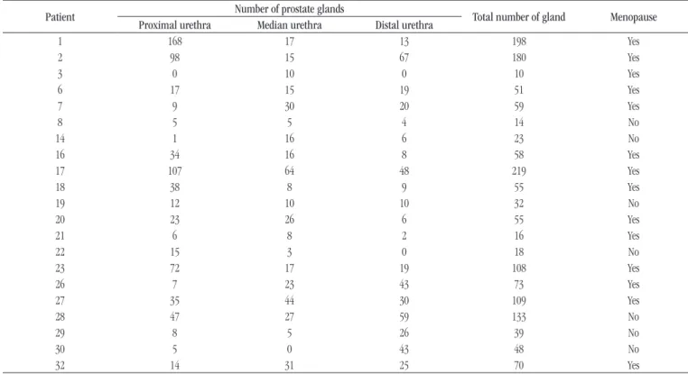
p = 0.032), suggesting the occurrence of an increase in anterior and distal structures when the median region structures are increased. The Kruskal-Wallis test revealed a predominance of the prostate glands in the median urethra of women who had reached menopause (H = 6.47, p = 0.011, adjusted for ties) (Table 3).
DiSCuSSion
Histological analysis of the autopsy material revealed glands composed of both squamous and columnar epithelium surrounding the urethra, with a dense stromal tissue around these structures, as also reported in the literature(9, 12-15).
According to Zaviačič (1998)(1), the female prostate can be
taBLE 3 − Features of prostate glands and menopause
Patient Number of prostate glands Total number of gland Menopause Proximal urethra Median urethra Distal urethra
1 168 17 13 198 Yes
2 98 15 67 180 Yes
3 0 10 0 10 Yes
6 17 15 19 51 Yes
7 9 30 20 59 Yes
8 5 5 4 14 No
14 1 16 6 23 No
16 34 16 8 58 Yes
17 107 64 48 219 Yes
18 38 8 9 55 Yes
19 12 10 10 32 No
20 23 26 6 55 Yes
21 6 8 2 16 Yes
22 15 3 0 18 No
23 72 17 19 108 Yes
26 7 23 43 73 Yes
27 35 44 30 109 Yes
28 47 27 59 133 No
29 8 5 26 39 No
30 5 0 43 48 No
32 14 31 25 70 Yes
taBLE 2 − Histological characteristics of prostate glands
Patient Age Number of prostate glands Total number of gland
Paraurethral inflammation
Paraurethral ectasia
Paraurethral bleeding
Cystic gland Proximal urethra Median urethra Distal urethra
1 96 168 17 13 198 Yes No No Yes
2 75 98 15 67 180 No No Yes Yes
3 85 0 10 0 10 No No No Yes
4 54 28 7 6 41 Yes Yes No No
5 51 28 10 20 58 Yes No No No
6 74 17 15 19 51 Yes Yes No No
7 60 9 30 20 59 No No No No
8 58 5 5 4 14 Yes Yes No No
14 47 1 16 6 23 Yes No No No
16 75 34 16 8 58 No No No Yes
17 71 107 64 48 219 No No No Yes
18 65 38 8 9 55 No Yes No Yes
19 45 12 10 10 32 Yes Yes No No
20 68 23 26 6 55 Yes Yes No No
21 96 6 8 2 16 Yes Yes No No
22 40 15 3 0 18 Yes Yes No No
23 81 72 17 19 108 No Yes No No
26 76 7 23 43 73 No Yes No No
27 64 35 44 30 109 Yes No No No
28 39 47 27 59 133 Yes No No No
29 42 8 5 26 39 No No No Yes
30 25 5 0 43 48 Yes Yes No No
31 53 6 11 24 41 Yes Yes No Yes
FigurE 1 − Histological panoramic view of prostate tissue
FigurE 2 −Glands and cystic structures surrounding the urethra. Magniication: 40×
FigurE 3 − Median urethral portion. Presence of surrounding bleeding. Magniication: 40×
FigurE 4 −Distal urethral portion. Inlammation surrounding the glands. Magniication: 40×
FigurE 5 −Correlation between number of prostate glands and age
FigurE 6 −Correlation between prostate glands in the proximal urethral portion and age
20 30 40 50 60 70 80 90 100 Scatter plot of total number of prostate glands versus age
Number of prostate glands
250
200
150
100
50
0
Age
Scatter plot of prostate glands in proximal urethra versus age
Number of prostate glands
180
160
140
120
100
80
60
40
20
0
Age
identiied, and is also formed by prostate tissue in the entire urethra (6% of cases).
No macroscopic prostate was visualized in this study. According to Zaviačič (1998)(1), the female prostate size and weight
corresponds to the entire female urethra and prostate combined, with an average size of 3.3 × 1.9 × 1 cm, and an average weight of 5.2 g. No size data were obtained in the present study. This study found nine cases of glands containing some cystic structures.
Very few studies in female prostate have been published. Wimpissinger et al. (2009)(16) used endoscopy and nuclear magnetic
resonance to evaluate seven women who had shown signs and symptoms of prostate problems such as ejaculation and chronic urethral pain. However, no histological study has been carried out.
Custodio et al. (2008)(6) studied the effect of aging on gerbil
prostate and reported that reduced levels of dehydroepiandrosterone and estradiol during senescence led to epithelial hypertrophy, metaplasia, neoplasia, and hyperplasia.
ConCLuSion
The number of prostate glands increases after menopause, with proliferative growth and spread from the median to proximal and distal urethra. These results suggest that a reduction in hormone levels can stimulate increase of the female prostate gland and induce further disorders in the woman. This study did characterize the paraurethral glands in women; however, due to the small number of patients studied, this data should be replicated in another cohort.
FigurE 8 − Correlation between prostate glands in the distal urethral portion and age
FigurE 7 − Correlation between prostate glands in the median urethral portion and age
rESuMo
Introdução: A próstata é uma glândula com papel importante no sistema reprodutor masculino e feminino. Dados sobre esse
órgão não foram completamente explorados em mulheres desde a sua primeira descrição, provavelmente por ser considerada uma glândula vestigial. Objetivo: Correlacionar a morfologia da próstata feminina com a idade em autópsias. Material e métodos:
Trinta e dois cadáveres do sexo feminino, sendo 31 adultos e um recém-nascido, tiveram sua região correspondente à próstata dissecada e avaliada por meio de histologia. A região uretral foi dividida em três partes: anterior, mediana e distal. As glândulas presentes nas amostras foram contadas. Dados clínicos foram coletados, incluindo idade e diagnóstico prévio de menopausa.
Resultados: Não foram observadas próstatas macroscopicamente. Análises morfológicas mostraram glândulas ao redor da uretra
com epitélio estratiicado, variando do tipo escamoso a colunar, com predomínio de células basóilas e algumas apresentando secreção em seu interior. Correlação signiicativa com tecido prostático foi detectada entre a uretra mediana e a proximal, assim como entre as uretras mediana e distal, sugerindo que quando as estruturas glandulares aumentam na região mediana, há também aumento nas estruturas anterior e distal. Além disso, o predomínio das glândulas na uretra mediana foi observado em mulheres pós-menopausa. Conclusão: Este estudo sugere que o número de glândulas prostáticas femininas aumenta após a menopausa, com disseminação e crescimento da região mediana para a proximal e distal.
Unitermos: próstata; mulheres; menopausa.
Scatter plot of prostate glands in median urethra versus age
Number of prostate glands
70
60
50
40
30
20
10
0
Age
Age
20 30 40 50 60 70 80 90 100
20 30 40 50 60 70 80 90 100 Scatter plot of prostate glands in distal urethra versus age
Number of prostate glands
70
60
50
40
30
20
10
rEFErEnCES
1. Zaviačič M, Sloboda, Jakubovský J, et al. Metastasizing adenocarcinoma of the female prostate (Skene’s paraurethral glands). Histological and immunohistochemical prostate marker studies and irst ultrastructural observation. Pathol Res Pract. 1998; 194: 129-36.
2. Santos FCA, Carvalho HF, Góes RM, et al. Structure, histochemistry, and ultrastructure of epithelium and stroma in the gerbil (Meriones unguiculatus) female prostate. Tissue Cell. 2003; 35: 447-57.
3. Santos FCA, Leite RP, Custódio AMG, et al. Testosterone stimulates growth and secretory activity of the female prostate in the adult gerbil (Meriones unguiculatus). Biol Reprod. 2006; 75: 370-9.
4. Santos FCA, Custodio AMG, Campos SGP, et al. Antiestrogen therapies affect tissue homeostasis of the gerbil (Meriones unguiculatus) female prostate and ovaries. Biol Reprod. 2008; 79: 674-85.
5. Custodio AMG, Góes RM, Taboga SR. Acid phosphatase activity in gerbil prostate: comparative study in male and female during postnatal development. Cell Biol Int. 2004; 28: 335-44.
6. Custodio AMG, Santo FCA, Campos SGP, et al. Aging effects on the Mongolian gerbil female prostate (Skene’s paraurethral glands): structural, ultrastructural, quantitative, and hormonal evaluations. Anat Rec. 2008; 291: 463-74.
7. Santos FCA, Taboga SR. Female prostate: a review about the biological repercussions of this gland in humans and rodents. Anim Reprod. 2006; 3: 3-18.
8. Perez AP, Biancardi MF, Goes RM, et al. Exposure to ethinylestradiol during prenatal development and postnatal supplementation with testosterone causes morphophysiological alterations in the prostate of male and female adult gerbils. Int . J. Exp. Pathol. 2011; 92: 121-30. 9. Zaviačič MZaviacic M, Ablin RJ. The female prostate and prostate-speciic antigen. Immunohistochemical localization, implications of this prostate marker in women and reasons for using the term “prostate” in the human female. Histol Histopathol. 2000 Jan; 15(1): 131-42. 10. Schubach G. The G-spot is the female prostate. Am J Obstet Gynecol. 2002 Apr; 186(4): 850.
11. Hines TM. The G-spot: a modern gynecologic myth. Am J Obstet Gynecol. 2001 Aug; 185(2): 359-62.
12. Dodson MK, Cliby WA, Keeney GL, et al. Skene’s gland adenocarcinoma with increased serum level of prostate-speciic antigen. Gynecol Oncol. 1994; 55: 304-7.
13. Dodson MK, Cliby WA, Pettavel PP, et al. Female urethral adenocarcinoma: evidence for more than one tissue of origin? Gynecol Oncol. 1995; 59: 352-7.
14. Ali SZ, Smilari TF, Gal D, et al. Primary adenoid cystic carcinoma of Skene’s glands. Gynecol Oncol. 1995; 57: 257-61.
15. McCluggage WG1, Ganesan R, Hirschowitz L, et al. Ectopic prostatic tissue in the uterine cervix and vagina: report of a series with a detailed immunohistochemical analysis. Am J Surg Pathol. 2006 Feb; 30(2): 209-15. 16. Wimpissinger F, Tscherney R, Stackl W. Magnetic resonance imaging of female prostate pathology. J Sex Med 2009; 6: 1704-11.
CorrESPonDing author
Thalita Cristina de Mello Costa