Fisioter. Mov., Curitiba, v. 30, n. 4, p. 821-830, Oct./Dec. 2017 Licenciado sob uma Licença Creative Commons DOI: http://dx.doi.org/10.1590/1980-5918.030.004.AO18
Dynamic physiological responses to the
incremental shuttle walk test in adults
Respostas isiológicas no incremental shuttle walk test em adultos
Evandro Fornias Sperandio, Ricardo Luís Fernandes Guerra, Victor Zuniga Dourado*
Universidade Federal de São Paulo (Unifesp), Santos, SP, Brazil
Abstract
Introduction: Understanding the normal dynamic physiological responses to the incremental shuttle walk test might enhance the interpretation of walking performance in clinical settings. Objective: To assess dynamic physiological responses to the incremental shuttle walk test and its predictors in healthy adults.
Methods: We assessed the simultaneous rates of changes of Δoxygen uptake/Δwalking velocity (ΔVO2/
ΔWV), Δheart rate/Δoxygen uptake (ΔHR/ΔVO2), Δventilation/Δcarbon dioxide production (ΔVE/ΔVCO2), and Δtidal volume/Δlinearized ventilation (ΔVT/ΔlnVE) during the incremental shuttle walk test in 100 men and women older than 40 years. Fat and lean body masses (bioimpedance) were also evaluated. Results: We
found that the dynamic relationships were not sex-dependent. Participants aged ≥ 70 presented declines in ΔVO2/ΔWV slope compared to those aged 40-49 (215 ± 69 vs. 288 ± 84
mL.min-1.km.h-1). Obese participants presented shallower slopes for ΔVO2/ΔWV (2.94 ± 0.90 vs. 3.84 ± 1.21 mL.min-1.kg-1.km.h-1) and ΔVT/ ΔlnVE (0.57 ± 0.20 vs. 0.67 ± 0.26). We found negative influence of fat body mass on ΔVT/ΔlnVE (R2 = 0.20) and positive influence of lean body mass on ΔVO2/ΔWV (R
2 = 0.31), ΔHR/ΔVO2 (R2 = 0.25), and ΔVT/ΔlnVE (R2 = 0.44). Conclusion: Dynamic relationships during walking were slightly influenced by age, but not
sex-dependent. Body composition played an important role in these indices. Our results may provide better interpretation of walking performance in patients with chronic diseases.
Keywords: Exercise. Walking. Physical Fitness.
822
Introduction
The cardiopulmonary exercise test (CPET) offers
the opportunity to appreciate precisely the complex in-teraction among musculoskeletal, cardiovascular and respiratory systems in a controlled environment (1). The peak physiological values obtained in an incre-mental protocol are generally used as indices of
car-diorespiratory fitness. Among these variables, peak oxygen uptake (peak VO2) and VO2 obtained at lactate
threshold are the most commonly used indices (2). Although the peak variables is valuable, the consideration of a single value for the variables of interest may be unsuitable as a frame of reference for interpreting continuous dynamic physiological
responses to the CPET. Moreover, the interpretation
of the dynamic relations does not require maximum effort, because the trend has a linear behavior. Thus, the dynamic relationships can be used in submaxi-mal physical tests. This strategy makes it possible to
improve the interpretation of physical fitness both
in patients and in healthy people. Furthermore, submaximal tests can be performed by any non-physician healthcare professionals able to do so.
The dynamic relationships such as Δoxygen uptake/
Δwork load (ΔVO2/ΔW), Δheart rate/Δoxygen up
-take (ΔHR/ΔVO2), Δventilation/Δcarbon dioxide production (ΔVE/ΔVCO2) and Δtidal volume/ Δlinearized ventilation relationship (ΔVT/ΔlnVE)
may improve the interpretation of exercise capac-ity (3). For example, the shallower rate changes in
ΔVO2/ΔW may indicate problems of mechanical efficiency. Similarly, a marked increase in slope of the ΔHR/ΔVO2 relationship may indirectly indi -cate cardiac abnormalities or peripheral muscle incapacity to use the oxygen. A steeper increase in
ΔVE/ΔVCO2 relationship indicates hyperventila -tion, increased dead space of breathing, or both. In
addition, lower values of ΔVT/ΔlnVE relationship
translate a tachypneic breathing pattern (4). To ana-lyze these dynamic relationships properly, a rapidly incremental exercise protocol is required (3).
Walking is the most popular form of physical activ-ity among middle-aged and older adults. It has also been widely used as a method of assessing functional exercise capacity in in patients with chronic diseases as well as in asymptomatic individuals (5 – 7). The incremental shuttle walk test (ISWT) was developed in the 90s in order to evaluate patients with respira-tory diseases. This test is performed with a speed
Resumo
Introdução: Compreender as respostas fisiológicas dinâmicas normais ao incremental shuttle walk test pode
melhorar a interpretação do desempenho da caminhada em ambientes clínicos. Objetivo: Avaliar as respostas
fisiológicas dinâmicas em detrimento do incremental shuttle walk test e seus preditores em adultos saudáveis.
Métodos: Foram avaliadas as taxas simultâneas de alterações de Δcaptação de oxigênio/Δvelocidade da cami
-nhada (ΔVO2 /ΔVC), taxa de Δfrequência cardíaca/Δcaptação de oxigênio (ΔFC/ΔVO2), Δventilação/Δliberação de gás carbônico (ΔVE/ΔVCO2 ), e o Δvolume corrente/Δventilação linearizada (ΔVC/ΔlnVE) durante o incre
-mental shuttle walk test em 100 homens e mulheres com mais de 40 anos. Massa gorda e a massa magra corporal (bioimpedância) também foram avaliadas. Resultados: Descobrimos que as relações dinâmicas não
eram dependentes de sexo. Os participantes com idade ≥ 70 apresentaram declínios na inclinação da relação ΔVO2 /ΔVC em comparação com aqueles com idade entre 40-49 (215 ± 69 vs 288 ± 84 mL/min/km/h). Os par
-ticipantes obesos apresentaram achatamento da inclinação ΔVO2 /ΔVC (2,94 ± 0,90 vs 3,84 ± 1,21 mL/min/kg/ km/h) e ΔVC/ΔlnVE (0,57 ± 0,20 vs 0,67 ± 0,26). Encontramos influência negativa da massa gorda corporal em ΔVC/ΔlnVE (R2 = 0,20) e a influência positiva da massa magra corporal sobre ΔVO
2 /ΔVC (R
2 = 0,31), ΔFC/ΔVO 2
(R2 = 0,25), e ΔVC/ΔlnVE (R2 = 0,44). Conclusão: Relações dinâmicas durante a caminhada foram ligeiramente
influenciada pela idade, mas não pelo sexo. Já a composição corporal desempenhou um papel importante nes -ses índices. Nossos resultados podem proporcionar uma melhor interpretação do desempenho da caminhada
em pacientes com doenças crônicas.
823
increase every minute imposed by audio signals until exhaustion. The normal peak physiological responses to the ISWT were recently described in adults older than 40 years (8). There is scarce information on dynamic physiological responses during walking. Considering that the ISWT is a rapidly incremental walking test, it would be possible to perform the dy-namic physiological analysis during this test. This knowledge might be useful for better interpretation of walking performance in middle-aged and older adults with or without chronic diseases. We therefore assessed the dynamic physiological responses to the ISWT and checked the age- and sex-related changes in physiological responses to the ISWT. In addition,
we verified the association between the main physi -ological variables and body composition in adults older than 40 years old.
Methods
Participants
One hundred participants were selected voluntarily for the present cross-sectional study. Inclusion criteria for this study were: be male or female aged 40 years old or above and being free from cardiovascular, respiratory, metabolic,
neuro-muscular or musculoskeletal affections. Exclusion
criteria were: having orthopedic problems, recent respiratory infections, unstable or stable angina in the last four weeks, recent myocardial infarction, an-gioplasty or cardiac surgery in the last three months.
Individuals with abnormal spirometry, defined as
forced expiratory volume in the 1st second (FEV 1) to forced vital capacity (FVC) ratio < 70%, as well as FEV1 and FVC < 80% of the predicted values (9),
with a BMI above of 35 kg/m2 and with high levels
of habitual physical activity (assessed by question-naire) and current smokers were also excluded. The volunteers were recruited through dissemination in social networks, folders displayed in the universities of the region, local magazines and newspapers. The participants were informed about the possible risks and discomforts of the procedures proposed in the
present study and signed a consent form. The Ethic Committee for Research in Humans of the Federal University of São Paulo (UNIFESP) approved this study by the protocol 186.796.
Protocol
Potential participants underwent a series of evaluations over the course of mornings of two days spaced within seven days of each other. On day 1 individuals: completed a physical activity readiness questionnaire (10), a face-to-face interview regard-ing history of respiratory diseases based on the American Thoracic Society (ATS) questionnaire (11);
and cardiovascular disease (CVD) risk stratification
was performed according to the American College of Sports Medicine (ACSM) (12). We investigated the
presence of self-reported major risk factors for CVD, including age (male ≥ 45 years; female ≥ 55 years);
family history of premature coronary heart disease
(CHD) (definite myocardial infarction before 55 years old in father or 65 years old in mother or other first-degree relative); systemic arterial hypertension;
diabetes; dyslipidemia and current cigarette smok-ing; and a questionnaire on physical activity (13). They also performed spirometry, and measurements of anthropometry and body composition. Those who met the eligibility criteria returned on day 2 and held three ISWT with 20 min apart.
We classified participants as sedentary by ques -tionnaire according to the American College of Sports Medicine recommendations (i.e., 30 minutes/day, 5days/wk of at least moderate-to-vigorous physical activity) (14). Participants were asked about the time spent in exercise activities per day and per week and the type and intensity of physical activities so that we could exclude those involved in very vigorous physi-cal activities.
Anthropometry and body composition
Body mass (kg) and height (m) were measured following a standardized technique with participants using light clothes and without shoes. Body mass was measured to the nearest 0.1 kg in a portable digital
scale (TBF 310GS, Tanita, Arlington Heights, IL, USA)
and height was determined to the nearest 0.5 cm with
a portable stadiometer (Wood, WCS, Curitiba, PR,
Brazil). The BMI was then calculated (kg.m-2). Body composition was assessed in a subsample of 69 par
-ticipants (38 females; 59 ± 10 years), using the same
824
(FBM) were calculated using the regression equations
developed for healthy individuals (16).
Spirometry
Spirometry was performed using a hand-held
spirometer (Spiropalm; COSMED, Pavona di Albano,
Italy) according to the criteria established by the
American Thoracic Society (17). FEV1, FVC and FEV1/FVC were quantified. In order to calculate the
predicted spirometric variables, Brazilian reference values (18) were used.
Incremental shuttle walk test
The ISWT was conducted in a 10-m long hallway at a progressive speed (increased by 0.17 m/s ev-ery minute). The walking speed was controlled by a series of sounds indicating the moment the subject
should change directions around the cone. Every 60
s, another sound indicated the moment at which the subject should increase the pace. The test was terminated when the patient indicated that he was unable to continue or the operator assessed that the patient was unable to sustain the speed and cover the distance to the cone prior to the beep sounding.
This was defined as the patient being 0.5 m away
from the cone when the bleep sounds on a second successive 10-m length. When the patients were just 0.5-m outside of the mark, they were advised to increase their speed of walking; if the patient fails to do so then the test was terminated and the dis-tance recorded. Furthermore, standardized verbal commands were given to the participants, as recom-mended (19). When the subject was unable to reach the closest cone (i.e. was > 0.5 m from the cone) by the time the signal sounded, the rater ended the test, which could also be terminated by the subject for any reason. Dyspnea and leg fatigue were
quan-tified before and after each test. Three tests were
performed with at least 20 minutes of rest among each of them. A new test was initiated only if the blood pressure, heart rate and perceived exertion had returned to baseline values. The incremental shuttle walk distance (ISWD) in meters obtained in
the third test was quantified and the walking veloc
-ity (WV), in km.h-1, was registered every minute. The
ISWT was developed to assess functional exercise
capacity in patients with lung disease and its origi-nal protocol consists of 12 stages (total distance,
1.020 m) (20). However, because we were applying
the test in healthy subjects, we extended it to 15 stages (1.500 m), in order to avoid the ceiling effect. We kept adding 0.17 m/s every minute beyond the 12th stage, as other studies have made (21).
Physiologic responses
During the third ISWT, expired gases were col-lected through a mask and assessed by a portable telemetric gas analyzer (K4b2, Cosmed, Pavona di
Albano, Italy). The participants breathed through a face mask with minimal dead space (0.30 mL). The
gas analyzer with a total weight < 1 kg was attached
on the trunk of the participants by straps over the shoulders, allowing free movement. The data were analyzed in a continuous on-line radio transmission and displayed on a personal computer located at one of the course extremities. The reference gas, 3 L syringe, and delay calibrations were performed fol-lowing the manufacturer’s recommendations. The metabolic, cardiovascular and ventilatory variables were analyzed breath by breath. All tests were per-formed under the same altitude, atmospheric pres-sure, and mild temperature. The following variables
were emphasized: VO2, VCO2, HR, VT, and VE. Data were filtered every 15 s and the arithmetic mean of
the variables was used for assessing dynamic physi-ological relationships for each participant.
The simultaneous rates of change of key variables were determined according to described by Neder
et al. (3): ΔVO2/ΔWV (ml.min-1.km.h-1 and ml.min-1.
kg-1.km.h-1) to characterize the mechanical efficiency; ΔHR/ΔVO2 (beat.min-1.L.min-1) as a cardiovascular efficiency; ΔVE/ΔVCO2 (L.min-1.L.min-1) as a ventila-tory efficiency, and ΔVT/ΔlnVE to characterize the
breathing pattern. Such dynamic relationships were obtained by simple linear regressions and the values
of the slope coefficient of the central trend line gen -erated by the equation was submitted to analysis.
Statistical analysis
825 magnitude of the effect (SME = magnitude of the ef
-fect/outcome standard deviation) for submaximal re-lations, considering the alpha at 0.05 and beta at 0.80. We note that the relationship with greater variability
and lower magnitude of effect was the ΔVE/ΔVCO2 with SME = 0.8 (2/2.5). We obtained the minimum value of 26 participants in each group. As for multiple
regression analysis, we considered 15 observations for each predictor included in the model, which resulted
in at least 60 participants (age, sex, height, and sex).
Data were evaluated descriptively and are
pre-sented as mean ± standard deviation. The sex-relat -ed differences of the main physiological variables
were firstly evaluated using the Student t test or
Mann-Whitney test, and two-way analysis of
vari-ance (ANOVA) was used to determine differences among age groups (i.e., 40-49, 50-59, 60-69, and ≥ 70 years) and possible interactions with sex.
Bivariate correlations were assessed by Pearson or
Spearman coefficients depending on the distribution of data and the size of the correlation coefficients were classified as follows: .00 to .30 = negligible;
.30 to .50 = low; .50 to .70 = moderate; .70 to .90 = high; .90 to 1.00 = very high (22).
A series of enter multiple regression analysis equations were developed using the main dynamic physiological responses as dependent variables. Multiple regression models were adjusted by age, body mass, height and sex. Multicollinearity was as-sessed before stating the regression procedures. The
alpha error for all tests was set at 5%.
Results
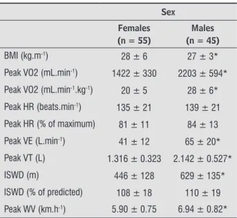
One hundred participants (55 females, 60 ± 10 years) composed the sample of the present study (Table 1).
Table 1 - General characteristics and peak values of the key physiological variables during the incremental shuttle walk test according to sex
Sex
Females (n = 55)
Males (n = 45)
Age (yr) 59 ± 11 59 ± 9
Body mass (kg) 73 ± 15 80 ± 12*
Height (m) 1.57 ± 0.07 1.71 ± 0.07*
Table 1 - General characteristics and peak values of the key physiological variables during the incremental shuttle walk test according to sex
Sex
Females (n = 55)
Males (n = 45)
BMI (kg.m-1) 28 ± 6 27 ± 3*
Peak VO2 (mL.min-1) 1422 ± 330 2203 ± 594*
Peak VO2 (mL.min-1.kg-1) 20 ± 5 28 ± 6*
Peak HR (beats.min-1) 135 ± 21 139 ± 21
Peak HR (% of maximum) 81 ± 11 84 ± 13
Peak VE (L.min-1) 41 ± 12 65 ± 20*
Peak VT (L) 1.316 ± 0.323 2.142 ± 0.527*
ISWD (m) 446 ± 128 629 ± 135*
ISWD (% of predicted) 108 ± 18 110 ± 19
Peak WV (km.h-1) 5.90 ± 0.75 6.94 ± 0.82*
Note: *p < 0.05 males vs. females; BMI = body mass index; VO2 = oxygen uptake; HR = heart rate; VE = minute ventilation; VT = tidal volume; ISWD = incremental shuttle walk distance; WV = walking velocity.
The dynamic relationships were not sex-dependent
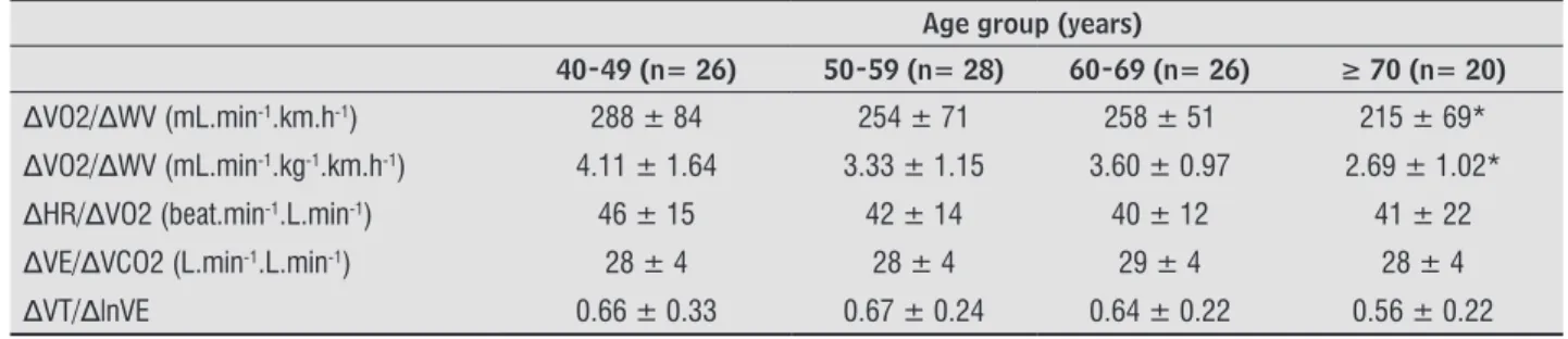
(Table 2). We found a significant decline in ΔVO2/ΔWV in participants aged ≥ 70 compared to those aged
40-49 (Table 3) with no interactions with sex.
Obese participants (n = 30) presented shallower slopes for ΔVO2/ΔWV and ΔVT/ΔlnVE as well as
lower values of peak VO2 (Table 4). The ΔVT/ΔlnVE relationship was negatively influenced by FBM and LBM presented positive influence on ΔVO2/ΔWV, ΔHR/ΔVO2, and ΔVT/ΔlnVE relationships (Figure 1).
Table 2 - Dynamic physiologic responses to the incremental shuttle walk test according to sex
Sex
Females (n = 55)
Males (n = 45) ΔVO2/ΔWV (mL.min-1.km.h-1) 258 ± 68 257 ± 68
ΔVO2/ΔWV (mL.min-1.kg-1.km.h-1) 3.69 ± 1.23 3.32 ± 1.11
ΔHR/ΔVO2 (beat.min-1.L.min-1) 40 ± 12 42 ± 16
ΔVE/ΔVCO2 (L.min-1.L.min-1) 29 ± 4 28 ± 4
ΔVT/ΔlnVE 0.63 ± 0.22 0.65 ± 0.27
Note: ΔVO2/ΔWV = mechanical efficiency; ΔHR/ΔVO2 =
cardio-vascular efficiency; ΔVE/ΔVCO2 = ventilatory efficiency; ΔVT/
ΔlnVE = linearized (ln) berthing pattern.
(To be continued)
826
Table 3 - Dynamic physiologic responses to the incremental shuttle walk test according to age
Age group (years)
40-49 (n= 26) 50-59 (n= 28) 60-69 (n= 26) ≥ 70 (n= 20)
ΔVO2/ΔWV (mL.min-1.km.h-1) 288 ± 84 254 ± 71 258 ± 51 215 ± 69*
ΔVO2/ΔWV (mL.min-1.kg-1.km.h-1) 4.11 ± 1.64 3.33 ± 1.15 3.60 ± 0.97 2.69 ± 1.02*
ΔHR/ΔVO2 (beat.min-1.L.min-1) 46 ± 15 42 ± 14 40 ± 12 41 ± 22
ΔVE/ΔVCO2 (L.min-1.L.min-1) 28 ± 4 28 ± 4 29 ± 4 28 ± 4
ΔVT/ΔlnVE 0.66 ± 0.33 0.67 ± 0.24 0.64 ± 0.22 0.56 ± 0.22
Note: *p < 0.05 vs. 40-49 years. ΔVO2 /ΔWV = mechanical efficiency; ΔHR/ΔVO2 = cardiovascular efficiency; ΔVE/ΔVCO2 = ventilatory efficiency; ΔVT/ΔlnVE = linearized (ln) berthing pattern.
Table 4 - Dynamic physiological responses and peak oxygen uptake to the incremental shuttle walk test in obese and non-obese participants
Body mass index (kg.m-2)
< 30 (n = 70) ≥ 30 up to 35 (n = 30)
ΔVO2 /ΔWV (mL.min-1.km.h-1) 263 ± 70 247 ± 64
ΔVO2/ΔWV (mL.min-1.kg-1.km.h-1) 3.84 ± 1.21 2.94 ± 0.90*
ΔHR/ΔVO2 (beat.min-1.L.min-1) 41 ± 15 44 ± 13
ΔVE/ΔVCO2 (L.min-1.L.min-1) 28 ± 4 30 ± 4
ΔVT/ΔlnVE 0.67 ± 0.26 0.57 ± 0.20*
Peak VO2 (mL.min-1.kg-1) 25 ± 6 19 ± 5*
Note: *p < 0.05 vs. non-obese participants;ΔVO2 /ΔWV = mechanical efficiency; ΔHR/ΔVO2 = cardiovascular efficiency; ΔVE/ΔVCO2 = ventilatory efficiency; ΔVT/ΔlnVE = linearized (ln) berthing pattern; VO2 = oxygen uptake.
100
80
60
40
20
30 40 50 60 70 80 90
0,0 0,2 0,4 0,6 0,8
FBM (%) LBM (kg)
LBM (kg) LBM (kg)
1,0 1,2 90
30 40 50 60 70 80 90
30 40 50 60 70 80 90
0 500
400
300
200
100
1,4
1,2
1,0
0,8
0,6
0,4
0,2
0,0 0
60
50
50
30
20
10
• r = -0.49
ΔHR/ΔVO2 = 81.5 (0.7 x LM); R² = 0.24; p <0.01
• r = -0.45
ΔVT/lnVE = 1.001 - (0.01 x FBM; R² = 0.20; p < 0.01 • r = -0.66
ΔVT/lnVE = -0.21 + (0,01 x LBM); R² = 0.43; p < 0.01
ΔV
T/lnVE
ΔV
T/lnVE
• r = -0.55
ΔVO2/ΔWV = 77,4 + (3,5 x LBM); R² = 0.30; p < 0.01
Δ
VO
2/ΔW
V = (mL.min
-¹.k
m.h
-1)
ΔHR/ΔV
O2
(b
ea
t.min
-¹.L.min -1)
Figure 1 - Significant correlations among fat (FBM) and lean (LBM) body masses, and mechanical efficiency (ΔVO2/ΔWV),
827 Peak VO2 at the end of the ISWT showed high
correlation with ΔVO2/ΔWV (r = 0.83 and R2 = 0.70; p < 0.001), weak correlation with ΔHR/ΔVO2 (r = -0.41 and R2 = 0.17; p < 0.001), and moderate correlation
with ΔVT/ΔlnVE (r = 0.62 and R2 = 0.40; p < 0.001).
After multiple regression analysis, only ΔVT/ΔlnVE
was determined for weight and height (R2 = 0.07).
Discussion
This study evaluated dynamic physiological re-sponses during walking at progressive velocity in healthy middle-aged and older men and women. Our results constitute a useful characterization of nor-mal responses for important physiological indices of exercise capacity in this age group considering the
lack of information in the literature regarding field
walking tests.
We observed the influence of age only on ΔVO2/ ΔWV. The decline in aerobic capacity seen in older age was associated with a decreased mechanical efficien -cy, especially in septuagenarians’. These changes may be due to a gradual reduction in muscle mass, muscle strength and aerobic capacitythat typically occurs in parallel with aging. We have also observed a high
positive correlation between peak VO2 at the end of
the ISWT and ΔVO2/ΔWV in the present study. Other
studies have reported weak correlations between VO2 and aerobic efficiency on the cycle ergometer (3).
There is some controversy in the literature on this issue. This dynamic relationship has been described as independent of age, sex and level of physical
ac-tivity. For example, Neder et al. (3) did not find any influence of age on ΔVO2/ΔWV in a randomized study
involving sedentary individuals in a wide age range (20 to 80 years). Our results stand in contrast to this,
however, on the other hand, reinforces the findings in the literature when CPET was performed on a tread -mill. Jones et al. (23) showed that the cost of walking was higher and usual walking velocity was lower in older women than in their younger counterparts. The higher muscle co-activation of agonist and antagonist muscles during the walking at various speeds may partially explain the elevated cost of walking (24). The higher energy costs in older individuals during exercise on a treadmill, in turn, results in reduced
cardiovascular efficiency of 8% according to the find -ings of Woo et al. (25). Moreover, aging has been as-sociated with a decrease in muscle capillarization and
mitochondrial enzyme activity. Reduced oxidative
capacity may lead to premature or excessive lactate accumulation and an increase in the oxygen cost of
ex-ercise (26). Additional explanations include changes in cardiac function, skeletal muscle blood flow from a
decrease in capillary content and density, nutritional status, and hormone levels in parallel with aging (27).
Our results reinforce the findings of the literature, although we were unable to find studies that evalu -ated the ΔVO2/ΔWV relationship during the ISWT.
In the study by Neder et al. (3) performed on a cycle ergometer, age-dependent changes in
cardio-vascular efficiency, ventilator efficiency and breathing
pattern were observed. This may be attributed to physiological differences between walking (ISWT)
and cycling. Although peak values of VO2, VE, and HR as well as HR and breathing reserves were described
as similar between ISWT and cycling in patients with chronic obstructive pulmonary disease and in healthy
subjects (8), the ISWT evoked a lower peak VCO2 and
blood lactate concentration than the incremental cy-cle ergometer test, suggesting a reduced contribution from nonaerobic metabolism to energy production. Besides that, the act of pedaling is not as familiar as walking, especially for older individuals. Thus, the
age-dependent changes in cardiovascular efficiency, ventilatory efficiency and breathing pattern observed in older individuals could be influenced by the exer -cise modality. Therefore, it is reasonable to assume that walking is more representative of daily life than cycling, and it could alter the outcome of the cycle ergometer test (28).
Also on the cycle ergometer test in a ramp proto-col, women presented less favorable values of ΔHR/
ΔVO2, ΔVE/ΔVCO2 and ΔVT/ΔlnVE (3). Our results
were different, considering that we did not observe any sex-related differences in the studied dynamic
relationships despite the lower values for peak VO2. Reinforcing our findings, Woo et al. (25) evaluated young (20 - 30 years) and older adults (65 to 79 years) in a ramp CPET performed on a treadmill and did not find any sex-related differences in exercise ef
-ficiency. Sex influenced the ΔVE/ΔVCO2 relationship in a ramp CPET performed on a cycle ergometer (3). However, in a multiple regression analysis, only age
remained an independent predictor for this rela-tionship, independent of sex (3). Davis et al. (29) evaluated twenty-eight healthy men and women that
828
difference between exercises for ventilatory efficien -cy but the opposite was observed in the women since different values were observed on the treadmill. The authors conclude that healthy women demonstrate an exercise type dependency for the ventilatory
ef-ficiency. The ventilatory response (VE) for the same
increase in metabolic rate is greater when the par-tial pressure of carbon dioxide in arterialized venous blood (PavCO2) is lower than normal (30). Thus, less
efficient ventilatory response in women, observed in incremental tests in cycle ergometer, can be justified
by their lower levels of PavCO2 at rest compared to
men (3). However, this exaggerated ventilatory re -sponse was not found in our study. The ISWT can be considered a submaximal test in many cases, as it is not allowed to run. For this reason, the so-called “ceil-ing effect” may occur, result“ceil-ing in decreased
produc-tion of lactate, thereby flattening the linear behavior
of ΔVE/ΔVCO2. But the test in cycle ergometer are symptom-limited, therefore, have no “ceiling effect”. Finally, we believe that this difference between the tests is the cause of the discrepancy of the results
regarding the ventilatory efficiency in women.
In the present study, obese participants present-ed worse values of ΔVO2/ΔWV and ΔVT/ΔlnVE. Our results are consistent with previous results widely reported in the literature (31). Obesity increases the oxygen demand for any given task. Beginning with the basal metabolism, which is increased in comparison with eutrophic subjects (32). The increased cardiovas-cular demand is not accompanied by increased heart, blood vessels, lungs and muscles. Consequently, there are higher cardiovascular and ventilatory demands in obese individuals (2). The ΔVO2/ΔW relationship has been described as displaced upward with no changes in the slope of the regression line (33). Our results were different. There wasn’t upward displacement of the ΔVO2/ΔW relationship, as no significant differ -ences in the y-intercept (data not shown) were
ob-served, however, there was a significant reduction in
the slope of the linear regression compared to normal weight and overweight participants. This difference may be attributed to the absence of weight bearing on the cycle ergometer. Certainly, during the walking at progressive velocity there is greater demand for anaerobic metabolism in obese participants. Since
the glycolytic fibers are less efficient in metabolizing
oxygen, there was a shallow ΔVO2/ΔWV relationship.
Additionally, the obese participants were less fit than the others, which contribute to the flattening of the
relationship behavior. The tachypneic breathing pat-tern observed for the obese participants reinforces this hypothesis. Instead, the LBM presented positively
influence on three of the four studied dynamic rela -tionships. These results are likely to be related to the well-known stroke volume–body mass relationship and to the underlying relationship between lean body mass and regular physical activity (34).
The present study has limitations that should be discussed. The dynamic relationships are more
clearly defined in ramp CPETs. It would be not op
-erationally easy to simulate a ramp protocol in a field test. However, the ISWT is a rapidly incremental test
and its load increments in stages up to a minute may generate physiological responses that allow the as-sessment of dynamic relationships (2). The original ISWT protocol was developed for patients with respi-ratory diseases and does not allow individualization. This may have generated very small load increments
for some participants. However, this should not have
happened oftentimes, as we excluded those involved in regular physical activity (14). In fact, an objective assessment of the level of physical activity in daily life would be very informative. Although sedentary and physically inactive individuals had been included in our study, we do not believe that the very low volume
of physical activity has significantly influenced the
submaximal physiological responses in our study. Thirty percent of our sample was composed of obese participants; however, they did not present cardio-vascular, pulmonary, metabolic or musculoskeletal co-morbidities.
The results of this study contribute to the phys-iotherapist clinical practice, they show that through ISWT is possible to perform the analysis of dynamic physiological dynamics in response to the test and this knowledge may be useful and allows better in-terpretation of the walking ability of patients with or without chronic diseases affecting exercise capacity in middle-aged and older adults.
Conclusion
We conclude that the decline in aerobic capacity seen with older age was associated with a decreased
mechanical efficiency, especially in septuagenarians’.
829
relationships. Body composition plays important role in the dynamic physiology of walking. In addition, our results provide values of dynamical physiologi-cal responses during walking in healthy adults, en-abling better interpretation of the walking ability of middle-aged and older adults with or without chronic diseases that affect exercise capacity.
References
1. ERS Task Force, Palange P, Ward SA, Carlsen KH, Casa
-buri R, Gallagher CG, et al. Recommendations on the use of exercise testing in clinical practice. Eur Respir
J. 2007;29(1):185-209.
2. Wasserman K, Hansen J, Sue DY, Whipp BJ, Casaburi
R. Principles of exercise testing and interpretation.
4th ed. Philadelphia: Lippincott Wiliams & Wilkins;
2005. 576 p.
3. Neder JA, Nery LE, Peres C, Whipp BJ. Reference values for dynamic responses to incremental cycle ergometry
in males and females aged 20 to 80. Am J Respir Crit Care Med. 2001;164(8 Pt 1):1481-6.
4. Sperandio EF, Alexandre AS, Yi LC, Poletto PR, Gotfryd
AO, Vidotto MC, et al. Functional aerobic exercise ca -pacity limitation in adolescent idiopathic scoliosis.
Spine J. 2014;14(10):2366-72.
5. Singh SJ, Morgan MD, Hardman AE, Rowe C, Bardsley PA. Comparison of oxygen uptake during a convention-al treadmill test and the shuttle wconvention-alking test in chronic
airflow limitation. Eur Respir J. 1994;7(11):2016-20. 6. Sperandio EF, Arantes RL, Matheus AC, Silva RP, Lau
-ria VT, Romiti M, et al. Intensity and physiological re
-sponses to the 6-minute walk test in middle-aged and
older adults: a comparison with cardiopulmonary
ex-ercise testing. Braz J Med Biol Res. 2015;48(4):349-53.
7. Sperandio EF, Arantes RL, Silva RPd, Matheus AC, Lau
-ria VT, Bianchim MS, et al. Screening for physical in -activity among adults: the value of distance walked in the six-minute walk test. A cross-sectional diagnostic
study. Sao Paulo Med J. 2016;134(1):56-62.
8. Dourado VZ, Guerra RL, Tanni SE, Antunes LCO, Godoy I. Physiological responses to the incremental shuttle
walk test in healthy adults. Eur Respir J. 2011;38(Sup -pl 55):2180.
9. Sociedade Brasileira de Pneumologia e Tisiologia. Di-retrizes para testes de função pulmonar. J Pneumol. 2002;28(Supl. 3):1-82.
10. Thomas S, Reading J, Shephard RJ. Revision of the
Physical Activity Readiness Questionnaire (PAR-Q).
Can J Sport Sci. 1992;17(4):338-45.
11. Ferris BG. Epidemiology Standardization Project
(American Thoracic Society). Am Rev Respir Dis. 1978;118(6 Pt 2):1-120.
12. Thompson PD, Arena R, Riebe D, Pescatello LS. ACSM’s new preparticipation health screening recommen-dations from ACSM’s guidelines for exercise testing
and prescription, ninth edition. Curr Sports Med Rep.
2013;12(4):215-7.
13. Barros MVG, Nahas MV. Reprodutibilidade (teste-reteste) do questionário internacional de atividade
física (QIAF-Versão 6): um estudo piloto com adultos no Brasil. Rev Bras Ciênc e Mov. 2000;8(1):23-6.
14. American College of Sports Medicine Position Stand. The recommended quantity and quality of exercise for developing and maintaining cardiorespiratory and
muscular fitness, and flexibility in healthy adults. Med Sci Sports Exerc. 1998;30(6):975-91.
15. Kyle UG, Bosaeus I, Lorenzo AD, Deurenberg P, Elia
M, Manuel Gomez J, et al. Bioelectrical impedance
analysis-part II: utilization in clinical practice. Clin
Nutr. 2004;23(6):1430-53.
16. Kyle UG, Genton L, Karsegard L, Slosman DO, Pichard
C. Single prediction equation for bioelectrical imped-ance analysis in adults aged 20--94 years. Nutrition. 2001;17(3):248-53.
17. Miller MR, Hankinson J, Brusasco V, Burgos F, Casaburi
R, Coates A, et al. Standardisation of spirometry. Eur Respir J. 2005;26(2):319-38.
18. Pereira CAC, Sato T, Rodrigues SC. Novos va
-lores de referência para espirometria forçada em
brasileiros adultos de raça branca. J Bras pneumol.
2007;33(4):397-406.
19. Singh SJ, Puhan MA, Andrianopoulos V, Hernandes NA,
Mitchell KE, Hill CJ, et al. An official systematic review of the European Respiratory Society/American Tho
-racic Society: measurement properties of field walk
830
20. Singh SJ, Morgan MD, Scott S, Walters D, Hardman AE. Development of a shuttle walking test of disability in patients with chronic airways obstruction. Thorax. 1992;47(12):1019-24.
21. Lanza FC, Zagatto EP, Silva JC, Selman JPR, Imperatori
TBG, Zanatta DJM, et al. Reference Equation for the
Incremental Shuttle Walk Test in Children and
Ado-lescents. J Pediatr. 2015;167(5):1057-61.
22. Mukaka MM. Statistics corner: A guide to
appropri-ate use of Correlation coefficient in medical research. Malawi Med J. 2012;24(3):69-71.
23. Jones LM, Waters DL, Legge M. Walking speed at self-selected exercise pace is lower but energy cost higher
in older versus younger women. J Phys Act Health. 2009;6(3):327-32.
24. Peterson DS, Martin PE. Effects of age and walking speed on coactivation and cost of walking in healthy
adults. Gait Posture. 2010;31(3):355-9.
25. Woo JS, Derleth C, Stratton JR, Levy WC. The influence
of age, gender, and training on exercise efficiency. J Am Coll Cardiol. 2006;47(5):1049-57.
26. Russ DW, Lanza IR. The Impact of Old Age on Skeletal Muscle Energetics: Supply and Demand. Curr Aging
Sci. 2011;4(3):234-47.
27. Lakatta EG. So! What’s aging? Is cardiovascular aging
a disease? J Mol Cell Cardiol. 2015;83:1-13.
28. Fotheringham I, Meakin G, Punekar YS, Riley JH, Cock
-le SM, Singh SJ. Comparison of laboratory- and
field-based exercise tests for COPD: a systematic review.
Int J Chron Obstruct Pulmon Dis. 2015;10:625-43.
29. Davis JA, Tyminski TA, Soriano AC, Dorado S, Costello
KB, Sorrentino KM, et al. Exercise test mode depen
-dency for ventilatory efficiency in women but not men. Clin Physiol Funct Imaging.. 2006;26(2):72-8.
30. Oren A, Wasserman K, Davis JA, Whipp BJ. Effect of CO2 set point on ventilatory response to
exer-cise. J Appl Physiol Respir Environ Exerc Physiol.
1981;51(1):185-9.
31. Arena R, Cahalin LP. Evaluation of cardiorespiratory
fitness and respiratory muscle function in the obese population. Prog Cardiovasc Dis. 2014;56(4):457-64.
32. Wilms B, Ernst B, Thurnheer M, Weisser B, Schultes B. Correction factors for the calculation of metabolic
equivalents (MET) in overweight to extremely obese
subjects. Int J Obes (Lond). 2014;38(11):1383-7.
33. Lafortuna CL, Agosti F, Busti C, Galli R, Sartorio A. The energy cost of cycling and aerobic
perfor-mance of obese adolescent girls. J Endocrinol Invest. 2009;32(8):647-52.
34. Dourado VZ, Guerra RL, Tanni SE, Antunes LC, Godoy I.
Reference values for the incremental shuttle walk test
in healthy subjects: from the walk distance to physio-logical responses. J Bras Pneumol. 2013;39(2):190-7.
Received in 03/22/2016
Recebido em 22/03/2016
Approved in 04/04/2017