The prevalence of swine enteropathogens in Brazilian grower and finish herds
A.M. Viott
1, A.P. Lage
2, E.C.C. Cruz Junior
3, R.M.C. Guedes
31
Laboratório de Patologia Veterinária, Universidade Federal do Paraná, Palotina, PA, Brazil. 2
Departamento de Medicina Veterinária Preventiva, Universidade Federal de Minas Gerais, Belo Horizonte, MG, Brazil.
3
Departamento de Clínica e Cirurgia, Universidade Federal de Minas Gerais, Belo Horizonte, MG, Brazil.
Submitted: September 10, 2011; Approved: July 02, 2012.
Abstract
Diarrhoea among growing and finishing pigs is an important problem in many herds. The prevalence of L. intracellularis, B. pilosicoli, B. hyodysenteriae, Salmonella spp., enterotoxigenic E. coli, Trichuris suisand the occurrence of mixed infection were investigated. Fecal samples for forty-six herds with diarrhea or a history of diarrhea were randomly collected in Minas Gerais state, Brazil. The enteric pathogens were detected by culture (E. coliandSalmonellasp.), PCR (L. intracellularis andBrachyspiraspp.) and eggs counts (T. suis). The overall herd prevalence ofL. intracellularis, Salmonella enterica serotype Typhimurium and enterotoxigenic E. coli were 19.56%, 6.52%, 10.86% respectively. Mixed infection was diagnosed in 30.43% of herds, andL. intracellularisand Salmonella entericaserotype Typhimurium are main pathogens association (10.87%).B. pilosicoli was diagnosed only in two herds, always associated with mixed infections.B. hyodysenteriaeandT. suiswere not demonstrated in any sample. These pathogens have been reported world-wide but stud-ies regarding epidemiology in Brazil are few. This study contributes to establish of prevention pro-grams for the control enteropathogens in grower finish herds in Brazil.
Key words:swine, enteropathogens, grower finish herds, prevalence, Brazil.
Introduction
According to research, Brazil is the fourth largest pro-ducer of pigs, behind China, the European Union and United States. The state of Minas Gerais is the fourth larg-est producer of pork in Brazil, and the growth of pig pro-duction in the state has been increasing year by year (Abipesc 2009). Because of this it is necessary to accurately detect and identify porcine pathogens in order to devise proper treatments and prevention programs (Baccaroet al., 2003).
The enteric infections are among the most frequent diseases in pig production, being responsible for significant losses and significant economic impact on industry. The damages are represented primarily by reducing weight gain, mortality and expenses with antibiotics (McOrist 2005). Further, some of these diseases can be transferred to humans (Weneger and Bager, 1997). Several agents have been suggested as possible causes of diarrhea in
grow-ing/finishing pigs. Infection byL. intracellularis causing porcine proliferative enteritis, swine salmonellosis caused bySalmonella entericaserotype Typhimurium, porcine in-testinal espirochetosis caused byB. pilosicoli,swine dysen-tery caused by B. hyodysenteriae, and Trichuris suis causing intestinal trichuriasis are among the most prevalent at this age (Batteet al., 1977; Thomsonet al., 1998; Stegeet al., 2000; Baccaroet al., 2003). Enterotoxigenic strains of Escherichia colimay also be present among growing pigs weighing 30-50 kg with or without symptoms (Stegeet al., 2000).
The risk of diarrhea increases when the herd is in-fected by two or more pathogens simultaneously (Stegeet al., 2000; Suh and Song, 2005). Usually the pigs with mixed infection have lesions more pronounced with lower development of the herd. Suh and Song (2005) analyzing 462 swine fecal samples observed the occurrence of mixed infections, represented by 3% of the samples
simulta-Send correspondence to: R.M.C. Guedes. Departamento de Clínica e Cirurgia, Universidade Federal de Minas Gerais, Belo Horizonte, MG, Brazil E-mail: guedes@vet.ufmg.br.
neously infected with Salmonella sp. and B. hyodysenteriae, 2.2% with L. intracellularis and Salmo-nella entericaspp. and 1.3% withL. intracellularisandB. hyodysenteriae.
These pathogens have been reported in world-wide but (Thomsonet al., 1998; Stegeet al., 2000 Jacobsonet al., 2005; Suh and Song, 2005; Laet al., 2006) the impor-tance and involvement of some of these organisms in clini-cal disease still remains to be clarified in the Brazilian swine herds. The purpose of this study was to determine the prevalence of the enteric pathogensL. intracellularis,B. pilosicoli,B. hyodysenteriae,Salmonella enterica, entero-toxigenicE. coli, T. suisand the occurrence of mixed infec-tions in Brazilian growing/finishing pigs from swine farms with diarrhea or a history of diarrhea in the state of Minas Gerais, Brazil.
Materials and Methods
Herds and animals
Farrow-to-finish herds with diarrhea or a history of diarrhea were randomly selected from four different geo-graphical regions of the state of Minas Gerais, Brazil [South and southwest (SSW), metropolitan area of Belo Horizonte (MBH), Zona da Mata (ZM) and Triângulo Mineiro e Alto Paranaiba (TMHP)]. The sample size was calculated at two levels, property and animals. The feces were collected between January 2008 and February 2009. To determine the number of herds, simple sampling was used with an estimated prevalence of 50% confidence level of 95% and an estimated error of 15% (Noordhuizenet al., 2001).
In each farm, between 10 and 15 fecal samples were randomly collected from 60 to 14 day-old pigs. Feces were collected directly from the rectum and shipped under re-frigeration to the Laboratory of Veterinary Pathology in the Federal University of Minas Gerais.
Microbiology investigation
For the isolation of Salmonella sp. 1 g of feces was mixed in 4 mL sterile phosphate buffer saline (PBS). For pre enrichment 1 mL of this solution was added to 9 mL of peptone water (DIFCO) (37 °C/18 h). After incubation ali-quots of 0.1 mL were seeded in selective broth Rappaport-Vassiliadis (DIFCO) (42 °C/24 h). Subsequently aliquots of the selective broth were plated on XLT-4 agar (DIFCO) and brilliant green agar (DIFCO). The suspected colonies were identified (Bergey and Holt, 1994). These colonies were grown in Brain Heart Infusion agar (BHI agar) (DIFCO) and submitted to the following biochemical tests: urea broth (DIFCO), lysine broth (DIFCO) and triple sugar iron agar (TSI) (DIFCO). Serology using anti “O” poly-valent antiserum (PROBAC®) was used to confirm the iso-lation ofSalmonella entericasp. Colonies confirmed were
selected for serotyping at the Institute Oswaldo Cruz, Rio de Janeiro, Brazil.
Aliquots of the feces mixed in PBS were plated in MacConkey agar (37 °C/24 h) for the isolation of lactose positive colonies (Lac+ colonies). These colonies were frozen in glycerol at -80 °C for later PCR multiplex for enterotoxigenicE. coli.
Detection ofL. intracellularis,B. hyodysenteriaeand
B. pilosicoli
The methods use for the diagnostic of L. intracellularis, B. hyodysenteriaeandB. pilosicoliand the sensitivity of detection are a multiplex –PCR for rapid de-tection (Laet al., 2006). The positive controls were kindly provided by department of Veterinary and Biomedical Sci-ences at University of Minnesota, EUA.
Extraction of DNA and multiplex PCR primers
DNA was extracted from all faeces using the QIAamp DNA Stool Mini Kit (QIAGEN) according to the manufac-turer’s instructions. The respective primer sequences were: H1 (5’-ACTAAAGATCCTGATGTATTTG-3’) and H2 (5’-CTAATAAACGTCTGCTGC-3’) targeting a 354 bp region on the nicotinamide adenine dionucleotide hybride (NADH) oxidase (nox) gene of B. hyodysenteriae; P1
(5’-AGAGGAAAGTTTTTTCGCTTC-3’) and P2
(5’-GCACCTATGTTAAACGTCCTTG-3’) targeting a 823 bp region of the 16S rRNA gene ofB. pilosicoli; and Lint-146F (5’-GATAATCTACCTTCGAGACGG-3’) and Lint -745R (5’-TGACCTCAGTGTCAGTTATCGT-3’) targeting a 655 bp region of 16S rRNA gene of L. intracellularis.
Multiplex PCR
The DNA was amplified in 25mL total volume. Am-plifications mixtures consisted of 1 X PCR buffer (contain-ing 1, 5 mmol L-1of Mg Cl2), 1.25 U of Taq DNA polymer-ase (Cenbiot, Porto Alegre, Brazil), 0.1 mmol L-1of each dNTP (Invitrogen, Carlsbad, CA), 0.2 mmol L-1 of each primer pair (H1 and H2, P1 and P2, 146F and Lint-745R) (Prodimol, Wisconsin, USA) and 2.5 mL chromo-somal template DNA. Cycling conditions involved an initial 5 min Taq DNA polymerase activation step at 95 °C, fol-lowed by 35 cycles of denaturation at 95 °C for 30 s, anneal-ing at 58 °C for 90 s, and a primer extension step at 68 °C for 2 min. A final 10 min extension step at 68 °C. The PCR prod-ucts were subjected to electrophoresis in 1% (w/v) agarose gels in 1 x TAE buffer, stained with ethidium bromide and viewed over UV light. The sensitivity of detection of the multiplex PCR was estimated (Laet al., 2003).
Multiplex PCR for detection ofEscherichia coli
enterotoxigenic virulent genes
controls for the factors of virulence, four reference samples ofE. coliwere kindly provided by the Veterinary Diagnos-tic Laboratory at the University of Minnesota.
Extraction of DNA
After thawing the Lac + colonies were plated on MacConkey agar. Isolated colonies were resuspended in microtube containing 500 mL sterile PBS. The bacteria were centrifuged at 7000 rpm for 10 min for sedimentation. Sediment was added 270 mL lysis solution (Tris HCl 1.0 M, pH 8.0; EDTA 0,5 M; NaCl 4.5 M; SDS 10%) and 200mL of High TE (Tris HCl 1.0 M, pH 8.0; EDTA 0.5 M). After mixing and vortex for 20 to 30 seconds, the mixture was incubated at 55 °C for at last 3 h. The lysate containing the bacterial DNA was subjected to DNA extracting by phenol-chloroform (Sambrooket al., 1989).
Multiplex PCR
The PCR technique used four pairs of primers for the toxins Stb, StaP, LT and Stx2e and five pairs for the fimbriae K99, F18, 987P, K88 and F41. Briefly the DNA was amplified in 10mL total volume containing 10 ng of bacterial DNA. Amplifications mixtures consisted of 0.2 mmol L-1of each dNTP (Prodimol, Wisconsin, USA), 1.5 mmol L-1 of MgCl2 (Cenbiot, Porto Alegre, Brazil), 0.5 mmol L-1of each primer (Invitrogen, Carlsbad, CA) and 5 U of TAQ polymerase (Cenbiot, Porto Alegre, Brazil). Cycling conditions involved an initial 1 min Taq DNA polymerase activation step at 94 °C, followed by 25 cycles of denaturation at 94 °C for 1 min, at 55 °C for 1 min, and a primer extension step at 70 °C for 2 min. A final 10 min ex-tension step at 72 °C. The PCR products were subjected to electrophoresis in 6% (w/v) polyacrylamide gels in 1 x TBE buffer, stained with silver.
Parasitological exam
For detection ofT. suis eggs in swine feces, fecal samples were individually examined by flotation technique (Willis-Mollay) with a hyper saturate solution of NaCl (Hoffmann 1987). The infection intensity was scored ac-cording the number of eggs per gram of feces: 0, none; 1-5 mild; 5-10, moderate; and more than 10 severe.
Statistics
For statistical analyses Fischer’s exact test (SAS In-stitute, Cary, North Caroline, USA 1999) was used to deter-mine the association between herd size and pathogen prevalence.
Results
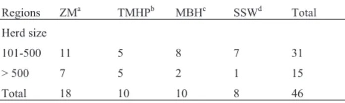
The study comprised 46 farrow-to-finish pig herds. Forty six farms were analyzed, 31 with 101-500 sows and 15 with > 500 sows. The number of herds collected from each region and is in Table 1. A total of 512 faecal samples were examined. The analysis by the Fisher exact test
showed no significant association between the pathogen and the size of the farm.
Salmonellaspp.
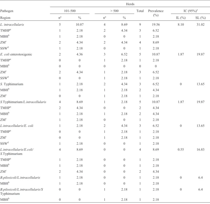
The only subspecies ofSalmonellaspp. found, poten-tially pathogenic for pigs, wasSalmonella enterica subspe-cies Typhimurium. The overall herd prevalence of Salmonella entericasubspecies Typhimurium was 6.52%. Salmonella enterica subspecies Typhimurium was diag-nosed in 3 herds and was detected in 31 (6.05%) samples. One herd with 101-500 sows and two herds with more than 500 sows were positive forSalmonella entericasubspecies Typhimurium (Table 2).
Others subspecies ofSalmonella enterica, not patho-genic for pigs were found in seven herds (15.2%). Salmo-nella entericasubspecies Agona was observed in one herd (2.17%) and two samples (0.4%) in a MBH herd with 101-500 sows. TheSalmonella enterica subspecies Pan-ama was diagnosed in three herds (6.52%) and 13 samples (2.54%). All herds were localized in ZM, two with 101-500 sows and one with > 500 sows. TheSalmonella enterica subspecies Schwarzenground was observed in one herd (2.17%) and two samples (0.4%) in a MBH herd with > 500 sows.Salmonella entericauntypable was observed in two herds (4.34%) and two samples (0.4%). The herds were lo-cated in MBH one with 101-500 sows and one with > 500 sows.
L. intracellularis, B. pilosicoliandB. hyodysenteriae
The limit of detection obtained in PCR was 103 cells per gram of faeces for B. pilosicoli and 104 for L. intracellularis and B. hyodysenteriae. The 104 cells per gram of feces were obtained when the three species were applied together in equal number in the fecal samples.
B. hyodysenteriaewas not isolated from any of the samples from the 46 piglet-producing herds. The overall herd prevalence ofB. pilosicoliwas 4.36%. This pathogen was demonstrated in two herds and in nine (1.75%) sam-ples, always in mixed infections (Table 2). L. intracellulariswas diagnosed in 9 herds (19.56%) and was detected in 59 samples (11.52%). This bacteria was more prevalent in ZM (8.69%) and TMHP (6.52%) regions (Ta-ble 2).
Table 1- Number of properties samples and herd size in each region of Minas Gerais.
Regions ZMa TMHPb MBHc SSWd Total
Herd size
101-500 11 5 8 7 31
> 500 7 5 2 1 15
Total 18 10 10 8 46
aZona da Mata” (ZM),bTriângulo Mineiro e Alto Paranaíba (TMHP), cMetropolitan area of Belo Horizonte (MBH), dSouth and southwest
EnterotoxigenicE. coli
The overall prevalence of enterotoxigenicE. coliwas 10.87%. The number of positive herds and samples was 5 and 26 (5.49%) respectively. The 512 stool collected only 473 samples showed growth of positive lactose colonies in MacConkey’s agar. The enterotoxigenic E. coli bacteria was more prevalent in ZM (6.52%) (Table 2).
The enterotoxigenic genotype more observed was the profile StaP-Stb-F18 with 8/26 (30.76%) positive samples, followed by genotype StaP-Stb-987P with 6/26 (23.07%) positive samples. Other genetic profiles found were:
LT-K88, StaP-Stb-K99, Stb-987p-K99, StaP-987p all with two samples each and K99-LT, STx2E-987p, LT-K88-STx2E and 987p-LT all with one positive sample detected.
Trichuris suis
T. suiseggs were not diagnosed in any of the samples from the 46 piglet-producing herds.
Mixed Infection
The overall herd prevalence of mixed infection was high 30.43%. Mixed infections were diagnosed in 14 herds
Table 2- Distribution of prevalence according to herd size, region and pathogen;Lawsonia intracellularis, enterotoxigenicE. coli,Salmonella
Typhimurium and mixed infection.
Herds
Pathogen 101-500 > 500 Total Prevalence
(%)
IC (95%)e
Region nº % nº % IL (%) SL (%)
L. intracellularis 5 10.87 4 8.69 9 19.56 8.10 31.02
TMHPa 1 2.18 2 4.34 3 6.52
MBHb 1 2.18 0 0 1 2.18
ZMc 2 4.34 2 4.34 4 8.69
SSWd 1 2.18 0 0 1 2.18
E. colienterotoxigenic 2 4.36 3 6.52 5 10.87 1.87 19.87
TMHPa 0 0 1 2.18 1 2.18
MBHb 0 0 0 0 0 0
ZMc 2 4.34 1 2.18 3 6.52
SSWd 0 0 1 2.18 1 2.18
S.Typhimurium 1 2.18 2 4.34 3 6.52 0 13.65
MBHb 1 2.18 1 2.18 2 4.34
ZMc 0 0 1 2.18 1 2.18
S.Typhimurium/L.intracellularis 4 8.69 1 2.18 5 10.87 1.87 19.87
TMHPa 2 4.34 0 0 2 4.34
MBHb 1 2.18 1 2.18 2 4.34
ZMc 1 2.18 0 0 1 2.18
L.intracellularis/E. coli 1 2.18 2 4.34 3 6.52 0 13.65
TMHPa 0 0 1 2.18 1 2.18
ZMc 0 0 1 2.18 1 2.18
SSWd 1 2.18 0 0 1 2.18
L.intracellularis/E.coli/ S.Typhimurium
4 8.69 0 0 4 8.69 0.55 16.83
TMHPa 1 2.18 0 0 1 2.18
MBHb 1 2.18 0 0 1 2.18
ZMc 2 4.34 0 0 2 4.34
B.pilosicoli/L.intracellularis 1 2.18 0 0 1 2.18 0 6.4
MBHb 1 2.18 0 0 1 2.18
B.pilosicoli/L.intracellularis/S
Typhimurium
0 0 1 2.18 1 2.18 0 6.4
MBHb 0 0 1 2.18 1 2.18
and 67 samples (13.08%). Nine herds were positive for two agents (19.56%), and five herds were positive for three agents (10.87%). There was a higher frequency of mix in-fection in herds with 101-500 sows, 10 in a total, compared with herds with more than 500 sows four (Table 2). The MBH region had the highest occurrence of mix infection, five herds. The most common mixed infection in herds was Salmonella enterica subspecies Typhimurium associated withL. intracellularispresent in 5 herds (10.87%) and 28 fecal samples (5.47%), following byL. intracellularis asso-ciated with enterotoxigenicE. coliandSalmonella enterica subspecies Typhimurium present in 4 properties (8.69%) and 20 samples (3.9%). The concomitant detection of enterotoxigenic E. coli and L. intracellularis was diag-nosed in 3 herds (6.52%) and 12 samples (2.34%). B. pilosicoli was detected concomitantly with L. intracellularisin 1 herd (2.18%) corresponding a four sam-ples (0.78%), and with bothL. intracellularisand Salmo-nella entericasubspecies Typhimurium,one herd (2.18%) and three samples (0.58%) (Table 2).
Discussion
Generally, studies on enteric disease have focused on a specific pathogen (Fellstromet al., 1996), decreasing the chances of a correct diagnosis of the sanitary problems that affect the herd. The prevalence of six enteropathogens with pathogenic potential to grower/finisher pigs was studied and this prevalence was associated with de herd size. It was been reported that outbreaks of L. intracellularis and Brachyspiraspp. occur more often in the large production units (Holyoakeet al., 1996), indicating an association be-tween herd size and disease outbreak. However, the results in this study did not show this.
The prevalence of hers infected with L. intracellulariswas 19.56%. This result is in agree with pre-viously reported in other countries, where the prevalence of L. intracellularisvaries between 15% and 93.7% (Thomp-sonet al., 1998; Stegeet al., 2000; Jensenet al., 2006; Biksi et al., 2007) and in Brazil. In Brazil works reported a preva-lence of 15% of positive faecal samples (146/971), and 30% of 203 herds from seven different regions (Morenoet al., 2002; Baccaroet al., 2003). The differences among the prevalence numbers observed in this and in others works may be due to regional variations, different swine breeding systems, use of antibiotics and sampled population (sick or healthy animals) and detection assay used.
Using detection techniques, the prevalence of B. pilosicoliandB. hyodysenteriaein different geographic re-gions has been estimated. In a study in DanishB. pilosicoli was isolated from 19% from 79 herds and B. hyodysenteriaein 2.5% (Stegeet al., 2000). In a survey conducted in U. K. between 1992 and 1996 that involved 85 pig farmsB. pilosicoli was identified on 21 (25%) units, andB. hyodysenteriaeon 6 (7%) units (Thomson et al., 1998). A recent study from Sweden revealed that the
pres-ence ofB. pilosicoliin 34 herds of 105 studied and the ab-sence ofB. hyodysenteriae, the presence oB. pilosicoliwas significantly associated with herds having poor perfor-mance in growing pigs and with mix infection (Thomsonet al., 1998; Jacobsonet al., 2005). In our survey the presence ofB. pilosicoliwas associated with mix infection and only in two herds. None positive samples forB. hyodysenteriae were detected. This lower prevalence may be explained by the use of additives in the feed, in many countries, includ-ing Brazil, the prevalence ofBrachyspirasp. might be con-cealed by the use of antimicrobial feed additives (Hampson 2000). In a study conducted in Brazil 38 farms were ran-domly selected and bacteriologically examined (22 from farms using medicated feed and 16 with non-medicated feed),B. hyodysenteriaeandB. pilosicoliwere isolated re-spectively from 0% and 6.25% in medicated herds and from 31.8% and 45.5% from non-medicated farms (Barcelloset al., 2003)
In addition, variations related to PCR techniques could be observed. There is showed that variations like the use of pooled samples and the DNA extraction protocol may interfere negatively with the sensitivity of the test (Chiribogaet al., 1999) resulting in false negative results. The practical limit of detection of the multiplex PCR when the three species were applied together in the faecal sam-ples was 104cells per gram of feces. These levels of detec-tion are consistent with than those previously reported (Joneset al., 1993; MFlleret al., 1998; Laet al., 2003). PCR inhibition is also a problem in the diagnosis of faecal samples prepared directly for PCR. Hence, inhibition might be a problem in the detection of clinical and subclinical car-riers (Jacobsonet al., 2004). It is known that bacterial isola-tion forBrachyspira sp. is more sensitive than the PCR technique (Komareket al., 2009), and this fact may have contributed to the low numbers observed.
The overall prevalence forSalmonella enterica sub-species Typhimurium (6.52%) and others subsub-species of Salmonella enterica (15.2%) was high when compared with results ofSalmonella entericain others countries like Danish (10.1%) and U.K (13%) (Thomson et al., 1998; Stege et al., 2000). This variation in prevalence can be caused by differences in pig housing (asymptomatic carri-ers) or feeding regimen (meat meal) which might affect the degree of transmission (Hampson 2000).Salmonella spp. were diagnosed as the primary agent in both ZM and MBH regions, it is believed that the highest density of pigs in such areas associated with older facilities have contributed to this finding.Salmonella entericasubspecies Typhimurium can cause diarrhea in pigs, but even more important is that Salmonellaspp. is a zoonotic agent (Weneger and Bager, 1997; Stegeet al., 2000).Salmonellaspp. is a key player in the contamination of pig meat and its derivatives (Weisset al., 1999).
2003). A survey observed 76.8% of 72 herds infected by hemolyticE. coliand other work demonstrated 24.1% of the herds with pathogenic E. coli (MFller et al., 1998; Stegeet al., 2000). Despite all these results the significance of the presence of E. coliin grower/finish herds has not been well clarified. EnterotoxigenicE. colihas been associ-ated with damage to small intestinal villi, loss of villi epi-thelium and diarrhea in weaned pigs (Macedoet al., 2007). However, this enteric pathogen may alter the ecological balance of the gut flora or change the environment condi-tions in the gut, which would favors some bacteria (Jacob-son et al., 2003). Normally the affected herds had case history of previous problems with post-weaning diarrhea, this may indicate that these herds suffer from a high patho-gen load, or that the post-weaning diarrhea might predis-pose to outbreaks of others enteric diseases (MFlleret al., 1998; Thompsonet al., 1998; Stegeet al., 2000; Jacobson et al., 2005). An uncharacteristic presentation of F18-posi-tiveE colienteritis in 11-weekold pigs, and according to this author, strains of E coli positive for the F18 pilus, enterotoxins, and Stx2e endotoxin should be added to the list of differential diseases that cause severe diarrhea and vomiting in grower-finisher pigs (Pittman 2010). Of the 512 samples tested 39 no showed growth for Lac + colo-nies, it is believed that the overuse of antibiotics in the growing and finishing phase may have inhibited the growth of these bacteria on MacConkey agar.
Interactions between pathogens may occur and it is often observed in herds that have or had history of severe diarrhea with dead loss of pigs, usually in this cases there is an increase of lesions in the intestine (Jacobsonet al., 2005; Suh and Song, 2005). Mixed infection was present in 30.43% of herds, and the main combination observed was Salmonella Typhimurium associated with L. intracellularis. This association was already reported (Suh and Song, 2005) 2.2% of the 462 samples positive for Sal-monellaspp andL. intracellularis. Other associations have already been reported in Sweden 58% of 320 fecal samples were concomitant infected by B. pilosicoli and L. intracellularis(Jacobsonet al., 2005).
T. suis once relatively common in swine industry (Batteet al., 1977), was not detected in any of the samples. The control in recent years, associated with technological improvements in the swine industry, have resulted in the re-duced incidence of helminth parasites.
In conclusion,L. intracellularis is the main swine enteropathogen observed in herds in the state of Minas Gerais following by enterotoxigenicE. coliandSalmonella entericasubspecies Typhimurium. There was a high occur-rence of mixed infections in herds with a close relationship ofL. intracellulariscombined with other agents. The oc-currence of a large number of herds positive forE. coli, sug-gest that this bacteria may be the cause of diarrhea in growing and finishing pigs.
Acknowledgments
The authors thank the swine producers and veterinari-ans who participated in this project Julio Maria R. Pupa, Alvimar Lana S. Jalles, Bruno Zinato Carraro, Antonio Rodrigues de Oliveira, Flavia Leite, Israel de Souza. For statistical analysis our special thanks to Msc. Cristiano Nunes Nesi. The authors gratefully acknowledge the finan-cial support of Fundação de Pesquisa de Minas Gerais (FAPEMIG) and from INCT-Pecuária. A.P. Lage and R.M.C. Guedes have a research fellowship from CNPq.
References
ABIPECS Relatório 2008. Available in:http://www.abipecs.org.br/relatorios/rela2008_P.pdf. Setember, 2009.
Baccaro MR, Moreno AM, Shinka LT, Dotto DS (2003) Identifi-cation of bacterial agents of enteric disease by multiplex PCR in growing-finishing pigs. Braz J Microbiol 34:225-229.
Barcellos DE, Razia LE, Borowski SM (2003) Ocorrência e identificação de espiroquetas intestinais em suínos em gran-jas de porte industrial de duas regiões criatórias do estado do Rio Grande do Sul, em relação a medicação da ração. Ciên-cia Rural 33:725-729.
Batte EG, McLamb RD, Muse KE, Tally SD, Vestal TJ (1977) Pathophysiology of Swine Trichuriasis. Am J Vet Res 38:1075-1079.
Bergey DH, Holt JG (1994) Facultative anaerobic Gram-negative rods. In: Bergey’s Manual of Determinative Bacteriology. 9thedition. Williams & Wilkins, Baltimore, pp 175-189. Biksi I, Lorincz M, Molnár B, Kecskés T, Takács N, Mirt D,
Cizek A, Pejsak Z, Martineau GP, Sevin JL, Szenci O (2007) Prevalence of selected enteropathogenic bacteria in Hungar-ian finishing pigs. Acta Vet Hungary 55:219-127.
Chiriboga AE, Guiraes WV, Vanetti MC, Araujo EF (1999) De-tection ofL. intracelularisin feces of swine from the main producing regions in Brazil. Can J Microbiol 45:230-234. Fellström C, Pettersson B, Johansson KE, Lundeheim N,
Gun-narsson A (1996) Prevalence ofSerpulinaspecies in relation to diarrhea and feed medication in pig-rearing herds in Swe-den. Am J Vet Res 57:807-811.
Hampson DJ (2000) The Serpulina Story. In: Cargill C, McOrist S (eds). Proceedings of the 16thInternational Pig Veterinary Society Congress, Australia, pp 1-5.
Hoffmann RP (1987) Diagnóstico de parasitismo veterinário. Porto Alegre: Editora SULINA; 156p.
Holyoake PK, Jones GF, Davies PR, Foss DL, Murtaugh MP (1996) Application of a polymerase chain reaction assay for detection of proliferative enteritis-affected swine herds. J Vet Diagn Invest 8:181-185.
Jacobson M, Aspan A, HeldtanderKonigsson M, Hard CS, Wal-lgren P, Fellström C, Jensen WM, Gunnarsson A (2004) Routine Diagnostics of L. intracellularis preformed by PCR, serological andpost mortenexamination, with special emphasis on sample preparation methods for PCR. Vet Microbiol 102:189-201.
micro-bial findings between animals from good and poor perfor-mance herds. Res Vet Scie 74:163-169.
Jacobson M, Löfstedt MG, Holmgren A, Lundeheim N, Fellström C (2005) The prevalences ofB.spp. andL. intracellularisin Swedish Piglet Production Herds and Wild Boar Population. J Vet Med 52:386-391.
Jensen TK, Christensen BB, Boye M (2006)L. intracellularis in-fection in the large intestines of pigs. Acta Pathol. Microbiol Immunol Scand 114:255-164.
Jones GF, Ward GE, Murtaugh MP, Lin GF, Gebhart CJ (1993) Enhanced detection of intracellular organism of swine pro-liferation enteritis, ileal symbiont intracelullaris in feces by polymerase chain reaction. J Clinic Microbiol 31:2611-2615.
Komarek V, Maderner A, Spergser J, Weissenböck H (2009) In-fections with weakly haemolitic Brachyspira species in pig with miscellaneous chronic diseases. Vet Microbiol 134:311-317.
La T, Collins AM, Phillips ND, Oksa A, Hampson DJ (2006) De-velopment of a multiplex-PCR for rapid detection of the en-teric pathogensL. intracellularis,B. hyodysenteriae, andB. pilosicoliin porcine faeces. Lett ApplMicrobiol 42:284-288.
La T, Phillips ND, Hampson DJ (2003) Development of a duplex PCR assay for detection of B. hyodysenteriae and B. pilosicoliin pig faeces. J Clinical Microbiol 41:3372-3375.
Macêdo NR, Menezes CPL, Lage AP, Ristow LE, Reis A, Guedes RMC (2007) Detecção de cepas patogênicas pela PCR mul-tiplex e avaliação da sensibilidade a antimicrobianos de Escherichia coli isoladas em leitões diarréicos. Arq Bras Med Vet Zootec 59:1117-1123.
McOrist S (2005) Defining the full costs of endemic porcine proliferative enterophaty. Vet J 170:8-9.
Moreno AM, Baccaro MR, Coutinho LL (2002)L. intracellularis detection in swine feces from important production regions in Brazil. Arq Inst Biol 69:5-8.
MFller K, Jensen TK, Jorsal SE, Leser TD, Carstensen B (1998) Detection ofL. intracellularis, weakly beta-haemolytic in-testinal spirochaetes,Salmonella enterica, and haemolitic Escherichia colifrom swine herds with and without diarrhea among growing pigs. Vet Microbiol 62:59-72.
Noordhuizen JPTM, Frankena K, Thrusfield MV, Graat EAM (2001) Application of Quantitative Methods in Veterinary Epidemiology, 2ndedition. Wageningen, Paperback, 429 pp. Pittman JS (2010) Enteritis in grower-finisher pigs caused by
F18-positiveEscherichia coli.J S H P 18:81-86.
Sambrook J, Fritsch EF, Maniatis T (1989) Molecular Cloning: A Laboratory Manual. 2ndedition. Cold Spring Harbor Labora-tory, Cold Spring Harbor Laboratory Press, 309 pp. Stege H, Jensen TK, MFller K, Bækbo P, Jorsal SE (2000)
Preva-lence of Intestinal Pathogens in Danish Finishing pig Herds. Prev Vet Med 46:279-292.
Suh DK, Song JC (2005) Prevalence ofL. intracellularis,B. hyo dysenteriae, and Salmonella in swine Herds. J Vet Scie 6:289-193.
Thomson JR, Smith WJ, Murray BP (1998) Investigations into field cases of porcine colitis with particular reference to in-fection withSerpulina pilosicoli. Vet Rec 142:235-239. Weiss LHN, Nonnig R, Cardoso MRI (1999) Occurrence of
Sal-monellain finishing pigs in south Brazil. In: International symposium on Epidemiology and ontrol of Salmonella in Pork, Washington, pp 184.
Weneger HC, Bager F (1997) Pork as a source of human Salmo-nellosis. In: Procedings of the Second International Sympo-sium on Epidemiology and Control of Salmonella in Pork, Copenhagen, Denmark, August, pp. 3-8.