451
Pediatric Cardiovascular Surgery Service in São José do Rio Preto – Hospital de Base – Medical School in São José do Rio Preto Correspondence: Ulisses Alexandre Croti
Hospital de Base – FAMERP – Avenida Brigadeiro Faria Lima, 5544. CEP 15090-000 – São José do Rio Preto – São Paulo.
Telefones: 17-97726560 / 32015025 / 32226450. E-mail: [email protected]
Ulisses Alexandre CROTI, Domingo Marcolino BRAILE, Moacir Fernandes de GODOY, Aírton Camacho MOSCARDINI
Braz J Cardiovasc Surg 2005; 20(4): 451-452
CLINICAL-SURGICAL CORRELATION
RBCCV 44205-787
Atriocavoplastia com tecido atrial no tratamento da comunicação interatrial tipo seio venoso superior
Atriocavoplasty with atrial tissue in the treatment
of upper sinus-venous type interatrial
communication
CLINICAL DATA
A 13-year-old white male patient from São José do Rio Preto presented with an episode of pulmonary infection at two years of age, which evolved totally asymptomatically until a recent hospitalization when the patient was in a physical and radiological state suggestive of pneumonia. On being referred to a specialist, a medial arch dilation was identified on the chest radiogram. The patient was in a good general health, acyanotic, eupneic and ruddy. His heart rhythm was regular with two clicks, split of the second noise and ejective systolic murmur ++/6 at the mid left sternal border. The lungs were symmetrical and without adventitious noises as evidenced by auscultation. The abdomen was unchanged. The pulses were symmetrical and peripheral saturation was 96%.
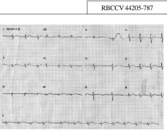
ELECTROCARDIOGRAM
The heart rhythm exhibited a sinusal pause with atrial escape at D2 and a rate of 85 beats per minute. The electrical axis of the QRS complex was indeterminate. Double positivity of the QRS was seen at V1, indicating a slight to moderate block in the right branch (Figure 1).
RADIOGRAM
The cardiac area was normal with an index of 0.45. A slight prominence of the pulmonary artery trunk with minor injury in the peribronchial space at the lung bases and signs of pulmonary vascular prominence were evidenced.
Article received in May, 2005 Article accepted in June, 2005
Case 8/2005
Fig. 1 - Preoperative echocardiogram
ECHOCARDIOGRAM
452
CROTI, UA ET AL - Atriocavoplasty with atrial tissue in the treatment of upper sinus-venous type interatrial communication - Case 8/2005
Braz J Cardiovasc Surg 2005; 20(4): 451-452
DIFFERENTIAL DIAGNOSIS Idiopathic dilation of the pulmonary trunk, aortopulmonary window, interatrial communication, partial defect of the atrioventricular canal, partial anomalous connection of the pulmonary veins and Scimitar Syndrome should all be considered.
DIAGNOSIS
With the aim of confirming the pulmonary venous connections in the left atrium and exclude other associations, a cineangiocardiographic study was indicated. This demonstrated that there was a concordant connection of the pulmonary veins in the left atrium. The right superior pulmonary vein was connected to the left atrium however its flow was towards the right atrium thus confirming the echocardiographic findings of superior sinus venosus interatrial communication.
OPERATION
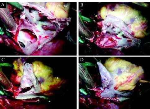
Medium transsternal thoracotomy was performed. A cardiopulmonary bypass was established under hypothermia of 32 ºC. Intermittent anterograde sanguineous cardioplegia at 4 ºC was utilized. The right atrium was opened using the “V-Y” atrioplasty technique with the aim of enlarging the superior vena cava with tissue from the right atrial wall. The posterior incision in the right atrium was continued towards the superior vena cava sideways, avoiding injuring the sinus node or its artery. After identifying the four pulmonary veins (Figure 2), a bovine pericardium patch was sutured isolating the pulmonary veins from the left atrium (Figure 2B). The superior vena cava was enlarged with tissue from the right atrium itself (atriocavoplasty) as is demonstrated in Figures 2C and 2D [1]. The cardiopulmonary bypass time was 80 minutes and myocardial ischemia time was 62 minutes. One year after the operation, the patient presented with heart rhythm similar to the preoperative period and total correction of the defect
BIBLIOGRAPHIC REFERENCES
1. DeLeon SY, Freeman JE, Ilbawi MN, Husayni TS, Quinones JA, Ow EP et al. Surgical techniques in partial anomalous pulmonary veins to the superior vena cava. Ann Thorac Surg. 1993;55(5):1222-6.
2. Dervanian P, Mace L, Puyo P, Folliguet TA, Abdelmoulah S, Santoro F et al. Techniques of correction of partial right abnormal pulmonary venous return associated with atrial septal defect. Arch Mal Coeur Vaiss. 1996;89(7):857-63.
Fig. 2 - A – right atrium open where the right pulmonary veins can be seen draining into the left atrium and the flow towards the superior sinus venosus interatrial communication. B – Interatrial connection closed with a bovine pericardial patch. C and D – Enlargement of the superior vena cava with tissue from the right atrium open in a “V-Y” shape.
A B
C D