Reduction of uncertainties in radiotherapy assessed
by Monte Carlo simulation: spectral analysis applied
to absorbed dose correction*
Redução de incertezas em radioterapia utilizando simulação Monte Carlo: análise espectral aplicada à correção de dose absorvida
Tatiana Marques1, Mirko Alva-Sánchez1, Patrícia Nicolucci2
OBJECTIVE: To calculate spectra of cobalt-60 beam at water depth and correction factors for absorbed dose measurements obtained with lithium fluoride thermoluminescent dosimeters using Monte Carlo simulation. MATERIALS AND METHODS: The simulations of secondary spectra of clinical cobalt-60 sources were performed with the PENELOPE Monte Carlo code at different water depths. Experimental measurements of deep doses were obtained with thermoluminescent dosimeters and ionization chamber under reference conditions for radiotherapy. Correction factors for the thermoluminescent dosimeters detectors were obtained through the ratio between the relative energy absorption for the low energy spectrum and the total spectrum. RESULTS: Deep spectral analysis has demonstrated the presence of secondary low-energy spectra responsible for a significant portion of the dose deposition. Discrepancies of 3.2% were observed among the doses measured with ionization chamber and thermoluminescent dosimeters. The adoption of correction factors has allowed a reduction in the discrepancy among absorbed doses to a maximum of 0.3%. CONCLUSION: Simulated spectra allow the calculation of correction factors for reading of thermoluminescent dosimeters utilized in the measurement of deep doses, contributing for the reduction of uncertainties associated with quality control of clinical beams in radiotherapy.
Keywords: Monte Carlo simulation; Radiotherapy; Spectrometry; Quality control; Thermoluminescent do-simetry; TLD.
OBJETIVO: Determinar, por simulação Monte Carlo, os espectros de feixes de cobaltoterapia em profundi-dade na água e fatores de correção para doses absorvidas em dosímetros termoluminescentes de fluoreto de lítio. MATERIAIS E MÉTODOS: As simulações dos espectros secundários da fonte clínica de cobalto-60 fo-ram realizadas com o código Monte Carlo PENELOPE, em diversas profundidades na água. Medidas experi-mentais de dose profunda foram obtidas com dosímetros termoluminescentes e câmara de ionização em condições de referência em radioterapia. Os fatores de correção para os dosímetros termoluminescentes foram obtidos através da razão entre as absorções relativas ao espectro de baixa energia e ao espectro total. RESULTADOS: A análise espectral em profundidade revelou a existência de espectros secundários de baixa energia responsáveis por uma parcela significativa da deposição de dose. Foram observadas discrepâncias de 3,2% nas doses medidas experimentalmente com a câmara de ionização e com os dosímetros termolu-minescentes. O uso dos fatores de correção nessas medidas permitiu diminuir a discrepância entre as doses absorvidas para, no máximo, 0,3%. CONCLUSÃO: Os espectros simulados permitem o cálculo de fatores de correção para as leituras de dosímetros termoluminescentes utilizados em medidas de dose profunda, contri-buindo para a redução das incertezas associadas ao controle de qualidade de feixes clínicos em radioterapia.
Unitermos: Simulação Monte Carlo; Radioterapia; Controle de qualidade; Espectrometria; Dosimetria termo-luminescente; TLD.
Abstract
Resumo
* Study developed at Hospital das Clínicas da Faculdade de Medicina de Ribeirão Preto da Universidade de São Paulo (HCFMRP-USP), Ribeirão Preto, SP, Brazil. Financial support: Conselho Nacional de Desenvolvimento Científico e Tecnológico (CNPq) and Coordenação de Aperfeiçoamento de Pessoal de Nível Superior (Capes).
1. PhD Fellow degree, Researchers at Departamento de Física e Matemática da Universidade de São Paulo (DFM-USP), Ribei-rão Preto, SP, Brazil.
2. PhD, Professor at Departamento de Física e Matemática da Universidade de São Paulo (DFM-USP), Ribeirão Preto, SP, Brazil.
Marques T, Alva-Sánchez M, Nicolucci P. Reduction of uncertainties in radiotherapy assessed by Monte Carlo simulation: spectral analysis applied to absorbed dose correction. Radiol Bras. 2010;43(2):119–123.
Mailing address: Tatiana Marques. Universidade de São Paulo. Avenida Bandeirantes, 3900. Ribeirão Preto, SP, Brazil, 14040-901. E-mail: [email protected]
Received March 26, 2009. Accepted after revision October 15, 2009.
INTRODUCTION
Quality assurance in radiotherapy must associate high accuracy in the planning and
prescribed doses with reproducibility of the planned technique, detailed documentation and a careful dosimetry of both the planned situation and treatment(1–3). Characteristics
with intensive chemotherapy and bone marrow transplantation. Small errors in the determination of depth doses may compro-mise vital organs, for example, the lungs(4,5). The determination of uncertainties as-sociated with radiotherapy is absolutely relevant for the assurance of efficiency of clinical treatments. A considerable fraction of such uncertainty resides in intrinsic char-acteristics of the methods and instrumen-tal apparatuses utilized in the determination of dosimetric parameters, which are di-rectly applied in the calculation for pre-scription of absorbed dose. In order to minimize the uncertainty associated with dosimetric parameters, it is possible to uti-lize the Monte Carlo simulation of ioniz-ing radiation interaction with matter in or-der to identify, quantify and correct inac-curacies from the dosimeters utilized in radiotherapy quality control.
Thermoluminescent dosimeters (TLDs) are among the dosimeters most frequently utilized in radiotherapy, with characteris-tics of effective atomic number (Zeffective = 8.2) close to that of water, high spatial de-tector resolution (0.9 mm) and low cost associated with their use(6–8). TLDs with
lithium fluoride (LiF) present energetic dependence for photons with energies < 662 keV(9), which implies the necessity to correct readings performed with such do-simeters as they are exposed to low energy radiation. In measurements of depth ab-sorbed dose(10), for example, those applied
in quality control of whole body irradia-tion(8), the spectrum of therapy beams at
depths of clinical interest is composed by high energy fractions, to which LiF pre-sents a linear response, and also by low energy fractions, in which there is energetic dependence(9).
The simulation of clinical treatments based on the Monte Carlo method has been widely utilized in radiotherapy mainly due to the accuracy associated with its use(11).
The Monte Carlo PENELOPE simulation code is a well established and efficient tool for spectrometry and clinical beam dosim-etry, allowing the faithful geometrical rep-resentation of several types of radiotherapy apparatuses(12). The PENELOPE code uses
material cross section libraries based on international standards(13), thus assuring a
faithful representation of characteristics interaction between materials of dosimet-ric interest.
The present study presents the energy spectra of a cobalt-60 (Co-60) clinical beam simulated in water depth, using the Monte Carlo PENELOPE simulation code. By means of quantitative analysis of dose con-tribution of primary and secondary compo-nents of such beam, it is possible to calcu-late correction factors for LiF-100 TLDs readings at several water depths. The con-tribution of the use of correction factors in absorbed doses in TLDs can be attained by comparison between the depth dose rate (DDR) curves built with experimental data collected with an ionization chamber and with TLDs, whose responses were cor-rected by these factors.
MATERIALS AND METHODS
Monte Carlo simulation
The computer simulations of ionizing radiations interaction with matter were performed by using the Monte Carlo PENELOPE simulation package, which comprises the PENELOPE particles trans-port code and the geometry package PENGEOM. In these simulations, the source was positioned at 80 cm from the phantom, with an aperture of 3.57°, which provided a radiation field of 10 × 10 cm2 on the surface of the phantom. The primary photon spectrum is composed by two peaks of equal occurrence probability, with 1.17 MeV and 1.33 MeV(12) energies,
respec-tively. The phantom was a homogeneous water cube with a 30 cm edge.
The photon spectra at depths were ob-tained by means of virtual impact detectors: virtually drawn pellets, simulating the TLDs experimental geometry at depth in-side the phantom, with 150 energy chan-nels uniformly distributed between 10 KeV and 1.33 MeV. The impact detectors have the function of counting the photons that reach the depth in which the detector is positioned, separating into channels and storing their respective energies(13). So, the
response generated at the end of each simu-lation, comprises the probability density function associated with the photon occur-rence, separated by energy, at each depth.
Based on the data in the response files, the total and low energy spectra were built at several depths of the phantom. Also, a simulated DDR curve in the central axis of the phantom was obtained, for determining the PENELOPE code accuracy in this type of simulation, by means of comparison with experimental data of depth dose.
Experimental data
The DDRs were experimentally ob-tained with a Farmer type 0.6 cm3 cylindri-cal ionization chamber with LiF-100 TLDs, both positioned at depth, within a cubic, homogeneous water phantom, with an edge of 50 cm, analogous to that in the computer simulation. The field projected on the sur-face of the phantom was 10 × 10 cm2, and was at a source-surface distance (SSD) of 80 cm.
The phantom was irradiated in a Si-emens Gammatron II S-80 cobalt therapy system, at the Radiotherapy Unit of Hos-pital das Clínicas da Faculdade de Medi-cina de Ribeirão Preto da Universidade de São Paulo. The TLDs were treated and read at the Center of Instrumentation, Dosimetry and Radioprotection of Universidade de São Paulo (Cidra-USP).
Determination of correction factors by energetic dependence
For each depth, the summatories of spectrum energies multiplied by the ratio between the respective LiF and water mass absorption coefficients (µen/ρ)(14) were cal-culated in two parts: one with energies in the 10 keV to 670 keV interval and another comprising all the energies in the spectrum, i.e., from 10 keV to 1.33 MeV. The calcu-lation of the correction factors was made as presented in equation 1.
RESULTS
Spectra were obtained at 24 different depths in the phantom, at 2 mm intervals in the first centimeter to observe the behav-ior at the build-upregion, and at every 1 cm in depth after the build-upregion, scanning the whole phantom extent. The total spec-trum simulated at a depth of 0.5 cm shown on Figure 1, presents a low energy spectral component with a peak at 220 keV (2.5%) with a total expression rate of 5.5%.
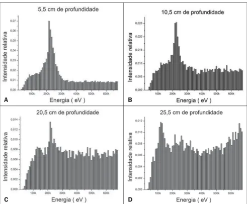
The secondary spectra simulated for the depths of 5.5 cm, 10.5 cm 20.5 cm and 25.5 cm along the phantom central axis are pre-sented on Figure 2. The relative intensities of the simulated spectra reach a maximum at the 5.5 cm depth, where the low energy spectra peak represents 7% of the total spectrum. After this depth, the contribution of the energy peak, centered at 220 keV, goes to 5% at 10.5 cm, decreasing to 1.4% and 1.2% at 20.5 cm and 25.5 cm respec-tively. However, the contribution in dose deposition increases, as the relative expres-sion of energies > 220 keV grows.
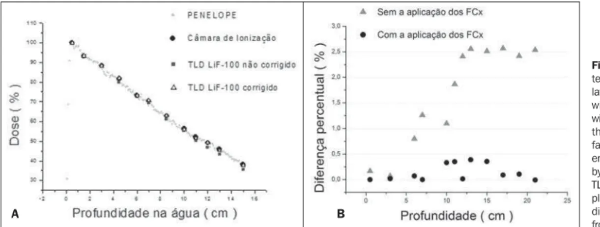
In order to obtain the FCx, the depths at which there were DDR data with both do-simeters (TLD and ionization chamber), were considered. Figure 3A graphically
The chart presented on Figure 3A dem-onstrates the PENELOPE code as appropri-ate and accurappropri-ate for simulating spectra in water depth, co-validating the spectral analysis presented in the present study. Based on the analysis of Figure 3, it is pos-sible to observe how much the readings with TLDs become less accurate at greater depths, because of the presence of low energy radiation, which confirms the need to apply the FCx. On Figure 3B, it is clear that the FCx approach the measurements performed with the TLDs to those per-formed with the ionization chamber, reduc-ing discrepancies from up to 3.3% to a maximum of 0.38%. The numerical data of the FCx can be observed on Table 1.
DISCUSSION
The clinical practice of radiotherapy with high quality indexes is primarily based on rigorous, periodic and reproducible ref-erence dosimetry(14,15) as corroborated by Equation 1. LiF-100 reading correction factors by contribution in dose of the secondary spectrum of a
Co-60 clinical beam in water depth.
Figure 1. Total energy spectrum at the depth of maximum dose of Co-60 in water.
presents the DDR values measured by the TLDs, with and without response correc-tion, together with the DDR measurements performed with the ionization chamber and the DDR curve calculated by means of in-tegration of simulated energy spectra with the PENELOPE code. The percentage dif-ferences between the absorbed dose read-ings with the TLDs, with and without the application of the FCx, and measurements with the ionization chamber are presented on Figure 3B.
Figure 2. Energy spectra at water depths, obtained by the Monte Carlo simulation, for a Co-60 beam on a 10 × 10 cm square field on the phantom surface. A: Secondary spectrum of the Co-60 clinical beam at a depth of 5.5 cm in water: 220 keV peak with 7% representativeness. B: Secondary spectrum of the Co-60 clinical beam at a depth of 10.5 cm in water: 220 keV peak with 2.4% representativeness and 600 keV peak with 0.6% representativeness. C: Secondary spectrum of the Co-60 clinical beam at a depth of 20.5 cm in water: 220 keV peak with 1.3% representativeness and 600 keV peak with 0.8% representativeness. D: Secondary spectrum of the Co-60 clinical beam at a depth of 25.5 cm in water: 220 keV peak and 600 keV peak, both with 0.95% representativeness.
B
D C
Depth dose rate and correction factors for LiF-100
Table 1 Comparison between the DDR values obtained with the TLDs corrected by response energetic dependence, and values measured with the ionization chamber.
X (cm) 0.5 3 6 7 10 11 12 13 15 17 19 21 IC* 100.0 88.4 73.2 70.6 56.4 52.0 49.2 45.8 38.1 32.5 29.3 27.0 TLD† 100.00 88.33 72.40 69.34 55.30 50.17 46.79 43.24 35.59 29.93 26.88 24.46 ∆% *,† 0.166 0.069 0.800 1.257 1.100 1.864 2.410 2.557 2.512 2.568 2.418 2.536 FCx 1.0017 1.0096 1.0101 1.0181 1.0199 1.0302 1.0412 1.0501 1.0606 1.0828 1.0860 1.1040 TLDcorrected ‡ 100.00 88.38 73.13 70.60 56.07 51.68 49.19 45.41 37.74 32.41 29.19 27.01 ∆% *,‡ 0.001 0.018 0.073 0.001 0.332 0.351 0.014 0.389 0.356 0.089 0.107 0.008
* IC, experimental measurements with ionization chamber; † TLD, experimental measurements with LiF-100
TLDs; ∆%, percentage difference; FCx, correction factors calculated with PENELOPE; ‡
TLDcorrected, measurement of absorbed dose in the TLD corrected by the FCx factors .
in the 100 to 300 keV interval, approxi-mately.
In all simulated spectra up to 15.5 cm within the phantom, the low energy peak is kept at 220 keV and the expression of portions with energies from 30 to 300 keV is predominant, which does not happen at depths below the middle of the phantom, at which the probability of occurrence of low energy particles are equally distributed in a larger interval, from 30 to 670 keV.
As the depth increases, the total spec-tra present a higher number of lower energy particles, which dislocates the mean energy of the simulated beam reaching that depth to a lower value than the mean energy of the incident Co-60 beam, that would be 1.25 MeV, commonly considered for the calculation of absorbed energy at water depth.
It is possible to observe that the area of secondary spectra with energies between 10 keV and 670 keV remains approxi-mately the same after the build-up depth, therefore dislocating the effective energy of the secondary spectrum which increases the dose in each detector. Such an increase is not duly observed in experimental mea-surements performed with LiF TLDs, as a function of their response energetic depen-dence in this energy range, which can be observed in the DDR curves obtained with these dosimeters (see Figure 3A).
In spite of the decrease in energy with depth be easily visualized from the obser-vation of simulated spectra, a more precise study of absorbed dose at a given depth must consider the (µen/ρ) corresponding to each discrete set of particles with a given energy, that are, by their turn, differently
Figure 3.A: DDR curve in wa-ter for the Co-60 beam simu-lated with PENELOPE, obtained with ionization chamber and with LiF TLDs before and after the application of correction factors. B: Percentage differ-ences between measurements by the ionization chamber and TLDs before and after the ap-plication of FCx. The percentage
difference interval is reduced from up to 2.6% to 0.3%..
A B
the utilization of TLDs as a complementary alternative to reference dosimetry, allowing more frequent quality control tests with relative simplicity, without excessive cost and interference in the clinical routine(16).
The reduction of uncertainties and er-rors associated with dosimetry in radio-therapy is the central theme of several sci-entific and clinical studies, all of them fac-ing the challenge the fact that precision and accuracy are not easily found in a single dosimeter(17). The use of Monte Carlo
simulation, combined with experimental measurements with TLDs for directly de-termining dosimetric parameters is a pre-cise and viable alternative to face the need of reducing uncertainty and errors in radio-therapy. With the correction factors pre-sented in the present study, it is possible to
utilize LiF TLDs for DDR measurements in water, with an error interval relative to the ionization chamber coherent with the indexes recommended by the reference lit-erature on quality control of radiotherapy beams.
The spectral analysis based on total and secondary photon spectra at water depth, simulated with the PENELOPE code(18,19),
absorbed by water and by the LiF, generat-ing different deposited doses in each one of these mediums.It is exactly by observ-ing the low energy portions and their sig-nificance at each depth that it is possible to relate the differences in absorption among the medium of interest, the water, and the dosimeter material, LiF, applying the FCx determined in the present study.
When compared, the PDDR curves ob-tained with an ionization chamber and with TLDs presented a percentage difference that increases with depth, from 0.2% to 3.2%, and remaining approximately con-stant, at around 2.5% for depths > 10 cm. Also, when observing that the relative probability of the low energy particles in-crease with depth in the phantom (analysis on Figure 2), and considering that the total number of simulated particles in each in-teraction remains the same, it is easy to observe that the FCx must increase with depth, as shown on Table 1.
CONCLUSION
The spectral analysis of a clinical Co-60 beam based on the Monte Carlo simu-lation allows the calcusimu-lation of satisfactory correction factors for the absorbed dose readings in LiF TLDs. The application of the correction factors FCx in TLD readings contributes for the increase in accuracy in the determination of dosimetric param-eters, intimately associated with the qual-ity control of clinical beams in radio-therapy.
The results of the present study may be extended for several clinical protocols, with the objective of making the use of TLDs more accurate and common.
Acknowledgments
Authors thanks to Setor de Radioterapia do Hospital das Clínicas da Faculdade de Medicina de Ribeirão Preto (HCFMRP-USP) and to Centro de Instrumentação, Dosimetria e Radioproteção da Universi-dade de São Paulo (CIDRA-USP) for essencial technical support.
REFERENCES
1. Kawa-Iwanicka A, Lobodziec W, Iwanicki T, et al. Dose uniformity in the total body irradiation technique using 15 MV photon beam. Physica Medica. 2004;20(Suppl 1):144–6.
2. International Atomic Energy Agency. Absorbed dose determination in external beam radiotherapy. An International Code of Practice for Dosimetry Based on Standards of Absorbed Dose to Water. Technical Reports Series No. 398. Vienna: Inter-national Atomic Energy Agency; 2000. 3. International Commission on Radiation Units and
Measurements. Measurement of absorbed dose in a phantom irradiated by a single beam of X or gamma rays. ICRU Report 23. Bethesda: Inter-national Commission on Radiation Units and Measurements; 1973.
4. Vrtar M. A dosimetric method of total body irra-diation. Cell Mol Biol Lett. 2002;7:337–40. 5. Zabatis Ch, Koligliatis T, Xenofos S, et al.
Dosim-etry in translation total body irradiation tech-nique: a computer treatment planning approach and an experimental study concerning lung spar-ing. J Buon. 2008;13:253–62.
6. Bloemen-van Gurp EJ, Mijnheer BJ, Verschueren TA, et al. Total body irradiation, toward optimal individual delivery: dose evaluation with metal oxide field effect transistors, thermoluminescence detectors, and a treatment planning system. Int J Radiat Oncol Biol Phys. 2007;69:1297–304.
7. Kron T. Thermoluminescence dosimetry and its applications in medicine–Part 1: Physics, mate-rials and equipament. Australas Phys Eng Sci Med. 1994;17:175–99.
8. Giordani AJ, Segreto HRC, Segreto RA, et al. Verificação das doses de radiação absorvidas durante a técnica de irradiação de corpo inteiro nos transplantes de medula óssea, por meio de dosímetros termoluminescentes. Radiol Bras. 2004;37:343–9.
9. Reyes FE. Procedimento de calibração de dosí-metros termoluminescentes em dose absorvida na água para fontes de irídio-192I de alta taxa de dose [dissertação de mestrado]. Rio de Janeiro: Insti-tuto de Radioproteção e Dosimetria; 2004.
10. Almeida CE, Affonseca M, Calcina CSG, et al. Rastreabilidade das referências metrológicas em dose absorvida na água do Programa de Qualida-de em Dosimetria. Radiol Bras. 2005;38:205–8.
11. Garnica-Garza HM. Monte Carlo-derived TLD cross-calibration factors for treatment verification and measurement of skin dose in accelerated par-tial breast irradiation. Phys Med Biol. 2009;54: 1621–31.
12. Panettieri V, Sempau J, Andreo P. Chamber-qual-ity factors in 60Co for three plane-parallel cham-bers for the dosimetry of electrons, protons and heavier charged particles: PENELOPE Monte Carlo simulations. Phys Med Biol. 2008;53: 5917–26.
13. International Commission on Radiation Units and Measurements. Tissue substitutes in radiation dosimetry and measurement. ICRU Report 44. Bethesda: International Commission on Radia-tion Units and Measurements; 1989.
14. Morávek Z, Rickhey M, Hartmann M, et al. Un-certainty reduction in intensity modulated proton therapy by inverse Monte Carlo treatment plan-ning. Phys Med Biol. 2009;54:4803–19. 15. Guckenberger M, Krieger T, Richter A, et al.
Potential of image-guidance, gating and real-time tracking to improve accuracy in pulmonary ster-eotactic body radiotherapy. Radiother Oncol. 2009;91:288–95.
16. McGarry CK, Cosgrove VP, Fleming VA, et al. An analysis of geometric uncertainty calculations for prostate radiotherapy in clinical practice. Br J Radiol. 2009;82:140–7.
17. Budrukkar A, Dutta D, Sharma D, et al. Compari-son of geometric uncertainties using electronic portal imaging device in focal three-dimensional conformal radiation therapy using different head supports. J Cancer Res Ther. 2008;4:70–6.
18. Salvat F, Fernández-Varea JM, Acosta E, et al. PENELOPE – a code system for Monte Carlo simulation of electron and photon transport. Paris: Nuclear Energy Agency; 2008.