J of Evolution of Med and Dent Sci/ eISSN- 2278-4802, pISSN- 2278-4748/ Vol. 4/ Issue 22/ Mar 16, 2015 Page 3859
EVALUATION OF CORONARY RISK FACTORS IN PATIENTS WITH ACUTE
MYOCARDIAL INFARCTION
Santosh R. G1, Rangaswamy R2
HOW TO CITE THIS ARTICLE:
Santosh R. G, Rangaswamy R. Evaluation of Coronary Risk Factors in Patients with Acute Myocardial Infarction. Journal of Evolution of Medical and Dental Sciences2015; Vol. 4, Issue 22, March 16;
Page: 3859-3864, DOI:10.14260/jemds/2015/555
ABSTRACT: INTRODUCTION: Cardiovascular disease is the commonest cause of death globally. Acute
myocardial infarction generally occurs when coronary blood flow decreases abruptly after thrombotic occlusion of a coronary artery causing focal or massive necrosis of cardiac muscle. The risk factor concept implies that a person with one risk factor is more likely to develop clinical atherosclerotic event and is more likely to do so earlier than a person with no risk factors. The presence of multiple risk factors further accelerates the atherosclerosis. Hence it is important to identify the major risk factors of coronary atherosclerosis in an individual with acute myocardial infarction so that further preventive measures can be taken in the form of lifestyle modification and pharmacotherapy.
MATERIALS AND METHODOLOGY: This is a hospital based study. This study comprises of 100 cases
of acute myocardial infarction admitted in ICCU under the department of medicine and 100 normal healthy controls in the age group of 29-85 years. Patients with the evidence of acute MI were diagnosed according to WHO criteria. Blood samples collected in vacutainers were analyzed for different biochemical parameters in the clinical biochemistry laboratory. RESULTS: Common risk factors have been evaluated in our study and we found that maximum MI patients were recorded in the age group of 51-60 years, with respect to other risk factors history like sex, majority of patients were males (82%), Sedentary life style (44%), Mixed dietary habits (84%), Family history of IHD (6%), Dyslipidemia and Smoking (46%), Hypertension (31%), Diabetes (37%), Obesity (18%). In our study we found that 81% of the patients of acute MI had multiple risk factors. CONCLUSION: Thus from the study we can conclude that risk factors play a major role in the genesis of coronary heart disease. Modification of these factors by pharmacotherapy, diet, physical exercises and behavioral therapy can improve the prognosis in these patients and also helps in reduction of incidence of CHD.
KEYWORDS: Acute MI (acute myocardial infarction), IHD (ischemic heart disease), CHD (coronary heart disease), Diabetes mellitus, Hypertension.
INTRODUCTION: Cardiovascular disease is the commonest cause of death globally. Acute myocardial
J of Evolution of Med and Dent Sci/ eISSN- 2278-4802, pISSN- 2278-4748/ Vol. 4/ Issue 22/ Mar 16, 2015 Page 3860 The earliest lesions are fatty streaks which consist of lipid engulfed macrophages (foam cells) and T- lymphocyte cells in the arterial intima. This progress to intermediate lesions composed of foam cells and smooth muscle cells migrating from intima to media. With time these develop into fibrous plaque with cap of connective tissue and smooth muscle cells overlying a core containing necrotic material and lipid, mainly cholesterol esters. Endothelial dysfunction is the first step in the development of atherosclerosis. Modified LDL-C, elevated plasma homocysteine concentration, diabetes mellitus, infections are the common cause of endothelial dysfunction. The dysfunctional endothelium undergoes a protective response that alters the normal homeostasis due to expression of adhesion molecules, growth promoting substances and activation of blood coagulation cascade. Monocytes and T-cells adhere to the activated endothelium and produce growth factors, cytokines, and chemo attractants etc., adherent cells migrate from media to intima. With repeated rounds of injury and repair, smooth muscle cells, foam cells, matrix proteins and T-cells accumulate to form atherosclerotic plaque. Rupture of plaque can trigger the thrombosis that precipitates clinical events. During the early phase plaque lesion grows away from the lumen, so affected vessel increase in diameter (compensatory enlargement) and will not cause flow-limiting stenosis and plaques of this type are not visible angiographically. When plaque size exceeds more than 40% of elastic lamina, artery cannot compensate by dilatation and the lesion begins to intrude into the arterial lumen thus becoming angiographically detectable.[1]
The risk factor concept implies that a person with one risk factor is more likely to develop clinical atherosclerotic event and is more likely to do so earlier than a person with no risk factors. Risk factor is a characteristic feature of an individual or population that is present early in life and is associated with an increased risk of developing future disease.[2] Coronary risk factors refer to
conditions that have been demonstrated by statistical procedures to increase the morbidity and mortality of coronary atherosclerosis. The management of risk factors can improve the coronary endothelial function and stop the progression of atherosclerosis, prevent disruption and thrombosis of the vulnerable atherosclerotic plaques and reduce CHD morbidity and mortality. When risk factors co-exist they multiply the risk of CHD several folds.[3] Identification and management of risk factors are
essential in preventing CHD in high risk asymptomatic individuals (primary prevention) and in preventing recurrent events in patients with established disease (secondary prevention). Coronary risk factors are broadly classified into classical (major) and novel risk factors. Classical risk factors have definite relationship with occurrence of CHD and are divided into non-modifiable and modifiable factors. Advancing age, sex (male) and genetic predisposition are non-modifiable and smoking, dyslipidemia, hypertension, diabetes mellitus, obesity and sedentary life style.[4]
Age is strong and independent risk factor and risk increases linearly upto 65 yrs. The increase risk of CAD in elderly persons should trigger more intense management of modifiable factors. CHD risk is more in males compared to females in the premenopausal age group, but after menopause the risk accelerates. Estrogen plays an important role in protecting premenopausal females. There is a association with CAD and history of first degree relative with early onset of CAD. Familial predisposition can be attributed to the inheritance of the risk factors such as hypertension, diabetes, platelet adhesiveness, increased thrombogenecity, and decreased fibrinolytic activity, endothelial and smooth muscle dysfunction.[5] Smoking accelerates the atherosclerosis process by enhancing oxidation
J of Evolution of Med and Dent Sci/ eISSN- 2278-4802, pISSN- 2278-4748/ Vol. 4/ Issue 22/ Mar 16, 2015 Page 3861 fibrinogen and also increases platelet aggregation and increases monocyte adhesion to endothelial cells.[6,7,8] Dyslipidemia may be inherited genetically or secondary to hypothyroidism, diabetes,
nephritic syndrome, chronic alcoholism and drug induced (thiazides, beta blockers, retinoic acid, HIV protease inhibitors). Dyslipidemia is characterized by elevated cholesterol, LDL-C, triglycerides, VLDL-C, small dense LDL-VLDL-C, chylomicrons with decreased HDL-C levels in the body.[5] Hypertension has
proinflammatory actions increasing the formation of hydrogen peroxide and free radicals which reduces the formation of nitric oxide by the endothelium and also increases the leukocyte adhesion and peripheral resistance.[9]
Patients with diabetes mellitus will have 3 to 5 fold increased risk of future cardiovascular events with even higher rates reported among diabetic women.[10,11] Persistent proteinuria is a strong
predictor of CHD in diabetic patients.[12] Insulin resistance stimulates increased glucose intake with
compensated hyperinsulinemia leading to metabolic derangements which acts as a base for the development of coronary risk factors. Hyperinsulinemia may raise BP through sympathetic stimulation and renal sodium retention. Metabolic syndrome is characterized by presence of abnormal obesity, peripheral insulin resistance, high blood pressure and dislipoproteinemia with elevated plasma triglycerides and decreased HDL-C levels. Individuals with this condition are prone for developing CHD.[1,13] Regular physical exercise reduces myocardial oxygen demand, increases the
myocardial efficiency and electrical stability, HDL-C levels and fibrinolysis, and also reduces BP, obesity, platelet aggregation, improves insulin sensitivity and endothelial function thus reducing the risk of CHD.[3] Obesity is state of excessive adipose tissue mass resulting from imbalance between
energy intake and expenditure which is measured by BMI (body mass index). BMI >30 is used as the threshold for obesity in both sexes. Specifically the intra-abdominal and abdominal wall fat has more significance than the subcutaneous fat present in other parts of the body. This distinction is easily made by determining the waist to hip ratio, with the ratio >0. 9 in female and >1in males being abnormal.[2]
MATERIALS AND METHODOLOGY: Thisis a hospital based study. This study comprises of 100 cases
of acute myocardial infarction admitted in ICCU under the department of medicine and 100 normal healthy controls in the age group of 29-85 years. Patients with the evidence of acute MI were diagnosed according to WHO criteria. Patients with coagulation disorders, collagen vascular disease, thyroid disorder, valvular heart disease and congenital diseases of heart were excluded from the study. After obtaining the consent from subjects and institutional ethical committee clearance, overnight fasting blood samples collected in vacutainers under sterile measures were analyzed for different biochemical parameters like blood sugar levels (FBS and PPBS by GOD-POD method),[14] lipid
profile (cholesterol,[15,16] triglycerides,[17,18,19] HDL-C[20,21,22] by precipitation method, LDL-C by
calculation method), cardiac enzymes (CK-MB[23] immune inhibition test, LDH[24] and SGOT[25] in the
clinical biochemistry laboratory.
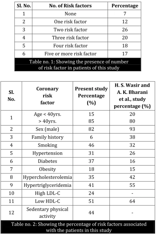
J of Evolution of Med and Dent Sci/ eISSN- 2278-4802, pISSN- 2278-4748/ Vol. 4/ Issue 22/ Mar 16, 2015 Page 3862 the age group of 51-60 years, with respect to other risk factors history like sex, majority of patients were males (82%), Sedentary life style (44%), Mixed dietary habits (84%), Family history of IHD (6%), Dyslipidemia and Smoking (46%), Hypertension (31%), Diabetes (37%), Obesity (18%). In our study we found that 81% of the patients of acute MI had multiple risk factors. This correlates with the observation made by H. S. Wasir and A. K. Bharani et al., study.[26]
Sl. No. No. of Risk factors Percentage
1 None 7
2 One risk factor 12
3 Two risk factor 26
4 Three risk factor 20
5 Four risk factor 18
6 Five or more risk factor 17
Table no. 1: Showing the presence of number of risk factor in patients of this study
Sl. No.
Coronary risk factor
Present study Percentage
(%)
H. S. Wasir and A. K. Bharani
et al., study percentage (%)
1 Age < 40yrs. > 40yrs.
15 85
20 80
2 Sex (male) 82 93
3 Family history 6 38
4 Smoking 46 32
5 Hypertension 31 26
6 Diabetes 37 16
7 Obesity 18 15
8 Hypercholesterolemia 35 42
9 Hypertriglyceridemia 41 55
10 High LDL-C 24 -
11 Low HDL-C 51 64
12 Sedentary physical
activity 44 -
Table no. 2: Showing the percentage of risk factors associated with the patients in this study
J of Evolution of Med and Dent Sci/ eISSN- 2278-4802, pISSN- 2278-4748/ Vol. 4/ Issue 22/ Mar 16, 2015 Page 3863
ACKNOWLEDGEMENT: We would like to acknowledge the Medicine department, the patients and
paramedical staff and to the institution for their kind support.
REFERENCES:
1. A. R. Neis, G. D. Smith et al., Ischemic heart disease; Oxford text book of Medicine: David A. W, Timothy M. C, 4th edition, vol. 2, Chapter 15. 4, 906-20 pg.
2. Peter Libby et al., Prevention and Treatment of atherosclerosis, Harrison’s principle of Internal
Medicine, Braunwald, Fauci, 15thedn, Vol. 1, Chapter 242, 1382-86 pg.
3. Russel Ross et al., Factors influencing the atherosclerosis, Hurst’s The Heart, Robert C. S, 9thedn,
Vol. 1, Chapter 39, 1139-59 pg.
4. Paul M R, Charles H. H et al., Plasma concentration of soluble intracellular adhesion molecule-1 and risks of future MI in apparently healthy men, Lancet, 1998, Vol. 351, 88-92 pg.
5. S. R Mittal, Monica Maheshwari et al., Cardiovascular risk assessment, Classic risk factors-present status, Medicine Update, 2004, Vol. 14, 22-77 pg.
6. Morrow J. D, Frie. B et al., Smoking has a cause of oxidative damage, NEJM, 1995, Vol. 332, 1198-1203 pg.
7. Meade T. W, Imeron. J et al., Effects of changes in smoking and other characteristics on clotting factors and the risk of IHD, Lancet, 1987, Vol. 2, 986-88 pg.
8. Wolfgang k, Malte. S et al CRP a sensitive marker of inflammation, Predicts future risks oc CHD in initially healthy middle aged men (MONICA Study), Circulation, 1999, Vol. 99, 237-48 pg. 9. Russel Ross et al., Atherosclerosis and inflammatory disease; NEJM, 1999, Vol. 340, No. 2,
115-23 pg.
10. Kannel W. B, Mc Gee D et al., Diabetes and Glucose tolerance as risk factor for cardiovascular disease- the Framingham study, Diabetes care, 1979, Vol. 2, 120-26 pg.
11. Manson J. E, Colditz G. A et al., A prospective study of maturity of onset of DM and risk of CAD and Stroke in women; Arch. Intern. Med. 1991 Vol. 151, 1141-47 pg.
12. Paul M, Ridkar, Peter Libby et al., Risk factors for atherosclerotic disease; Heart disease, Braunwald, P. Libby, 6thedn, Chapter 31, 1010-39 pg.
13. Grundy S. M et al., Small LDL, AtherogenicDyslipidemia and Metabolic syndrome; Circulation, 1998 Vol. 95, 1-4 pg.
14. Trinder, P.; Ann. Clin., Biochem. 6, (1969) 24.
15. Allain C. C., Poon L. S., Chan C. S. G., Richmond. W and Fu P., Clin. Chem., 20 (470) 1974. 16. Roeschlau P., Bernt E., and Gruber W. A., Clin. Chem. ClinBiochem 12 (226), 1974. 17. Trinder, P., (1969) Ann Clin. Biochem. 6:24.
18. Bucolo, G., David, H., (1973) Clin. Chem. 19:476. 19. Fossati, P., Prencipe, L., (1982) Clin. Chem. 28:2077.
20. Expert Panel on Detection, Evaluation and Treatment of High Blood Cholesterol in Adults (Adult Treatment Panel III), Executive Summary of the Third Report of the National Cholesterol Education Program (NCEP), JAMA, (2001), 285, 2486.
21. Tietz, N. W., Clinical guide to laboratory tests, 3rd Ed., (W. B Saunders eds. Philadelphia USA),
(1995), 334.
J of Evolution of Med and Dent Sci/ eISSN- 2278-4802, pISSN- 2278-4748/ Vol. 4/ Issue 22/ Mar 16, 2015 Page 3864 23. Neumeier, D., Prellwitz, W., Wurzburg, C., et al., Determination of creatine kinase activity. Isoenzyme MB activity in serum using immunological inhibition of creatine kinase M subunits. Clin. Chim. Acta 73, 445-451 (1976).
24. Bergmeyer. H. U., Bernt, E, (1974a), Lactate-dehydrogenase. UV-assay with pyruvate and NADH. In Bergmeyer, H. U. (ed.) Methods of enzymatic analysis, Vol 2. Academic press, New York, p. 574-579.
25. Burtis, C A., Ashwood, E. R., editors. Tietz Textbook of Clinical Chemistry, 2nd ed. Philadelphia,
W. B. Saunders Company. 1994, p. 790-795.
26.
H. S Wasir, A. K. Bharani, M. L. Bhatia et al., Correlation of risk factors with coronary angiographic findings in patients of IHD; JAPI, 1987, Vol. 36, No. 7, 483-87 pg.AUTHORS:
1. Santosh R. G. 2. Rangaswamy R.
PARTICULARS OF CONTRIBUTORS:
1. Assistant Professor, Department of Medicine, Kannur Medical College, Kannur.
2. Assistant Professor, Department of Biochemistry, Kannur Medical College, Kannur.
FINANCIAL OR OTHER
COMPETING INTERESTS: None
NAME ADDRESS EMAIL ID OF THE CORRESPONDING AUTHOR:
Dr. Rangaswamy R, Assistant Professor,
Department of Biochemistry, Kannur Medical College, Kannur, Kerala.
E-mail: rangaswamyr79@yahoo.com