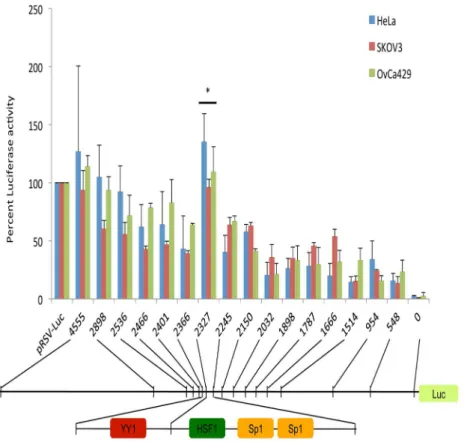
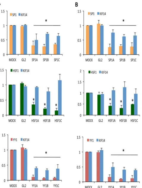
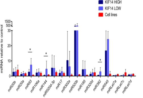
Transcriptional and epigenetic regulation of KIF14 overexpression in ovarian cancer.
Texto
Imagem


Documentos relacionados
In the present study, we developed a machine learning approach to predict the cell lines response to natural products, based on gene expression of cancer cell lines
Gene expression analysis of SSTR subtypes in primary prostate cancer tissues versus normal adjacent prostate tissues shows increased SSTR4 (p = 0.0236) and confirms lower
To understand the expression of Oct4 in OSCC cell lines (OSCCs), the endogenous protein level of Oct4 in nine established OSCC cell lines and one normal oral epithelial cell line SG
While the expression of MUC16 protein in 3T3 cells was clearly linked to hallmarks of trans- formation, some fully transformed ovarian cancer cell lines lack MUC16 expression
IL-1ß downregulates the expression of MITF-M and upregulates the expression of miR-155 in 2 melanoma cell lines.. The result was normalized to ß-actin expression (means ± SD for 3 or
C ; Representative in-cell western assay and summary data of the TRPC3 expression (normalized to CD14 expression used as an internal reference) in monocytes from normotensive
Quantitative analysis of p85α immunoblotted protein samples from bladder cancer cell lines normalized to tubulin and shown relative to pooled normal human urothelial
In this study, we assessed the potential tumor/metastasis suppressor functions of PG in OVCAs, using the normal ovarian cell line IOSE-364 and OVCA cell lines OV-90 (PG and