Treatment of Triple-Negative Breast Cancer
Yanfeng Qi1, Xueqi Fu2, Zhenggang Xiong3, Haitao Zhang3,4, Steven M. Hill1, Brian G. Rowan1, Yan Dong1,4*
1Departments of Structural and Cellular Biology, Tulane Cancer Center, Tulane University School of Medicine, New Orleans, Louisiana, United States of America,2College of Life Sciences, Jilin University, Changchun, Jilin, China,3Department of Pathology and Laboratory Medicine, Tulane Cancer Center, Tulane University School of Medicine, New Orleans, Louisiana, United States of America,4National Engineering Laboratory for AIDS Vaccine, College of Life Sciences, Jilin University, Changchun, Jilin, China
Abstract
A major challenge in breast cancer therapy is the lack of an effective therapeutic option for a particularly aggressive subtype of breast cancer, triple-negative breast cancer. Here we provide the first preclinical evidence that a second-generation selenium compound, methylseleninic acid, significantly enhances the anticancer efficacy of paclitaxel in triple-negative breast cancer. Through combination-index value calculation, we demonstrated that methylseleninic acid synergistically enhanced the growth inhibitory effect of paclitaxel in triple-negative breast cancer cells. The synergism was attributable to more pronounced induction of caspase-mediated apoptosis, arrest of cell cycle progression at the G2/M checkpoint, and inhibition of cell proliferation. Treatment of SCID mice bearing MDA-MB-231 triple-negative breast cancer xenografts for four weeks with methylseleninic acid (4.5 mg/kg/day, orally) and paclitaxel (10 mg/kg/week, through intraperitoneal injection) resulted in a more pronounced inhibition of tumor growth compared with either agent alone. The attenuated tumor growth correlated with a decrease in tumor cell proliferation and an induction of apoptosis. Thein vivostudy also
indicated the safety of using methylseleninic acid in the combination regime. Our findings thus provide strong justification for the further development of methylseleninic acid and paclitaxel combination therapy for the treatment of triple-negative breast cancer.
Citation:Qi Y, Fu X, Xiong Z, Zhang H, Hill SM, et al. (2012) Methylseleninic Acid Enhances Paclitaxel Efficacy for the Treatment of Triple-Negative Breast Cancer. PLoS ONE 7(2): e31539. doi:10.1371/journal.pone.0031539
Editor:Pranela Rameshwar, University of Medicine and Dentistry of New Jersey, United States of America
ReceivedNovember 16, 2011;AcceptedJanuary 12, 2012;PublishedFebruary 14, 2012
Copyright:ß2012 Qi et al. This is an open-access article distributed under the terms of the Creative Commons Attribution License, which permits unrestricted use, distribution, and reproduction in any medium, provided the original author and source are credited.
Funding:This work was supported by the National Cancer Institute grant K01CA114252 (YD), American Cancer Society grant RSG-07-218-01-TBE (YD), Mary Kay Foundation grant 019-11 (YD), Louisiana Cancer Research Consortium Start-up Fund (YD and HZ), Tulane Cancer Center Developmental Fund (YD), and Departmental Seed Fund from the Department of Structural and Cellular Biology, Tulane University School of Medicine (YD). The funders had no role in study design, data collection and analysis, decision to publish, or preparation of the manuscript.
Competing Interests:The authors have declared that no competing interests exist.
* E-mail: ydong@tulane.edu
Introduction
Triple-negative breast cancer (TNBC) refers to a subtype of breast cancer that is negative for expression of estrogen receptor and progesterone receptor, and lacks HER2 overexpression. The majority of TNBCs bear the gene expression profile of basal-like phenotype [1]. This subtype accounts for,10–15% of all types of
breast cancer. TNBC is particularly aggressive, associated with rapid relapse following therapy, increased prevalence of distant metastases, and shorter survival comparing with other breast cancer subtypes [2]. It is more prevalent in Hispanic and African American women than in other ethnic groups, with the majority of the cases occurring in premenopausal women [3,4].
Due to the ‘‘triple-negative’’ nature, patients with TNBC are not candidates for endocrine therapy or targeted therapy against HER2. TNBC patients are usually managed with standard treatments, and chemotherapy is the primary, if not the only, choice of systemic therapy. Taxanes, such as paclitaxel, alone or in combination with anthracycline reagents and/or alkylation reagents are the first-line chemotherapeutic drugs being used for TNBC [2,5]. Although patients generally have a favorable initial response to taxane regimens, rapid development of resistance to taxanes is prevalent [5]. Therefore, developing agents that could
effectively enhance the efficacy of taxanes and overcome resistance is urgently needed for the management of TNBC.
Selenium is an effective anticancer agent with a low toxicity profile [2]. The anticancer efficacy depends on the form and dosage of selenium administered [6–11]. Methylseleninic acid (MSA) is a potent second-generation selenium compound. It has very different biological and pharmacological activity than selenomethionine, the form of selenium used in the Selenium and Vitamin E Chemoprevention Trial [7,9–13]. MSA has been shown to enhance the efficacy of chemotherapeutic drugs in cultured estrogen-receptor-positive breast cancer cells as well as cultured prostate cancer cells and prostate tumor xenografts [14– 17]. However, the effectiveness of MSA alone or in a combination regime for treating TNBC has never been evaluated. In this report, we describe a series of experiments that were designed to determine the potential of using MSA to improve the therapeutic outcome of paclitaxel for the treatment of TNBC.
Materials and Methods
TNBC Cell Lines and Reagents
and MDA-MB-157 cells were cultured in DMEM medium, and BT-549 cells in RPMI 1640 medium. Both media were supplemented with 10% fetal bovine serum, 2 mM glutamine, 100 units per ml penicillin, and 100mg/ml streptomycin. Paclitaxel and MSA were purchased from Sigma.
Cell viability, Apoptosis, and Cell Proliferation Detection Cell viability was determined by using either the Sulforhoda-mine B (SRB) assay as described [18] or direct cell counting with trypan blue staining. Cell proliferation was measured by using the BrdU Cell Proliferation ELISA kit (Roche), and apoptosis of cultured cells detected by using the Cell Death Detection ELISAPLUSKit (Roche) as described [19,20]. The apoptotic cells on tumor sections were detected by the TUNEL assay using theIn SituCell Death Detection Kit-TMR red (Roche) per manufactur-er’s protocol. The TUNEL slides were counterstained with DAPI for nuclear staining. The images were taken by using a Nikon Eclipse 80i fluorescence microscope, and quantitated with the use of the Image J software.
Western blot analysis
The procedure was described previously [21]. Immune-reactive bands were quantitated by densitometry using the Licor Odyssey software and normalized to glyceraldehyde-3-phosphate dehydro-genase (GAPDH). The antibodies for the active forms of caspase-3 (Catalog Number 9664) and caspase-7 (Catalog Number 9491), and cleaved PARP (Catalog Number 5625) were obtained from Cell Signaling. The GAPDH antibody was from Millipore (Catalog Number Mab374).
Cell Cycle Analysis
Cells were detached from culture dishes by using 15 mM EDTA-Na2, and then fixed in 70% ethanol. The cell pellet was
washed with PBS twice and suspended in propidium iodide staining solution (0.1% (v/v) Triton X-100, 0.2 mg/ml DNase-free RNase A, and 20mg/ml propidium iodide). After incubation at room temperature for 30 min, DNA content was analyzed by using the BD LSRII flow cytometer. The Modfit LTTMsoftware was used to analyze the data.
Caspase-3/7 Activity Assay
Caspase-3/7 activity was measured by using the Caspase 3/7-Glo assay kit (Promega) according to manufacturer’s manual. The reading was measured by using the Fluostar Optima fluorescent plate reader.
MDA-MB-231 Tumor Xenograft Model
Female SCID hairless out-bred mice were obtained from Charles River at 8 weeks of age. The mice were kept in vivarium for two weeks to adapt before any procedure. One million MDA-MB-231 cells in PBS were inoculated into both sides of the mammary fat pad with Matrigel. When tumors were palpable,
,4–6 weeks after the inoculation, the mice were randomly
assigned to four groups. Control mice were given weekly intraperitoneal saline solution. The MSA group was treated with 3 mg selenium in the form of MSA per kg body weight daily by an oral route [22]. Paclitaxel, 10 mg/kg, was given by intraperitoneal injection weekly. The combination group was treated with daily oral MSA and weekly intraperitoneal paclitaxel. Body weights and tumor dimensions were monitored weekly. Tumor volume was calculated as0.524 x width2x length. At the end of the experiment, mice were anesthetized, and tumors removed, weighed, and fixed in 10% formalin for paraffin embedding and histological analyses.
All animal procedures were approved by the Tulane University Institutional Animal Care and Use Committee.
Immunohistochemical (IHC) Analysis
Paraffin-embedded blocks were sectioned, deparaffinized, rehydrated, and quenched by immersing in 1% H2O2. The slides
were boiled in 10 mM citrate buffer (pH 6.0) for 20 min for antigen retrieval, and pre-incubated with 5% blocking serum to prevent non-specific binding. Ki-67 antibody incubation was applied at room temperature for 1 hr. Ki-67 reaction was visualized by using the VECTASTAIN ABC system (Vector), with diaminobenzidine as the chromogen. Sections were counter-stained with hematoxylin. As the negative control, the primary antibody was replaced with a non-immune IgG at the same concentration, and no reactivity was detected. The IHC images were analyzed by using the ImmunoRatio online analysis tools [23].
Statistical Analysis
Mean activities were calculated from at least three independent experiments performed in triplicate. The Student’s two-tailedttest assuming unequal variance was used to determine significant differences between two groups.
Results
MSA synergistically enhances the growth-inhibitory efficacy of paclitaxel in MDA-MB-231 cells
We chose the most commonly-used and well-studied TNBC cell line, MDA-MB-231 [24–28], as the primary model for investigat-ing the combinatory effect of MSA and paclitaxel. MDA-MB-231 was derived from pleural effusion of a breast cancer from a Caucasian patient [29]. Cells were treated with pharmacologically achievable concentrations of MSA, paclitaxel, or the two drugs in combination for 72 hr. Cell viability was first determined by the SRB assay. The cell viability in all combination groups was lower than that in the respective single-agent treatment groups (Table 1). To determine whether these combinatory effects were synergistic, the data were analyzed with the Calcusyn software (Biosoft), which calculates combination index (CI) values using the median-effect principle [30] to delineate the interaction between two drugs. A CI value of ,1, 1, or .1 denotes synergism, additivity, or antagonism, respectively. All the combinations, except 2.5mM MSA and 10 nM paclitaxel, produced a CI value of less than 1, suggesting a synergy between MSA and paclitaxel in inhibition of cell growth (Table 2). We also assessed cell viability by direct cell counting with trypan blue staining, and the data (Fig. 1) are congruent with the SRB results.
MSA enhances paclitaxel-induced G2/M arrest
We next assessed the effect of the combination treatment on cell cycle distribution. The experiment was performed at the 24-hr time point in MDA-MB-231 cells. As depicted in Table 3, paclitaxel decreased the proportion of cells in G0/G1 and arrested cells in G2/M phase. At the 3.2mM concentration, MSA alone didn’t affect cell cycle distribution. However, it enhanced the paclitaxel effect by further decreasing the fraction of cells in G0/ G1 phase and increasing G2/M arrest.
MSA enhances paclitaxel-induced apoptosis
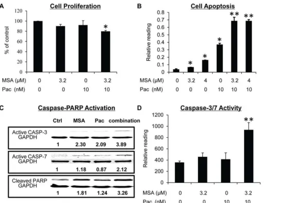
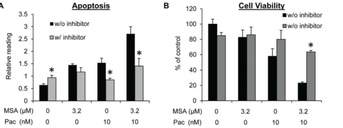
G2/M arrest induced by paclitaxel in cancer cells generally leads to extensive apoptosis [31]. We therefore evaluated apoptosis in MDA-MB-231 cells. As shown in Fig. 2B, an increase in apoptosis was observed after treatment of cells with either MSA or paclitaxel, and the increase was more pronounced as a result of the combination treatment. Caspase-PARP activation is the major pathway involved in apoptosis. We measured caspase-3 and -7 activation and PARP cleavage in MDA-MB-231 cells by Western blotting. Consistent with the apoptosis data, the combination treatment caused an enhanced activation of caspase-3 and -7 and PARP cleavage comparing to either MSA or paclitaxel alone (Fig. 2C). We also performed caspase-3/7 activity assay, and the activity data correlated well with the Western result (Fig. 2D). In addition, pre-treatment of cells with a pan-caspase inhibitor (Z-VAD-FMK) almost completely blocked apoptosis induction (Fig. 3A) and inhibition of cell viability (Fig. 3B) by paclitaxel, alone or in combination with MSA, demonstrating the caspase-PARP pathway as the major pathway involved.
MSA enhances paclitaxel-induced inhibition of
proliferation and increase of apoptosis in other TNBC cell models
To further demonstrate the universality of the combinatory effects, we determined cell proliferation and apoptosis in two other TNBC cell models, MDA-MB-157 and BT-549. MDA-MB-157
was derived from pleural effusion of a breast cancer from an African-American patient, and BT-549 from a primary breast cancer from a Caucasian patient [29]. In both models, the combination treatments led to a more robust induction of apoptosis and inhibition of proliferation than treatment with MSA or paclitaxel alone (Fig. 4).
MSA enhances paclitaxel efficacy in vivo
To evaluate thein vivoefficacy of MSA and paclitaxel, female SCID-hairless mice implanted with MDA-MB-231 cells were divided into four groups receiving saline solution as control, 3 mg selenium in the form of MSA per kg body weight daily, 10 mg/kg paclitaxel weekly, or the two drugs in combination. To be more consistent with the patient treatment schedule, all treatments were stopped after 4 doses of paclitaxel, and mice were maintained for an additional 4 weeks. As shown in Fig. 5A, MSA alone had no effect on tumor growth. In the paclitaxel group, tumor growth was suppressed until two weeks after termination of treatment, and tumor re-growth was observed on Day 42. The re-growth was greatly inhibited by the combination treatment. At the conclusion of the study, the average tumor weight in the paclitaxel group was significantly lower than that in the control group, and the combination treatment caused a more pronounced reduction of tumor weight (Fig. 5B). The combination treatment did not appear to cause more toxicity than paclitaxel alone since mice in the two groups had similar body weights (Fig. 5C). We then measured apoptosis induction by using the fluorescent TUNEL assay and proliferation index by Ki-67 IHC in the xenograft tumors. As shown in Fig. 6, the combination treatment led to a significant induction of apoptosis and inhibition of cell proliferation in the tumors.
Discussion
In the present study, we evaluated the efficacy of MSA in combination with paclitaxel for the treatment of TNBC. Our data showed that MSA could significantly enhance thein vitroandin vivo antitumor efficacy of paclitaxel in TNBC cells. Through CI value calculation for MSA and paclitaxel, each at three different concentrations, we demonstrated that the combinatory effect on cell growth, except that of the lowest dose combination, was Figure 1. MSA enhances the efficacy of paclitaxel in inhibiting the growth of MDA-MB-231 cells.Cells were treated with 3.2 or 4mM MSA, 10 nM paclitaxel, or the combinations for 72 hr. Live cells were counted with trypan blue staining.
doi:10.1371/journal.pone.0031539.g001 Table 1.Effect of MSA and/or paclitaxel on cell viability*.
Paclitaxel (nM) MSA (mM)
0 2.5 3.2 4
0 100 90.0613.2 80.3613.2 60.7610.5
10 47.865.7 44.760.8 35.361.3 29.761.7
20 42.662.8 35.862.3 30.361.7 26.163.8
40 38.762.0 37.763.3 28.160.4 25.062.6
*Results are expressed as % of vehicle control (mean
6SEM). doi:10.1371/journal.pone.0031539.t001
Table 2.Combination Index values for MSA and paclitaxel.
Paclitaxel (nM) MSA (mM)
2.5 3.2 4
10 1.163 0.752 0.761
20 0.796 0.696 0.73
40 0.812 0.726 0.758
synergistic. The synergism was attributable to more pronounced induction of caspase-mediated apoptosis, arrest of cell cycle progression at the G2/M checkpoint, and inhibition of cell proliferation. Our in vivo study also showed the safety of using MSA in the combination regime. The current study is the first to show thein vivoeffectiveness of MSA in enhancing chemothera-peutic efficacy in breast cancer. The focus on TNBC could be of more clinical importance considering the limited availability of effective treatment options for this deadly disease. An interesting observation that needs to be pointed out is that when MSA was combined with paclitaxel at their respective lowest tested concentration, the combination produced antagonistic effect
(CI = 1.163). The clinical relevance of this observation requires furtherin vivotesting. If proven to be true, the information could be helpful in selecting the effective doses of the two drugs for future clinical trials.
MSA, at the dose used in ourin vivostudy, has been shown to inhibit the growth of prostate tumor xenografts [14]. However, no growth inhibition was observed in our study with the MDA-MB-231 xenograft model. This discrepancy could be due to the difference in the model system. It could also result from the difference in the treatment protocols. In the prostate tumor studies, MSA treatment was initiated right after cell inoculation and continued until the end of the experiments. However, in our study, MSA supplementation was not started until the tumors became palpable. The mice were administered MSA for only 4 weeks, and were maintained drug-free thereafter. Although there was a slight trend of growth inhibition during MSA treatment, this inhibition was not significant because the tumors were rather small. Whether MSA treatment, in a long term or at a higher dose, could inhibit the growth of TNBC tumors awaits to be determined. In our study, paclitaxel alone dramatically inhibited the growth of the xenograft tumors. However, this was not accompanied by a significant change in apoptosis or proliferation. This could again arise from our treatment and recovery experimental protocol.
Previously, we reported that MSA can induce FADD expression in MCF-7 breast cancer cells [16]. This boost of FADD expression leads to recruitment of caspase-8, resulting in a robust apoptosis induction when combined with chemotherapeutic reagents. FADD has been shown to play an important role in G2/M arrest Figure 2. MSA enhances the efficacy of paclitaxel in inhibiting cell proliferation and inducing apoptosis, caspase activation, and PARP cleavage in MDA-MB-231 cells.Cells were treated with 3.2mM MSA, 10 nM paclitaxel, or the combination for 16 hr. Cells were then analyzed for proliferation (A), apoptosis (B), the levels of active form of caspase-3 and -7, and cleaved PARP by Western blotting (C), or Caspase-3/7 activity (D). The BrdU, apoptosis, and caspase-3/7 activity readings were normalized by the SRB reading in cells set up in parallel. Error bars, SEM. *, statistically significant (P,0.05) from the control group. **, statistically significant (P,0.05) from both single–agent-treated samples and the control
sample. Pac, paclitaxel.
doi:10.1371/journal.pone.0031539.g002
Table 3. Effect of MSA and/or paclitaxel on cell cycle distribution#.
Treatment Group G0/G1 S G2/M
Control 58.364.5 27.762.8 14.061.7
MSA 59.164.9 24.264.1 16.760.9
paclitaxel 33.565.6* 29.960.4 36.765.2*
combination 26.466.5** 27.364.4 46.3610.9**
#Results are expressed as mean % of cells in a specific phase of cell cycle
6
SEM.
*Significantly different compared to the control group (P
,0.05). **Significantly different compared to both single-agent-treated and control
groups (P,0.05).
and cell growth suppression induced by paclitaxel in breast cancer cells [32,33]. These findings suggest that increasing FADD expression by MSA may lower the threshold for G2/M arrest induced by paclitaxel treatment, consequently leading to increased sensitivity to paclitaxel. FADD activation also may result in activation of caspase-3 in TNBC cells, which in turn leads to activation of caspase-8 or -9 and apoptosis. The FADD-caspase-3
axis should be tested in future studies to further elucidate the mechanism underlying the combinatory effect of MSA and paclitaxel.
When compared with other types of breast cancer, TNBC has an increased rate of pathologic complete response to chemother-apy. However, TNBC is associated with more prevalent and earlier-onset recurrence and shorter overall survival [2]. Failure of Figure 3. Inhibiting caspase activity attenuates apoptosis induction and growth suppression by MSA and paclitaxel in MDA-MB-231 cells.Cells were pre-treated with 50mM pan-caspase inhibitor Z-VAD-FMK prior to treatment with 3.2mM MSA, 10 nM paclitaxel, or the combination. Apoptosis was determined at the 16-hr time point by Cell Death ELISA (A), and cell viability at 72 hr by SRB (B). Error bars, SEM. *, statistically significant (P,0.05) from the corresponding control sample not treated with the caspase inhibitor. Pac, paclitaxel.
doi:10.1371/journal.pone.0031539.g003
Figure 4. MSA enhances the effect of paclitaxel in inhibiting cell proliferation and inducing apoptosis in MDA-MB-157 and BT-549 cells.Cells were treated with MSA, paclitaxel, or the combination for 48 hr, and then subjected to analysis for proliferation (A, B) or apoptosis (C, D). The BrdU and apoptosis readings were normalized by the SRB reading in cells set up in parallel. Error bars, SEM. *, statistically significant (P,0.05)
from the control group. **, statistically significant (P,0.05) from both single-agent-treated sample and the control sample. Pac, paclitaxel.
G2/M arrest, as a result of mitotic slippage, abnormal regulation of G2/M transition genes, polyploidy induction, or multi-nucleation, has been shown to be the major mechanism underlying acquired resistance to paclitaxel [34]. Our finding that MSA could enhance paclitaxel-mediated G2/M arrest suggests the potential of using MSA to overcome paclitaxel resistance. In fact, we generated a paclitaxel-resistant MDA-MB-231 subline by long-term treatment of MDA-MB-231 cells with low doses of paclitaxel.
We found that MSA could reverse the resistance of the cells to paclitaxel (data not shown). This further demonstrates the benefit of using MSA in combination with paclitaxel for the management of TNBC.
According to the 2010 cancer statistics report from the American Cancer Society [35], cancer deaths have declined for both Caucasian and African Americans living in the United States. However, African Americans continue to suffer the greatest Figure 5. MSA enhances paclitaxel efficacy in vivo.(A) Tumor growth curve. Data are presented as tumor volumes in each group (n = 6 tumors/group). (B) Average tumor weight in each group at the end of the experiment. (C) Average body weight of the mice in each group. Error bars, SEM. *, statistically significant (P,0.05) from the control group. **, statistically significant (P,0.05) from both single-agent-treated sample and
the control sample. Pac, paclitaxel. doi:10.1371/journal.pone.0031539.g005
Figure 6. Induction of apoptosis and inhibition of tumor proliferation by paclitaxel and MSA in MDA-MB-231 xenograft.(A) TUNEL staining. Data are presented as mean number of TUNEL-positive cells in each image. (B) Ki-67 IHC. Data are presented as mean % of Ki-67-positive cells. Error bars, SEM. *, statistically significant (P,0.05) from both single-agent-treated sample and the control sample. Pac, paclitaxel. Right panels:
burden for each of the most common types of cancer. American Caucasian women have the highest incidence rate for breast cancer, although African American women are most likely to die from the disease [35]. The disparity could be attributable to many factors, such as genetic, socioeconomic, diet, and culture differences [3]. TNBC is the only subtype of breast cancer that displays racial disparity [2]. Selenium concentration has been shown to be inversely associated with the incidence of a number of cancers, including breast cancer [36,37]. Serum selenium concentration in African Americans were found to be significantly lower than that in Caucasians [38]. It is therefore possible that the clinical benefit of MSA could be more evident in African Americans with TNBC.
Acknowledgments
We are very grateful to Latonia Carrier (Department of Structural and Cellular Biology, Tulane University), Mary Price (Louisiana Cancer Research Consortium Flow Cytometry Core Facility), and Dina Gaupp (Tulane Center for Gene Therapy Histology Core Facility) for their excellent technical assistance.
Author Contributions
Conceived and designed the experiments: YD YQ XF SMH BGR. Performed the experiments: YQ. Analyzed the data: ZX HZ. Contributed reagents/materials/analysis tools: ZX HZ. Wrote the paper: YQ XF HZ SMH BGR YD.
References
1. Kreike B, van KM, Horlings H, Weigelt B, Peterse H, et al. (2007) Gene expression profiling and histopathological characterization of triple-negative/ basal-like breast carcinomas. Breast Cancer Res 9: R65.
2. Cleator S, Heller W, Coombes RC (2007) Triple-negative breast cancer: therapeutic options. Lancet Oncol 8: 235–244.
3. Bauer KR, Brown M, Cress RD, Parise CA, Caggiano V (2007) Descriptive analysis of estrogen receptor (ER)-negative, progesterone receptor (PR)-negative, and HER2-negative invasive breast cancer, the so-called triple-negative phenotype: a population-based study from the California cancer Registry. Cancer 109: 1721–1728.
4. Morris GJ, Naidu S, Topham AK, Guiles F, Xu Y, et al. (2007) Differences in breast carcinoma characteristics in newly diagnosed African-American and Caucasian patients: a single-institution compilation compared with the National Cancer Institute’s Surveillance, Epidemiology, and End Results database. Cancer 110: 876–884.
5. Gluz O, Liedtke C, Gottschalk N, Pusztai L, Nitz U, et al. (2009) Triple-negative breast cancer–current status and future directions. Ann Oncol 20: 1913–1927. 6. Li H, Stampfer MJ, Giovannucci EL, Morris JS, Willett WC, et al. (2004) A prospective study of plasma selenium levels and prostate cancer risk. J Natl Cancer Inst 96: 696–703.
7. Li GX, Lee HJ, Wang Z, Hu H, Liao JD, et al. (2008) Superior in vivo inhibitory efficacy of methylseleninic acid against human prostate cancer over selenome-thionine or selenite. Carcinogenesis 29: 1005–1012.
8. Reid ME, Stratton MS, Lillico AJ, Fakih M, Natarajan R, et al. (2004) A report of high-dose selenium supplementation: response and toxicities. Journal of Trace Elements in Medicine and Biology 18: 69–74.
9. Ip C, Thompson HJ, Zhu Z, Ganther HE (2000) In vitro and in vivo studies of methylseleninic acid: evidence that a monomethylated selenium metabolite is critical for cancer chemoprevention. Cancer Res 60: 2882–2886.
10. Ip C (1998) Lessons from basic research in selenium and cancer prevention. J Nutr 128: 1845–1854.
11. Wang L, Bonorden MJ, Li GX, Lee HJ, Hu H, et al. (2009) Methyl-selenium compounds inhibit prostate carcinogenesis in the transgenic adenocarcinoma of mouse prostate model with survival benefit. Cancer Prev Res (Phila Pa) 2: 484–495.
12. Lippman SM, Klein EA, Goodman PJ, Lucia MS, Thompson IM, et al. (2009) Effect of selenium and vitamin E on risk of prostate cancer and other cancers: the Selenium and Vitamin E Cancer Prevention Trial (SELECT). JAMA 301: 39–51.
13. Ohta Y, Kobayashi Y, Konishi S, Hirano S (2009) Speciation analysis of selenium metabolites in urine and breath by HPLC- and GC-inductively coupled plasma-MS after administration of selenomethionine and methylsele-nocysteine to rats. Chem Res Toxicol 22: 1795–1801.
14. Hu H, Li GX, Wang L, Watts J, Combs GF, Jr., et al. (2008) Methylseleninic acid enhances taxane drug efficacy against human prostate cancer and down-regulates antiapoptotic proteins Bcl-XL and survivin. Clin Cancer Res 14: 1150–1158.
15. Hu H, Jiang C, Ip C, Rustum YM, Lu J (2005) Methylseleninic acid potentiates apoptosis induced by chemotherapeutic drugs in androgen-independent prostate cancer cells. Clin Cancer Res 11: 2379–2388.
16. Li S, Zhou Y, Dong Y, Ip C (2007) Doxorubicin and selenium cooperatively induce fas signaling in the absence of Fas/Fas ligand interaction. Anticancer Res 27: 3075–3082.
17. Li S, Zhou Y, Wang R, Zhang H, Dong Y, et al. (2007) Selenium sensitizes MCF-7 breast cancer cells to doxorubicin-induced apoptosis through modula-tion of phospho-Akt and its downstream substrates. Mol Cancer Ther 6: 1031–1038.
18. Vichai V, Kirtikara K (2006) Sulforhodamine B colorimetric assay for cytotoxicity screening. Nat Protoc 1: 1112–1116.
19. Liu S, Zhang H, Zhu L, Zhao L, Dong Y (2008) Kruppel-like factor 4 is a novel mediator of selenium in growth inhibition. Mol Cancer Res 6: 306–313. 20. Liu S, Qi Y, Ge Y, Duplessis T, Rowan BG, et al. (2010) Telomerase as an
important target of androgen signaling blockade for prostate cancer treatment. Mol Cancer Ther 9: 2016–2025.
21. Dong Y, Zhang H, Hawthorn L, Ganther HE, Ip C (2003) Delineation of the Molecular Basis for Selenium-induced Growth Arrest in Human Prostate Cancer Cells by Oligonucleotide Array. Cancer Res 63: 52–59.
22. Jiang W, Jiang C, Pei H, Wang L, Zhang J, et al. (2009) In vivo molecular mediators of cancer growth suppression and apoptosis by selenium in mammary and prostate models: lack of involvement of gadd genes. Mol Cancer Ther 8: 682–691.
23. Tuominen VJ, Ruotoistenmaki S, Viitanen A, Jumppanen M, Isola J (2010) ImmunoRatio: a publicly available web application for quantitative image analysis of estrogen receptor (ER), progesterone receptor (PR), and Ki-67. Breast Cancer Res 12: R56.
24. Adams LS, Kanaya N, Phung S, Liu Z, Chen S (2011) Whole Blueberry Powder Modulates the Growth and Metastasis of MDA-MB-231 Triple Negative Breast Tumors in Nude Mice. J Nutr.
25. Fost C, Duwe F, Hellriegel M, Schweyer S, Emons G, et al. (2011) Targeted chemotherapy for triple-negative breast cancers via LHRH receptor. Oncol Rep 25: 1481–1487.
26. Prud’homme GJ, Glinka Y, Toulina A, Ace O, Subramaniam V, et al. (2010) Breast cancer stem-like cells are inhibited by a non-toxic aryl hydrocarbon receptor agonist. PLoS One 5: e13831.
27. Yamanaka K, Nakata M, Kaneko N, Fushiki H, Kita A, et al. (2011) YM155, a selective survivin suppressant, inhibits tumor spread and prolongs survival in a spontaneous metastatic model of human triple negative breast cancer. Int J Oncol 39: 569–575.
28. Zeng Q, Yang Z, Gao YJ, Yuan H, Cui K, et al. (2010) Treating triple-negative breast cancer by a combination of rapamycin and cyclophosphamide: an in vivo bioluminescence imaging study. Eur J Cancer 46: 1132–1143.
29. Neve RM, Chin K, Fridlyand J, Yeh J, Baehner FL, et al. (2006) A collection of breast cancer cell lines for the study of functionally distinct cancer subtypes. Cancer Cell 10: 515–527.
30. Chou TC, Talaly P (1977) A simple generalized equation for the analysis of multiple inhibitions of Michaelis-Menten kinetic systems. J Biol Chem 252: 6438–6442.
31. Bhalla KN (2003) Microtubule-targeted anticancer agents and apoptosis. Oncogene 22: 9075–9086.
32. Matsuyoshi S, Shimada K, Nakamura M, Ishida E, Konishi N (2006) Bcl-2 phosphorylation has pathological significance in human breast cancer. Pathobiology 73: 205–212.
33. Matsuyoshi S, Shimada K, Nakamura M, Ishida E, Konishi N (2006) FADD phosphorylation is critical for cell cycle regulation in breast cancer cells. Br J Cancer 94: 532–539.
34. Rieder CL, Maiato H (2004) Stuck in division or passing through: what happens when cells cannot satisfy the spindle assembly checkpoint. Dev Cell 7: 637–651. 35. Jemal A, Siegel R, Xu J, Ward E (2010) Cancer statistics, 2010. CA Cancer J Clin
60: 277–300.
36. Suzana S, Cham BG, Ahmad RG, Mohd RR, Fairulnizal MN, et al. (2009) Relationship between selenium and breast cancer: a case-control study in the Klang Valley. Singapore Med J 50: 265–269.
37. Kotsopoulos J, Chen Z, Vallis KA, Poll A, Ghadirian P, et al. (2010) Toenail selenium status and DNA repair capacity among female BRCA1 mutation carriers. Cancer Causes Control 21: 679–687.