from
Angelonia
x
angustifolia
and Heterologous
Expression as GST Fusion Protein in
Escherichia coli
Christian Gosch1, Karthik Mudigere Nagesh1, Jana Thill1, Silvija Miosic1, Sylvia Plaschil2,
Malvina Milosevic1,3, Klaus Olbricht4, Shaghef Ejaz5, Annette Rompel3, Karl Stich1, Heidi Halbwirth1* 1Vienna University of Technology, Institute of Chemical Engineering, Vienna, Austria,2Julius Ku¨hn-Institut, Institute for Breeding Research on Horticultural Crops, Quedlinburg, Germany, 3University of Vienna, Department of Biophysical Chemistry, Vienna, Austria, 4Humboldt University Berlin, Institute of Agriculture and Horticulture, Berlin, Germany,5Bahauddin Zakariya University, Department of Horticulture, Multan, Pakistan
Abstract
Blue Angelonia 6 angustifolia flowers can show spontaneous mutations resulting in white/blue and white flower
colourations. In such a white line, a loss of dihydroflavonol 4-reductase (DFR) activity was observed whereas chalcone synthase and flavanone 3-hydroxylase activity remained unchanged. Thus, cloning and characterization of a DFR of
Angelonia flowers was carried out for the first time. Two full length DFR cDNA clones, Ang.DFR1 and Ang.DFR2, were obtained from a diploid chimeral white/blueAngelonia6angustifoliawhich demonstrated a 99% identity in their translated
amino acid sequence. In comparison toAng.DFR2,Ang.DFR1was shown to contain an extra proline in a proline-rich region at the N-terminus along with two exchanges at the amino acids 12 and 26 in the translated amino acid sequence. The recombinant Ang.DFR2 obtained by heterologous expression in yeast was functionally active catalyzing the NADPH dependent reduction of dihydroquercetin (DHQ) and dihydromyricetin (DHM) to leucocyanidin and leucomyricetin, respectively. Dihydrokaempferol (DHK) in contrast was not accepted as a substrate despite the presence of asparagine in a position assumed to determine DHK acceptance. We show that substrate acceptance testing of DFRs provides biased results for DHM conversion if products are extracted with ethyl acetate. Recombinant Ang.DFR1 was inactive and functional activity could only be restored via exchanges of the amino acids in position 12 and 26 as well as the deletion of the extra proline.E. colitransformation of the pGEX-6P-1 vector harbouring theAng.DFR2and heterologous expression inE. coliresulted in functionally active enzymes before and after GST tag removal. Both the GST fusion protein and purified DFR minus the GST tag could be stored at280uC for several months without loss of enzyme activity and demonstrated identical substrate specificity as the recombinant enzyme obtained from heterologous expression in yeast.
Citation:Gosch C, Nagesh KM, Thill J, Miosic S, Plaschil S, et al. (2014) Isolation of Dihydroflavonol 4-Reductase cDNA Clones fromAngeloniaxangustifoliaand Heterologous Expression as GST Fusion Protein inEscherichia coli. PLoS ONE 9(9): e107755. doi:10.1371/journal.pone.0107755
Editor:Keith R. Davis, Indiana University, United States of America
ReceivedMay 11, 2014;AcceptedAugust 15, 2014;PublishedSeptember 19, 2014
Copyright:ß2014 Gosch et al. This is an open-access article distributed under the terms of the Creative Commons Attribution License, which permits unrestricted use, distribution, and reproduction in any medium, provided the original author and source are credited.
Data Availability:The authors confirm that all data underlying the findings are fully available without restriction. Relevant acccession numbers are included throughout the paper. Data are also available within the paper and its Supporting Information files.
Funding:These investigations were supported by a grant from the Austrian Science Fund (FWF): Project P 25399-B16. The funders had no role in study design, data collection and analysis, decision to publish, or preparation of the manuscript.
Competing Interests:The authors have declared that no competing interests exist.
* Email: hhalb@mail.zserv.tuwien.ac.at
Introduction
The genusAngeloniais a perennial plant which originates from South America. It belongs to the family Plantaginaceae (formerly Scrophulariaceae) [1], which comprises about 30 species, display-ing a great diversity in form and colour rangdisplay-ing from blue to violet, white and pink (Figure 1) [2,3]. The background of existing cultivar species is neither correctly reported nor investigated so far. Angelonia salicariifoliaHumb. et Bonpl. (syn.Angelonia salicar-iaefolia Humb. et Bonpl., syn. Angelonia gardnerii Hook.), Angelonia angustifolia Benth., Angelonia grandifloraC. Morr., Angelonia integerrima Spreng. are currently considered as ancestral species [4–6]. We therefore use the termAngelonia6 angustifolia. A. 6angustifolia is sold as a flowering pot and bedding plant. It also has a high potential as a cut flower [7]. Spontaneous mutations occur which result in bicoloured flowers segregating in white and blue genotypes [2]. The bicolourness in
Angelonia flowers is caused histogenetically (chimeral flower patterns) [2]. This makes it an interesting object for studies on the underlying molecular mechanisms.
the structural gene leading to a loss of functional activity or regulatory processes at the transcriptional or post-transcriptional level [17,18]. Post-transcriptional regulation processes are less understood, but micro RNAs seem to be largely involved [19–24]. At the transcriptional level, a variety of factors may result in lacking gene expression including mutations or transposable element insertions in structural genes and promoter regions, differentially expressed transcriptional factors and gene methyla-tion.
The establishment of flower colour can also be influenced by the substrate specificity of the enzymes, particularly by DFR [25,26]. DFR is an oxidoreductase (EC 1.1.1.219) catalyzing the NADPH dependent stereospecific reduction of (+)-(2R,3R)-dihydroflavonols
to the respective (2R,3S,4S)-flavan-2,3-trans-3,4-cis-diols (leu-coanthocyanidins), as well as the reverse reaction in the presence of NADP+
[27,28]. DFR is the first of the so-called ‘late’ enzymes of the flavonoid pathway which shows a major impact on the formation of anthocyanins, flavan 3-ols and flavonols. As a rule, DFRs accept dihydroflavonols independently from their hydrox-ylation pattern in the B-ring. Dihydrokaempferol (DHK, one hydroxyl group), dihydroquercetin (DHQ, two hydroxyl groups) and dihydromyricetin (DHM, 3 hydroxyl groups) are thus converted into leucopelargonidin, leucocyanidin and leucomyr-icetin, respectively. This provides bright-red pelargonidin, dark-red/pink cyanidin and blue/violet delphinidin possessing one, two and three hydroxyl groups, respectively in theB-ring (Figure 2). The DFR of some plants, however, show substrate specificity by accepting only dihydroflavonols showing at least two hydroxyl groups in theB-ring for example DFR fromPetuniax hybrida, Nicotiana tabacumandCymbidium hybrida[16,29].
We analyzed cyanic and acyanic flowers ofAngeloniafor the absence of enzymes from the anthocyanin pathway and demon-strated that the acyanic line is DFR deficient. For future studies into the molecular basis behind flower colour variegation in Angeloniawe isolated for the first timeDFRcDNA clones of this plant species. This study provides important clues for the investigation of DFR characteristics in general.
Results and Discussion
Enzyme activity inAngelonia6angustifoliawith divergent flower colour
Three key enzymes of the flavonoid pathway were tested in chimeral pink/blue, chimeral white/blue and homohistic white flowers of A.6angustifolia: CHS/CHI, FHT and DFR. ANS
preparations failed so far [10] (and unpublished results) and were, therefore, only studied with recombinant and/or purified enzymes [30]. Two different protocols [31] for the preparation of enzyme extracts from the flowers were tested. All three enzymes could be detected with preparations from flowers which were obtained according to a relatively simple extraction procedure with quartz sand and buffer, although with only moderate activity (Table 1). CHS and DFR activity were slightly higher when the preparations were obtained via a protocol particularly developed for polyphenol rich tissues [31,32] (data not shown). Whereas CHS/CHI and FHT activity was comparable in all three flower colour types, no DFR activity could be measured in white flowers in contrast to the white/blue and pink/blue flowers (Table 1). Heat inactivated enzyme preparations did not show any enzyme activity.
Cloning of DFR from Angelonia6angustifolia
Based on theDFR sequence information for the most closely related species available in the NCBI database, Antirrhinum pallida, Mimulus aurantiacus, Forsythia 6 intermedia and Solenostemon scutellarioides, degenerated primers (Table S1) were designed in conserved regions, which were used to isolate a fragment of a putative DFR cDNA clone from blue flowering petals of A. 6 angustifolia. Full sequence information was obtained by 59 and 39 RACE techniques (rapid amplification of cDNA ends) and specific primers were created (DFRfullF, A-DFRfullR, Table S1) which resulted in the isolation of two full size cDNA clones from the same plant material.Ang.DFR1(Accession No. KJ817183) consisted of 1317 bp with an open reading frame of 438 deduced amino acids and Ang.DFR2 (Accession No. KF285561) of 1314 bp with an open reading frame of 437 deduced amino acids. Several attempts to isolate aDFR cDNA clone from the white line failed despite the common genetic background. This correlates with the absence of DFR activity in white lines and supports our hypothesis that DFR deficiency could be based on a lack ofDFRexpression.
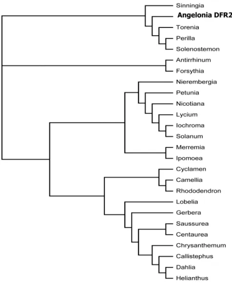
The two cDNA clones shared a sequence identity of 99% at the nucleotide level. In comparison toAng.DFR1,Ang.DFR2showed 4 exchanges and a deletion of 3 bp, which resulted in an exchange of two amino acids and a deletion of a proline in a proline-rich region at the N-terminus (Figure 3). Compared to many other DFRs, the cDNA clones fromAngeloniawere approximately 50 to 100 amino acids longer, but the length was comparable to those of the DFRs from the closely related species Antirrhinum majus (Accession X15536) andPerilla frutescens(Accession AB002817). In the phylogenetic analysis, the Angelonia DFRs clustered together with DFRs from Torenia, Perilla, Solenostemon and Sinningia, which all belong to the order Lamiales (Figure 4).
Heterologous expression in yeast and characterization of the recombinant enzymes
The cDNA clones were transferred into the pYES2.1 vector and heterologously expressed in yeast (Saccharomyces cerevisiae). The recombinant Ang.DFR2 was functionally active catalyzing the NADPH dependent conversion of dihydroflavonols into leu-coanthocyanidins (Table 2, grey-shaded). The enzyme demon-strated substrate specificity as described for a number of plants [25,26] and accepted DHQ and DHM as substrates but did not efficiently convert DHK. Johnson et al. [25] demonstrated that substrate specificity of DFR is determined in the amino acid region 132–157 in theGerbera hybridaDFR, which corresponds to amino acids 141–168 in Ang.DFR2. The presence of an aspartic acid in position 134 (position 143 of Ang.DFR2) of Petunia hybrida contrasts with the highly conserved asparagine found in many Figure 1.Angelonia6angustifoliaflowers.
doi:10.1371/journal.pone.0107755.g001
specificity [25]. However, Ang.DFR2 showed the same substrate specificity as the P. hybrida DFR despite the presence of an asparagine in this position. Ang.DFR1 in contrast did not show functional activity with any dihydroflavonol tested (Table 2, grey-shaded). The recombinant Ang.DFR2 was characterized in detail and the result is summarized as a standard enzyme assay in the Material and Method section and in Table 3.
Testing DFR activities
Substrate specificity of DFR has been intensively studied in various plants [25,26,28,33–41]. Substrate acceptance of DFR is either studiedin vitrousing recombinant enzymes, using enzyme preparations from plant sources, or in planta by monitoring changes in the flower colour of genetically modified plants. When substrate acceptance is comparedin vitro, several sources of error exist. Firstly, it is essential to consider the stereospecificity of DFR, which accepts only (+)-(2R,3R)-dihydroflavonols as substrates Figure 2.a: Chemical structure of dihydrokaempferol showing ring denotation and atom numbering. b: Simplified overview of the anthocyanin pathway. abbrev: ANS: anthocyanidin synthase, CHI: chalcone isomerase, CHS: chalcone synthase, DFR: dihydroflavonol synthase, FHT: flavanone 3-hydroxylase, F39H: flavonoid 39-hydroxylase, F3959H: flavonoid 39,59-hydroxylase.
doi:10.1371/journal.pone.0107755.g002
Table 1.Specific activities [nmol/s*kg] of chalcone synthase/chalcone isomerase (CHS/CHI), flavanone 3-hydroxylase (FHT) and dihydroflavonol 4-reductase (DFR) in genotypes ofAngelonia6angustifoliaflowers showing divergent flower colour.
Colour of Angelonia6angustifolia flower Specific enzyme activity [nmol/s*kg]
CHS/CHI FHT DFR
pink/blue 3463 6363 260615
white/blue 5664 4462 125610
white 6465 4864 0
[42]. Commercially available dihydroflavonols are frequently racemic mixtures and may occur in various qualities. The presence of impurities or the wrong stereoform may significantly hamper the reaction. Incubation of Ang.DFR2 with equimolar amounts of (+)-DHQ and a racemic mixture ((6)-taxifolin hydrate), respectively, reduced conversion rates of (6)-taxifolin by approximately 72% compared to (+)-DHQ. This is significantly higher than the 50% one would have expected from the fact that only one of the two stereoforms is converted. For comparison of different dihydroflavonols, it is therefore essential to use all of the same quality, stereoform and in equimolar amounts (same nmol, notmg). Another problem stems from the instability of leucoantho-cyanidins, which are rapidly oxidised and polymerized and may react with proteins [43–49]. With an increasing number of hydroxyl groups, compounds show a higher sensitivity towards oxidation and polymerization. Losses may thus vary for the different substrates and this could bias conclusions on substrate preference. Long incubation times (hours) should therefore be avoided.
As a rule, substrates and products are extracted with ethyl
TLC plates [37,38,50] or HPLC [33]. However solubility of leucoanthocyanidins in organic solvents varies [45] with their polarity, which increases with the number of hydroxyl groups. Compared to leucoanthocyanidins, dihydroflavonols are more easily extracted by ethyl acetate (Table S2). For DHK and DHQ this causes an acceptable error in conversion rates of approxi-mately 5%. Solubility of leucodelphinidin decreases dramatically however when compared to DHM and the other leucoanthocya-nidins (Table S2). As a result biased results are obtained for the conversion of DHM (Table 4). When leucodelphinidin was extracted with ethyl acetate, recovery rates were low in both organic and aqueous solvents (Table S2). We assume that this is a result of unspecific interactions with proteins in the interlayer after oxidation of the phenolic groups. Thus we recommend the separation of whole assays on paper stripes as described for glycosyltransferases [51]. This enables exhaustive recovery and reliable conversion rates for DHM.
Some authors demonstrate functional activity of DFRs by conversion of the formed leucoanthocyanidins into anthocyanidins via acidic treatment and subsequent anthocyanin quantification Figure 3. Alignment of the DFRs isolated from petals ofAngelonia6angustifolia.
doi:10.1371/journal.pone.0107755.g003
Figure 4. Phylogenetic tree of amino acid sequences of DFRs from different plant species.The following sequences were used (Accession numbers in parentheses):Angelonia6angustifolia Ang.DFR2 from the present study (KF285561),Antirrhinum majus (X15536),Perilla frutescens
(AB002817),Forsythia6intermedia(Y09127),Solenostemon scutellarioides(EF522155),Sinningia cardinalis(AY332536),Nierembergia sp.(AB078510),
Torenia hybrida(AB012924),Camellia sinensis(AB018686),Petunia6hybrida(EU189078),Nicotiana alata(FJ969389),Iochroma cyaneum(GU595064),
Solanum pinnatisectum(AY954035), Lycium ruthenicum (JN849097), Lobelia erinus (AB221076),Saussurea medusa (EF600682), Merremia dissecta
(EU189077),Ipomoea purpurea(AF028601),Rhododendron simsii(AJ413278),Centaurea maculosa(FJ376591),C. chinensis(Z67981),Dahlia variabilis
(FJ216425),Chrysanthemum6morifolium cultivar Shenyun(JF346164),Helianthus annuus(EU095849),Cyclamen graecum(AB517921),Gerbera hybrid
cv. ‘Terra Regina’ (Z17221).
doi:10.1371/journal.pone.0107755.g004
Table 2.Functional activity, substrate specificity and influence of mutations in the N-terminus on the functional activity of DFR fromAngelonia6angustifolia, varying positions between the two DFR types are marked in bold.
% Formation of
Mutation LPg LCy LDp
Ang.DFR1 PPPPPPSAAAVPATVCVTRA 0 0 0
G21 PPPPPPSAAAVPATVCVTGA 0 0 0
G23 PPPPSSAAAVPATVCVTRA 0 0 0
G24 PPPPSSAAAVPATVCVTGA 0 5362 4763
Ang.DFR2 PPPPSSAAAVPATVCVTGA 0 5064 4663
G3 PPPPSSAAAVPATVCVTRA 0 0 0
G4 PPPPPPSAAAVPATVCVTGA 0 0 0
G128 PPPPPPSAAAVPATVCVTRA 0 0 0
conversion is not quantitative and strongly depends on various factors [45]. Measurements of the converted anthocyanidin are therefore an acceptable tool for demonstrating functional activity of DFRs. It remains questionable however whether this provides reliable results for the comparison of substrates with divergent hydroxylation patterns.
Heterologous expression as glutathione S-transferase (GST) fusion protein inE. coli
Heterologous expression ofDFRis frequently performed inS. cerevisiae [50], however, this is time-consuming in comparison with expression in E. coli. Only a few recombinant DFRs were successfully expressed in E. coli thus far [28,33,40,41,53] with instability after His-tag removal reported. We therefore tested whether heterologous expression as a GST fusion protein inE. coli could be a viable option to rapidly produce highly purified, active recombinant DFR. TheDFRs were subcloned from pYES2.1 into the pGEX-6P-1 vector and heterologously expressed in E. coli. Purification of the recombinant proteins was performed by using glutathione SepharoseTM4B in either a batch procedure or by using GST Spin Traps. When the GST Spin Traps were used, a rapid loss of enzyme activity within two days was observed unless the enzyme was stabilized with BSA (2 mg/ml) and kept in Protein LoBind tubes. Both the resulting GST fusion protein and the purified enzyme after GST removal showed functional activity and no difference was observed in either the substrate acceptance of the fusion protein, the purified protein after GST removal or the recombinant enzyme produced in yeast. The purified recombinant
DFR was stable and could be kept at280uC for several months without loss of activity.
Restoring the functional activity of Ang.DFR1 by site-directed mutagenesis
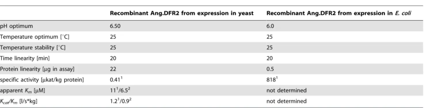
Although the two cDNA clones differed only in three amino acids, no functional activity could be observed with recombinant Ang.DFR1. To identify the decisive residues, we mutated the two DFRcDNA clones by site-directed mutagenesis and investigated the resulting recombinant proteins for functional activity. Neither the deletion of the extra proline, along with an exchange of serine to proline in the Ang.DFR1 sequence nor an exchange of the arginine 26 with glycine alone was able to restore the functional activity of Ang.DFR1. Only simultaneous mutation of all three positions resulted in a highly active recombinant Ang.DFR1. In the same way, exchange of glycine to arginine in Ang.DFR2 or insertion and exchange of an additional proline with serine at position 12 led to a complete loss of Ang.DFR2 activity (Table 2). The proline-rich area in theAngelonia DFRis closely located to the N-terminus and contains a unique structural feature (Figure S1), which is also not found in theVitisDFR (Swiss-Prot Accession Number P93799_VITVI) and therefore not covered by the crystal structure [28]. It is thus difficult to explain why differences in the three amino acids are essential for functional activity. The region however, is closely located to a glycine-rich motif constituting the NADPH binding site [28]. As proline often acts as a structural disruptor [54] we theorize that the presence of two additional prolines and the large arginine residue in position 26 of Table 3.Characterization of recombinant DFR fromAngelonia6angustifoliaobtained from heterologous expression in yeast (left)
andE. coli(right).
Recombinant Ang.DFR2 from expression in yeast Recombinant Ang.DFR2 from expression inE. coli
pH optimum 6.50 6.0
Temperature optimum [uC] 25 25
Temperature stability [uC] 25 25
Time linearity [min] 20 20
Protein linearity [mg in assay] 22 0.5
specific activity [mkat/kg protein] 0.411 8181
apparentKm[mM] 111/6.52 not determined
Kcat/Km[l/s*kg] 1.21/0.92 not determined
1DHQ as a substrate, 2DHM as a substrate.
doi:10.1371/journal.pone.0107755.t003
Table 4.Dependence of product formation through sample handling after stopping the enzyme reactions.
% Formation of
Sample handling LPg LCy LDp
no extraction, sample transferred on paper stripes 4262 7765 2161
separation of ethyl acetate extracts on TLC plates 4064 7763 763
remaining aqueous phase after ethyl acetate extraction transferred on paper stripes n.d. 7764 5363*
separation of 1-butanol extracts on TLC plates 4362 7562 962
The substrates were converted with recombinant dahlia DFR (Accession FJ216425). Substrates and products were separated on cellulose in chloroform/acetic acid/water (10:9:1, v:v:v). n.d: not detected.
*total amount of radioactivity was very low.
Ang.DFR1, which contrasts with the small glycine in Ang.DFR2, has an negative impact on protein folding or disturbs proper NADPH binding.
Conclusions
This study focused on the isolation and characterization ofDFR from Angelonia x angustifolia which seems to play an essential role in the formation of chimeral patterns resulting in the formation of white/blue flowers. The DFR was used as model cDNA clone to demonstrate the suitability for heterologous expression as a GST fusion protein in E. coli. In addition, we demonstrated possible sources for biased results in substrate acceptance studies with DFRs.
Materials and Methods
Chemicals
(2-14C)-Malonyl-coenzyme A (55 mCi/mmol) was purchased from New England Nuclear Corp. GmbH (Vienna, Austria). (14 C)-Labeled dihydroflavonols, (+)-DHK, (+)-DHQ, and (+)-DHM, were synthesized as described before [27,38] using recombinant F3959H from Sollya heterophylla and F39H from Tagetes erecta. (6)-Taxifolin hydrate was purchased from Sigma-Aldrich (Mu-nich, Germany).
Plant material
The studies were performed on pink/blue and white/blue flowering chimeras and white genotypes (arisen after somatic segregation of the white/blue chimera type) of Angelonia 6angustifolia plants (Figure 1) which were previously described [2]. The plants were grown in the greenhouse at the Julius Ku¨hn-Institut in Quedlinburg (Germany). Flowers were harvested, shock-frozen in liquid nitrogen and stored at280uC until use.
Enzyme preparation
Enzyme preparations were obtained using protocols 1 and 2 as described [31]. To remove low molecular compounds, crude enzyme preparations were passed through a gel chromatography column (Sephadex G25, GE Healthcare, Freiburg, Germany). Protein content was determined by a modified Lowry procedure [55] using crystalline bovine serum albumin as a standard.
Enzyme assays
Assays with enzyme preparations fromAngelonia petals were performed according to [31] using naringenin as a substrate for FHT and DHQ as a substrate for DFR. CHS cannot be measured independently from CHI because the latter does not need any cofactor and rapidly converts naringenin chalcone formed by CHS into naringenin. Therefore, a combined assay was used as described previously [31]. For the standard enzyme assay with the recombinant DFR obtained from yeast, the reaction contained in a final volume of 50mL: 15mL enzyme preparation (22mg), 0.048 nmol (14C)-dihydroflavonol, 0.25 nmol NADPH, 30mL 0.1 M KH2PO4/K2HPO4 buffer pH 6.5 containing 0.4% Na ascorbate. The amount of enzyme was chosen within a range to ensure that the maximum conversion rate of the best substrate was around 50% (linear range of the reaction). The reaction mixture was incubated for 15 min at 25uC. Assays using DHK and DHQ as substrates were stopped by addition of 10mL 100% acetic acid with products and substrates being extracted twice with 50mL ethyl acetate. The combined organic phases were transferred to a pre-coated thin-layer cellulose plates without fluorescence indica-tor (Merck, Darmstadt, Germany) and developed in chloroform/
acetic acid/water (10:9:1, v:v:v). The evaluation was carried out on a Berthold LB 2842 TLC Linear Analyzer (Wilbad, Germany) by integration of the peak areas. Assays using DHM as substrates were stopped with the addition of 10mL 100% acetic acid and 50mL methanol. The mixture was chromatographed on 20 cm61 cm stripes of paper (Schleicher Schuell, 2043 b, Germany) in chloroform/acetic acid/water (10:9:1, v:v:v). Con-version rates were determined using a Typhoon 8600 and the software ImageQuant 5.1 (GE Healthcare, Munich, Germany).
Characterization of the recombinant DFR
All data represents an average of at least three independent experiments. Determination of the pH optimum was carried out as described for the standard DFR assay, but using 0.2 M McIlvaine buffers with pH values between 4.5 and 9.0.
Cloning ofDFR cDNAs fromAngelonia6angustifolia
mRNA was isolated from Angeloniapetals using the mMACS mRNA Isolation Kit (Miltenyi Biotec, Germany). cDNA was prepared using the SuperScript II Reverse Transcriptase (Invitro-gen, Carlsbad, CA) and the oligo(-dT) anchor primer GAC-CACGCGTATCGATGTCGAC(T)16V. Based on information available in the NCBI-GenBank, nucleotide or amino acid sequences ofDFRs from other plants from the order Lamiales, were aligned (Accessions X15536; EU305680, Y09127, EF522156) and conserved regions in the N-terminal region adopting the Rossmann fold [28] were used for the design of degenerated primers. The obtained cDNA fragments were isolated, ligated into the vector pCR2.1-TOPO (Invitrogen, Carlsbad, CA) and transformed in E. coli strain TOP10 (Invitrogen, Carlsbad, CA). Fragments were sequenced by a commercial supplier (Microsynth Austria AG, Vienna, Austria) and sequences were used for the design of specific 59- and 39 -primers for the amplification of the ends of theDFR by RACE techniques, using the SMART RACE cDNA amplification kit (Clontech, Takara Bio Europe, Saint-Germain-en-Laye, France) according to the manufacturer’s instructions. Proofreading ampli-fication of the complete open reading frame was carried out using specific forward and reverse primers (Table S1) and theTaq/Pwo Expand High Fidelity PCR System (Roche, Mannheim, Ger-many).
Phylogenetic analysis
Multiple alignments were performed with ClustalW [56,57]. The phylogenetic tree was conducted and bootstrapped with MEGA version 5 [58] using the neighbor-joining method and 1000 replicates.
Heterologous expression inSaccharomyces cerevisiae Proofreading cDNA amplicons were ligated into the pYES2.1/ V5-His-TOPO vector (Invitrogen, Paisley, UK). Sense constructs were isolated and confirmed by sequencing. The vectors harbouring the DFR cDNAs Ang.DFR2 and Ang.DFR1 were transformed into yeast strain INVSc1 using the Sc. EasyComp Transformation Kit (Invitrogen, Carlsbad, CA). Preparation of the protein fractions was performed using a modified protocol according to Pompon et al. [59]. Protein fractions were shock frozen in liquid nitrogen and stored at280uC.
Heterologous expression inE.coli
double digestion withBamHI andEcoRI (Fermentas, Germany). Sticky endDFRinserts were generated using sticky end PCR [60] withPfuDNA polymerase (Fermentas, Germany) and the primers listed in Table S1. For each of theDFRs, two PCR reactions (PCR 1: A-DFR-FL, A-DFR-RS; PCR2: A-DFR-FS, A-DFR-RL) were performed with diluted pYES2.1 plasmids harbouring the Ang.DFR2 and Ang.DFR1 as templates. The PCR products were eluted and purified from the agarose gel after electrophoresis. Denaturation and reannealing of an equimolare mixture of the PCR products from the PCR 1 and 2 resulted theoretically in 25% double stranded dihydroflavonol 4-reductase sequences with sticky BamHI (GATC) andEcoRI (AATT) ends for direct ligation with T4 DNA ligase (Promega) into linearized pGEX-6P-1. After transformation into E. coli strain TOP 10 and isolation of the plasmids, integrity of the vector construct harbouring the dihydroflavonol 4-reductases (pGEX-6P-1::Ang.DFR2 and pGEX-6P-1::Ang.DFR1) was confirmed by sequencing. Single colonies harbouring parent DFRs and mutants were used for heterologous expression inE. colias described [61].
Purification of GST fusion proteins and removal of GST tag by enzymatic cleavage
GST fusion proteins were purified from the cell extract using Sepharose 4B (GE Healthcare, Munich, Germany) in either a batch procedure or by using GST Spin Traps according to the manufacturer’s instructions The DFR was liberated from GST by PreScission protease according to the GST Gene Fusion System Manual (GE Healthcare, Munich, Germany).
Site-directed mutagenesis
Mutants were generated from pGEX-6P-1 vector constructs harbouring theDFRsby use of Q5 Site-Directed Mutagenesis Kit
(NewEngland Bioloabs, Vienna Austria). Primer were designed using the NEBase ChangerTM v 1.01 provided at http:// nebasechanger.neb.com. The sequences are given in table S1. The integrity of the constructs was confirmed by commercial sequencing (Microsynth Austria AG, Vienna Austria).
Supporting Information
Figure S1 Alignment of the Ang.DFR2 amino acid sequence with DFRs from different plant species. Red letters indicate maximal consensus.
(DOC)
Table S1 List of primers used. (DOC)
Table S2 Recovery rates estimated by quantification of radioactivity at the scintillation counter after extraction of 10mM (14C)-labelled dihydroflavonols and leu-coanthocyanidins.
(DOC)
Acknowledgments
The authors kindly acknowledge excellent technical assistance from Renate Paltram and Ju¨rgen Greiner and the undergraduate student Stefan Hauer. Thanks to Luke McLaughlin for critically reading the manuscript. This work was performed within the frame of COST FA1006 (PlantEngine).
Author Contributions
Conceived and designed the experiments: HH KS AR. Performed the experiments: KMN SM SP MM SE. Analyzed the data: JT CM SP CG KMN SM MM KO SE AR HH. Contributed reagents/materials/analysis tools: KO SP AR CM HH KS. Wrote the paper: HH CG KS SP KO.
References
1. Albach D, Meudt H, Oxelman B (2005) Piecing together the ‘‘new’’ Plantaginaceae. American Journal of Botany 92: 297–315.
2. Plaschil S, Olbricht K (2008) Histogenetic variation in flowers ofAngelonia
Humb. et Bonpl. Journal of Applied Botany and Food Quality 82: 41–46. 3. Kampny CM (1995) Pollination and flower diversity in Scrophulariaceae. The
Botanical Review 61: 350–366.
4. Bailey LH (1947) Standard cyclopedia of horticulture. New York, London: Macmillan.
5. Moˆro FV, Pinto AC, dos Santos JM, Damia˜o Filho CF (2001) A scanning electron microscopy study of the seed and post-seminal development in
Angelonia salicariifoliaBonpl. (Scrophulariaceae). Annals of Botany 88: 499– 506.
6. Souza VC (2001) A new species of Bacopa Aubl. (Scrophulariaceae) from South America. Acta Bot Bras 15: 57–61.
7. Armitage AM (1998) The influence of light and temperature on flowering and growth ofAngelonia angustifoliaBenth. HortScience 33: 508–508. 8. Holton TA, Cornish EC (1995) Genetics and biochemistry of anthocyanin
biosynthesis. The Plant Cell 7: 1071.
9. Stich K, Eidenberger T, Wurst F, Forkmann G (1992) Enzymatic conversion of dihydroflavonols to flavan-3, 4-diols using flower extracts of Dianthus caryophyllusL. (carnation). Planta 187: 103–108.
10. Halbwirth H, Muster G, Stich K (2008) Unraveling the biochemical base of dahlia flower coloration. Natural Product Communications 3: 1259–1266. 11. Schwinn KE, Markham KR, Giveno NK (1993) Floral flavonoids and the
potential for pelargonidin biosynthesis in commercial chrysanthemum cultivars. Phytochemistry 35: 145–150.
12. Onozaki T, Mato M, Shibata M, Ikeda H (1999) Differences in flower color and pigment composition among white carnation (Dianthus caryophyllus L.) cultivars. Scientia Horticulturae 82: 103–111.
13. Miosic S, Knop K, Ho¨lscher D, Greiner J, Gosch C, et al. (2013) 4-Deoxyaurone formation inBidens ferulifolia(Jacq.) DC. PloS one 8: e61766.
14. Ohno S, Hosokawa M, Hoshino A, Kitamura Y, Morita Y, et al. (2011) A bHLH transcription factor, DvIVS, is involved in regulation of anthocyanin synthesis in dahlia (Dahlia variabilis). Journal of Experimental Botany 62: 5105– 5116.
15. Forkmann G, Stotz G (1984) Selection and characterisation of flavanone 3-hydroxylase mutants ofDahlia,Streptocarpus,VerbenaandZinnia. Planta 161:
16. Davies KM, Schwinn KE, Deroles SC, Manson DG, Lewis DH, et al. (2003) Enhancing anthocyanin production by altering competition for substrate between flavonol synthase and dihydroflavonol 4-reductase. Euphytica 131: 259–268.
17. Mol J, Grotewold E, Koes R (1998) How genes paint flowers and seeds. Trends in Plant Science 3: 212–217.
18. Koes R, Verweij W, Quattrocchio F (2005) Flavonoids: a colorful model for the regulation and evolution of biochemical pathways. Trends in Plant Science 10: 236–242.
19. Zhou X, Wang G, Zhang W (2007) UV-B responsive microRNA genes in
Arabidopsis thaliana. Molecular Systems Biology 3: 103.
20. Vaucheret H (2006) Post-transcriptional small RNA pathways in plants: mechanisms and regulations. Genes & Development 20: 759.
21. Filipowicz W, Bhattacharyya SN, Sonenberg N (2008) Mechanisms of post-transcriptional regulation by microRNAs: are the answers in sight? Nature Reviews Genetics 9: 102–114.
22. Deguchi A, Ohno S, Hosokawa M, Tatsuzawa F, Doi M (2013) Endogenous post-transcriptional gene silencing of flavone synthase resulting in high accumulation of anthocyanins in black dahlia cultivars. Planta: 1–11. 23. Almeida R, Allshire RC (2005) RNA silencing and genome regulation. Trends in
Cell Biology 15: 251–258.
24. Gou J-Y, Felippes FF, Liu C-J, Weigel D, Wang J-W (2011) Negative regulation of anthocyanin biosynthesis in Arabidopsis by a miR156-targeted SPL transcription factor. The Plant Cell Online 23: 1512–1522.
25. Johnson ET, Ryu S, Yi H, Shin B, Cheong H, et al. (2001) Alteration of a single amino acid changes the substrate specificity of dihydroflavonol 4-reductase. The Plant Journal 25: 325–333.
26. Johnson ET, Yi H, Shin B, Oh BJ, Cheong H, et al. (1999)Cymbidium hybrida
dihydroflavonol 4-reductase does not efficiently reduce dihydrokaempferol to produce orange pelargonidin-type anthocyanins. The Plant Journal 19: 81–85. 27. Halbwirth H, Kahl S, Jager W, Reznicek G, Forkmann G, et al. (2006) Synthesis
of (14
C)-labeled 5-deoxyflavonoids and their application in the study of dihydroflavonol/leucoanthocyanidin interconversion by dihydroflavonol 4-reductase. Plant Science 170: 587–595.
28. Petit P, Granier T, d’Estaintot BL, Manigand C, Bathany K, et al. (2007) Crystal structure of grape dihydroflavonol 4-reductase, a key enzyme in flavonoid
29. Winkel-Shirley B (2001) Flavonoid biosynthesis. A colorful model for genetics, biochemistry, cell biology, and biotechnology. Plant Physiology 126: 485. 30. Saito K, Kobayashi M, Gong Z, Tanaka Y, Yamazaki M (1999) Direct evidence
for anthocyanidin synthase as a 2-oxoglutarate-dependent oxygenase: molecular cloning and functional expression of cDNA from a red forma of Perilla frutescens. The Plant Journal 17: 181–189.
31. Halbwirth H, Waldner I, Miosic S, Ibanez M, Costa G, et al. (2009) Measuring flavonoid enzyme activities in tissues of fruit species. Journal of Agricultural and Food Chemistry 57: 4983–4987.
32. Claudot A-C, Drouet A (1992) Preparation and assay of chalcone synthase from walnut tree tissue. Phytochemistry 31: 3377–3380.
33. Xie D-Y, Jackson LA, Cooper JD, Ferreira D, Paiva NL (2004) Molecular and biochemical analysis of two cDNA clones encoding dihydroflavonol-4-reductase fromMedicago truncatula. Plant Physiology 134: 979–994.
34. Trabelsi N, Petit P, Manigand C, Langlois d’Estaintot B, Granier T, et al. (2008) Structural evidence for the inhibition of grape dihydroflavonol 4-reductase by flavonols. Acta Crystallographica Section D: Biological Crystallography 64: 883– 891.
35. Peters DJ, Constabel CP (2002) Molecular analysis of herbivore-induced condensed tannin synthesis: cloning and expression of dihydroflavonol reductase from trembling aspen (Populus tremuloides). The Plant Journal 32: 701–712. 36. Thill J, Regos I, Farag MA, Ahmad AF, Kusek J, et al. (2012) Polyphenol
metabolism provides a screening tool for beneficial effects ofOnobrychis viciifolia
(sainfoin). Phytochemistry 82: 67–80.
37. Fischer D, Stich K, Britsch L, Grisebach H (1988) Purification and characterization of (+)-dihydroflavonol (3-hydroxyflavanone) 4-reductase from flowers ofDahlia variabilis. Archives of Biochemistry and Biophysics 264: 40– 47.
38. Fischer TC, Halbwirth H, Meisel B, Stich K, Forkmann G (2003) Molecular cloning, substrate specificity of the functionally expressed dihydroflavonol 4-reductases from Malus domestica and Pyrus communis cultivars and the consequences for flavonoid metabolism. Archives of Biochemistry and Biophysics 412: 223–230.
39. Polashock JJ, Griesbach RJ, Sullivan RF, Vorsa N (2002) Cloning of a cDNA encoding the cranberry dihydroflavonol-4-reductase (DFR) and expression in transgenic tobacco. Plant Science 163: 241–251.
40. Hua C, Linling L, Shuiyuan C, Fuliang C, Feng X, et al. (2013) Molecular cloning and characterization of three Genes Encoding dihydroflavonol-4-Reductase fromGinkgo bilobain anthocyanin biosynthetic pathway. PloS one 8: e72017.
41. Shimada N, Sasaki R, Sato S, Kaneko T, Tabata S, et al. (2005) A comprehensive analysis of six dihydroflavonol 4-reductases encoded by a gene cluster of theLotus japonicusgenome. Journal of Experimental Botany 56: 2573–2585.
42. Heller W, Forkmann G, Britsch L, Grisebach H (1985) Enzymatic reduction of (+)-dihydroflavonols to flavan-3, 4-cis-diols with flower extracts fromMatthiola incanaand its role in anthocyanin biosynthesis. Planta 165: 284–287. 43. Deffieux D, Gaudrel-Grosay S, Grelard A, Chalumeau C, Quideau S (2009)
Stable solid-supported leucoanthocyanidin variants for flavanoid biosynthesis elucidation. Tetrahedron Letters 50: 6567–6571.
44. Ito S, Joslyn M (1965) Apple leucoanthocyanins. Journal of Food Science 30: 44–51.
45. Smathers RU, Charley H (1967) Problems in measuring leucoanthocyanin content of pears. Journal of Food Science 32: 310–314.
46. Stafford HA, Lester HL, Porter LJ (1985) Chemical and enzymatic synthesis of monomeric procyanidins (leucocyanidins or 39, 49, 5, 7-tetrahydroxyflavan-3, 4-diols) from (2R,3R)-dihydroquercetin. Phytochemistry 24: 333–338. 47. Hsia C, Claypool L, Abernethy J, Esau P (1964) Leucoanthocyan material from
immature peaches. Journal of Food Science 29: 723–729.
48. Halbwirth H, Fischer TC, Roemmelt S, Spinelli F, Schlangen K, et al. (2003) Induction of antimicrobial 3-deoxyflavonoids in pome fruit trees controls fire blight. Journal of BioSciences 58c: 765–770.
49. Pfeiffer J, Ku¨hnel C, Brandt J, Duy D, Punyasiri P, et al. (2006) Biosynthesis of flavan 3-ols by leucoanthocyanidin 4-reductases and anthocyanidin reductases in leaves of grape (Vitis viniferaL.), apple (MalusxdomesticaBorkh.) and other crops. Plant Physiology and Biochemistry 44: 323–334.
50. Martens S, Teeri T, Forkmann G (2002) Heterologous expression of dihydroflavonol 4-reductases from various plants. FEBS letters 531: 453–458. 51. Stich K, Halbwirth H, Wurst F, Forkmann G (1997) UDP-glucose: flavonol
7-O-glucosyltransferase activity in flower extracts of Chrysanthemum segetum. Zeitschrift fu¨r Naturforschung C, Journal of Biosciences 52: 153.
52. Des Marais DL, Rausher MD (2008) Escape from adaptive conflict after duplication in an anthocyanin pathway gene. Nature 454: 762–765. 53. Winefield CS, Lewis DH, Swinny EE, Zhang H, Arathoon HS, et al. (2005)
Investigation of the biosynthesis of 3-deoxyanthocyanins inSinningia cardinalis. Physiologia Plantarum 124: 419–430.
54. Hotzy J, Schneider N, Kovermann P, Fahlke C (2013) Mutating a conserved proline residue within the trimerization domain modifies Na+ binding to excitatory amino acid transporters and associated conformational changes. Journal of Biological Chemistry 288: 36492–36501.
55. Sandermann H, Strominger JL (1972) Purification and properties of C55-isoprenoid alcohol phosphokinase from Staphylococcus aureus. Journal of Biological Chemistry 247: 5123–5131.
56. Thompson JD, Higgins DG, Gibson TJ (1994) CLUSTAL W: improving the sensitivity of progressive multiple sequence alignment through sequence weighting, position-specific gap penalties and weight matrix choice. Nucleic Acids Research 22: 4673–4680.
57. Thompson JD, Gibson T, Higgins DG (2002) Multiple sequence alignment using ClustalW and ClustalX. Current Protocols in Bioinformatics: 2.3. 1–2.3. 22.
58. Tamura K, Peterson D, Peterson N, Stecher G, Nei M, et al. (2011) MEGA5: molecular evolutionary genetics analysis using maximum likelihood, evolution-ary distance, and maximum parsimony methods. Molecular Biology and Evolution 28: 2731–2739.
59. Pompon D, Louerat B, Bronine A, Urban P (1996) [6] Yeast expression of animal and plant P450s in optimized redox environments. Methods in Enzymology 272: 51–64.
60. Zeng G (1998) Sticky-end PCR: new method for subcloning. BioTechniques 25: 206–208.
![Table 1. Specific activities [nmol/s*kg] of chalcone synthase/chalcone isomerase (CHS/CHI), flavanone 3-hydroxylase (FHT) and dihydroflavonol 4-reductase (DFR) in genotypes of Angelonia 6 angustifolia flowers showing divergent flower colour.](https://thumb-eu.123doks.com/thumbv2/123dok_br/18325104.350208/3.918.190.732.94.648/activities-flavanone-hydroxylase-dihydroflavonol-genotypes-angelonia-angustifolia-divergent.webp)