Sevoflurane Inhibits Nuclear Factor-
B
Activation in Lipopolysaccharide-Induced
Acute Inflammatory Lung Injury via Toll-Like
Receptor 4 Signaling
Xi Jia Sun, Xiao Qian Li, Xiao Long Wang, Wen Fei Tan, Jun Ke Wang*
Department of Anesthesiology, First Affiliated Hospital, China Medical University, Shenyang 110001, Liaoning, China
Abstract
Background
Infection is a common cause of acute lung injury (ALI). This study was aimed to explore whether Toll-like receptors 4 (TLR4) of airway smooth muscle cells (ASMCs) play a role in
li-popolysaccharide (LPS)-induced airway hyperresponsiveness and potential mechanisms.
Methods
In vivo: A sensitizing dose of LPS (50µg) was administered i.p. to female mice before anes-thesia with either 3% sevoflurane or phenobarbital i.p. After stabilization, the mice were challenged with 5µg of intratracheal LPS to mimic inflammatory attack. The effects of sevo-flurane were assessed by measurement of airway responsiveness to methacholine, histo-logical examination, and IL-1, IL-6, TNF-αlevels in bronchoalveolar lavage fluid (BALF). Protein and gene expression of TLR4and NF-κB were also assessed.In vitro: After
pre-sensitization of ASMCs and ASM segments for 24h, levels of TLR4and NF-κB proteins in
cultured ASMCs were measured after continuous LPS exposure for 1, 3, 5, 12 and 24h in presence or absence of sevoflurane. Constrictor and relaxant responsiveness of ASM was measured 24 h afterwards.
Results
The mRNA and protein levels of NF-κB and TLR4in ASM were increased and maintained at
high level after LPS challenge throughout 24h observation period, bothin vivoandin vitro. Sevoflurane reduced LPS-induced airway hyperresponsiveness, lung inflammatory cell in-filtration and proinflammatory cytokines release in BALF as well as maximal isometric con-tractile force of ASM segments to acetylcholine, but it increased maximal relaxation response to isoproterenol. Treatment with specific NF-κB inhibitor produced similar
protec-tions as sevoflurane, including decreased expressions of TLR4and NF-κB in cultured
OPEN ACCESS
Citation:Sun XJ, Li XQ, Wang XL, Tan WF, Wang
JK (2015) Sevoflurane Inhibits Nuclear Factor-κB Activation in Lipopolysaccharide-Induced Acute Inflammatory Lung Injury via Toll-Like Receptor 4 Signaling. PLoS ONE 10(4): e0122752. doi:10.1371/ journal.pone.0122752
Academic Editor:Bernhard Ryffel, French National
Centre for Scientific Research, FRANCE
Received:January 9, 2015
Accepted:February 13, 2015
Published:April 14, 2015
Copyright:© 2015 Sun et al. This is an open access
article distributed under the terms of theCreative Commons Attribution License, which permits unrestricted use, distribution, and reproduction in any medium, provided the original author and source are credited.
Data Availability Statement:All relevant data are
within the paper.
Funding:Funding for this project was provided by
National Natural Science Foundation of China, No. 30371376. The funders had no role in study design, data collection and analysis, decision to publish, or preparation of the manuscript.
Competing Interests:The authors have declared
ASMCs and improved pharmacodynamic responsiveness of ASM to ACh and isoproterenol.
Conclusions
This study demonstrates the crucial role of TLR4activation in ASMCs during ALI in
re-sponse to LPS. Sevoflurane exerts direct relaxant and anti-inflammatory effectsin vivoand in vitrovia inhibition of TLR4/NF-κB pathway.
Introduction
Airway smooth muscle (ASM) is the primary effector tissue for regulating bronchomotor tone. ASM damage occurs in several pulmonary diseases such as asthma and chronic obstructive pulmonary disease (COPD) [1,2]. ASM cells (ASMCs) are highly plastic and their contractility and regulatory functions are altered by cytokines and chemokines released in response to flammatory stimuli. Recent studies have shown that ASMCs are highly activated during in-flammation and lung tissue damage with engagement of Toll-like receptors (TLRs) [3,4]. TLRs are transmembrane pattern recognition receptors. As a part of the innate immune system, they are key elements in recognizing viral and bacterial components [5,6]. To date, 11 different TLRs have been identified in humans. All TLRs possess a common intracellular TIR domain for initiating a signal following activation by bacterial components or conserved pathogen-as-sociated molecular patterns of microbes [7,8]. Detection of microbes by TLRs evokes an in-flammatory response. TLR4can be specifically activated by presenting a pathogen-derived antigen to naive T cells when sensing lipopolysaccharide (LPS), a common constituent in the cell wall of gram-negative bacteria, to initiate an immune response [6,9–12]. Collectively, these data support an emerging concept that the quantity of TLR4expressed on ASMCs will elicit constrictor and relaxant responses of ASM secondary to the autocrine actions of cytokines re-leased by the sensitized ASM itself [2,13,14].
It has been well documented that volatile anesthetics contribute to immunosuppression in the postoperative period, especially when they are applied at higher concentrations or for lon-ger times [15]. Volatile anesthetics are used to treat status asthmaticus; thus, the effects of vola-tile anesthetics on lung tissue have been a focus of attention. Some studies reported that volatile anesthetics delivered by mechanical ventilation alleviated lung inflammation by reduc-ing TNF-αand nitric oxide release in rats that had received intratracheal LPS [16–20]. Sevo-flurane is commonly used to sedate patients prior to intubation for mechanical ventilation because of its good controllability [21]. Recently, additional advantages of sevoflurane have been reported, including dilation of bronchioles and reduction of bronchial hyperresponsive-ness in patients with asthma, indicating that it has lung protective effects [21–23]. However, the mechanism of these effects remains unclear. Reversible inhibition of voltage-dependent cal-cium channels and decreased intracellular calcal-cium mobilization may result in a decreased con-centration of intracellular calcium in response to volatile anesthetics [21,24].
lung inflammation and airway hyperresponsiveness in a series ofin vivoandin vitro
experi-ments. This may lead to new and safe therapeutic methods.
Materials and Methods
In vivo
Ethics statement. Female C57BL/6 mice, weighing 20–30 g, aged 8–12 weeks, were ob-tained from the Animal Facility at the China Medical University. Mice were bred in micro-iso-lator cages (24°C) under a 12:12 h light–dark cycle with free access to regular chow and water. Animal care and experimental procedures were approved by the Animal Care and Use Com-mittee of the China Medical University.
Animals and experimental protocols. At 42–56 days of age, all mice were anesthetized with phenobarbital (20 mg/kg, i.p.) 4 hours after treatment with 50μg LPS (Escherichia coli
se-rotype 055:B5; Sigma-Aldrich) for sensitization. After stabilization, the mice were randomized to one of four groups: PENTO + Normal saline (NS) group (PN group, n = 8), PENTO + LPS group (PL group, n = 8), SEV + NS group (SN group, n = 8), and SEV + LPS group (SL group, n = 8), and continuously maintained under anesthesia with 3% sevoflurane (Abbott, Wiesba-den, Germany) or with additional bolus doses of phenobarbital, if necessary. Anesthesia depth was ensured by absent pedal reflexes throughout the experiments. Tracheas were exposed with a neck midline incision under sterile conditions, cannulated with a 20-gauge, 1-inch-long cath-eter, and sutured. Animals were ventilated at a tidal volume of 20 ml/kg with 100 breaths per minute. The end-tidal sevoflurane concentration was measured continuously with a gas analyz-er (Capnomac Ultima; Datex, Helsinki, Finland). Aftanalyz-er the end-tidal concentration of sevoflur-ane was stable for 15 min, airway responsiveness to a methacholine (Mch) challenge was evaluated and the mice were euthanized to harvest tissue samples. During surgical and experi-mental procedures, oxygen saturation, heart rate, and rectal temperature were continuously monitored and maintained within physiologic ranges (Fig 1A).
Airway responsiveness determination. Airway responsiveness to methacholine (Mch, acetyl-ß-methylcholine chloride, Sigma, St. Louis) was assessed with a plethysmographic chamber (Buxco Electronics, Sharon, CT) sized for mice and connected to a pressure transduc-er (PT-5; Grass Instruments, West Warwick, RI). Mice wtransduc-ere exposed to increasing concentra-tions of nebulized Mch (2.5μg/kg, 25μg/kg, 50μg/kg, 100μg/kg, and 200μg/kg) by an aerosonic ultrasonic nebulizer (DeVilbiss). The initial values were taken as baseline and the ex-piratory resistance (Re) and dynamic lung compliance (Cldyn) values for each Mch challenge dose were recorded with AniRes2003 software. The time interval of administration was at least 5 min to ensure the pressure returned to baseline.
Harvesting and analysis of BALF. Lung inflammation was evaluated by cell counts and protein contents in bronchoalveolar lavage fluid (BALF). The left lung was ligated and the right lung was lavaged three times with 1 mL of PBS. The amount of BALF retrieved was approxi-mately 80% of the instilled volume. After centrifugation for 10min at 1000r/min, the pellet was resuspended in PBS. Total cell numbers were counted using a hemocytometer, and the num-bers of eosinophils, neutrophils, macrophages, and lymphocytes determined on the basis of morphologic criteria. At least 200 cells were counted per sample. The supernatants were col-lected and immediately frozen on dry ice and stored at -80°C for cytokine measurements using ELISA Kits (Quantikine M; R&D Systems Europe) for IL-1, IL-6, and TNF-α.
according to the standard procedure. The sections were deparaffinized, hydrated gradually, and stained with hematoxylin and eosin. The slices were examined by light microscopy.
Immunohistochemical analysis for the expression of TLR4. The localization of TLR4
was determined by immunocytochemistry. Paraffin histologic sections were dewaxed with xy-lene and then rehydrated in graded concentrations of ethanol. Sections were incubated with rabbit anti-mouse TLR4antibodies (eBioscience, San Diego, CA) overnight at room tempera-ture. Secondary swine anti-rabbit antibodies (DAKO, Copenhagen, Denmark) conjugated to Fig 1. Schematic diagram of experimental design.(A) Experimental protocolin vivo. (B) Experimental protocolin vitro.
streptavidin-biotin horseradish peroxidase were added for 30 minutes. Staining was revealed using a HRP-Dab Staining Kit (Vector Laboratories, Burlingame, CA). Densitometric analysis was performed using MetaMorph/Evolution MP 5.0/BX51 Image analytical system (Beijing, China).
Western blot analysis for TLR4and NF-κB expression. The expression of TLR4and NF-κB was determined by western blot analyses. Cytosolic proteins and nuclear proteins for TLR4
or NF-κB were isolated from the lung tissue using a modification of the method described by Dong [30]. The proteins were probed with primary TLR4 antibodies and NF-κB antibodies (Santa Cruz Biotechnology) overnight at 4°C. Then, alkaline phosphatase–conjugated second-ary antibodies were added to enhance chemiluminescence detection (NBT/BCIP, Promega, ShangHai, China). Blots were compared with reference to the findings for GAPDH and quanti-fied by densitometric analysis with Lab works software (UVP Upland, CA, USA).
Real-time quantitative PCR for TLR4mRNA detection. Total RNA was isolated with
TRIzol reagent and purified from ASM by the RNeasy Mini Kit (Qiagen, West Sussex, U.K.) according to the manufacturer’s instructions. Quantitative real-time PCR for TLR4mRNA was carried out using an ABI Prism 7000 (Applied Biosystems, Foster City, CA) under the follow-ing amplified cyclfollow-ing conditions: 2 min at 50°C for Uralic-DNA glycosylase incubation; 10-min denaturation at 95°C; 40 cycles of 15-s annealing at 95°C and elongation for 1 min at 60°C with SYBR Green PCR Master Mix Reagent (Applied Biosystems). The primer sequences used for PCR were as follows: TLR4: Forward AGGTCGGTGACTTCAAGAC-3'; Reverse 5'-CCACCTCTGTTTTA-3' NF-κB: Forward CCTGCTTCTGGAGGGTGATG-3'; Reverse 5'-TCCGGCCGCTATATGCA-3'. To confirm appropriate amplification, the size of the PCR products was verified on gels. The quantity of gene expression was assessed by the comparative Ct method and expression of GAPDH was used as a loading control. All reactions were per-formed in triplicate.
In vitro
ASMC culture and protocols of sevoflurane and LPS exposure. In order to avoid the complexity ofin vivomodels and to better understand the underlying mechanisms, we have
simulated the interventions described above in anin vitromodel. ASMCs were purchased from
PriCells (MIC-CELL-0005, PriCells, WuHan, China). They were cultured and expanded in Dulbecco’s modified Eagle’s medium (DMEM) supplemented with 10% fetal bovine serum ac-cording to the manufacturer’s instructions. Cells at passages 4–6 were used for experiments.
Immunoblot analysis of TLR4and NF-κB. TLR4and NF-κB in lysates isolated from cul-tured ASMCs were detected using the ECL system (Amersham Biosciences, US) followed by exposure to autoradiography film. The protein band intensities were quantified by densitome-try as previously described.
Real-time quantitative PCR for TLR4mRNA detection. TLR4 mRNA in purified nuclear
samples from cultured ASMCs as assessed by real-time quantitative PCR performed as above.
Preparation and treatment of mouse ASM tissues
After general anesthesia with intramuscular injections of phenobarbital (20 mg/kg) and exsan-guination, tracheas were dissected via open thoracotomy and divided into eight ring segments, 6–8 mm in length, as described previously [31]. The ASM segments were cultured in Krebs-Henseleit buffer at room temperature for 24 h and then treated according to the same protocols as cultured ASMCs above.
Pharmacodynamic studies of ASM constrictor and relaxant responsiveness. After 24-h incubation, ASM segments were placed in organ baths containing Krebs-Henseleit buffer aerat-ed with 5% CO2in oxygen, and the tissues were attached to force transducers (Trading, Aar-hus, Denmark) from which isometric tension was continuously displayed on a multichannel recorder. Tracheal segments were mounted on two L-shaped metal prongs, with a resting ten-sion of 0.8 mN, as previously described [32]. The maximal isometric contractile force (Tmax) of ASM segments to cumulative administration of acetylcholine (ACh) in final bath and subse-quently maximal relaxation (Rmax) to isoproterenol were assessed as previously described [32]. The relaxation responses to isoproterenol were elicited after half-maximal contraction by an ED50dose of ACh and analyzed in terms of % Rmax from the initial contracted level. Con-centrations of ACh from10-9to 10–3M and of isoproterenol from 10–9to 10–4M were used in this experiment.
Statistical analysis
Data are presented as mean ± standard error of mean (SEM). Comparisons between groups were made using the Student’s t-test (two-tailed) or ANOVA with Tukey’s post-test analysis, where appropriate. All test results were considered significant atP<0.05.
Results
Airway responsiveness determination
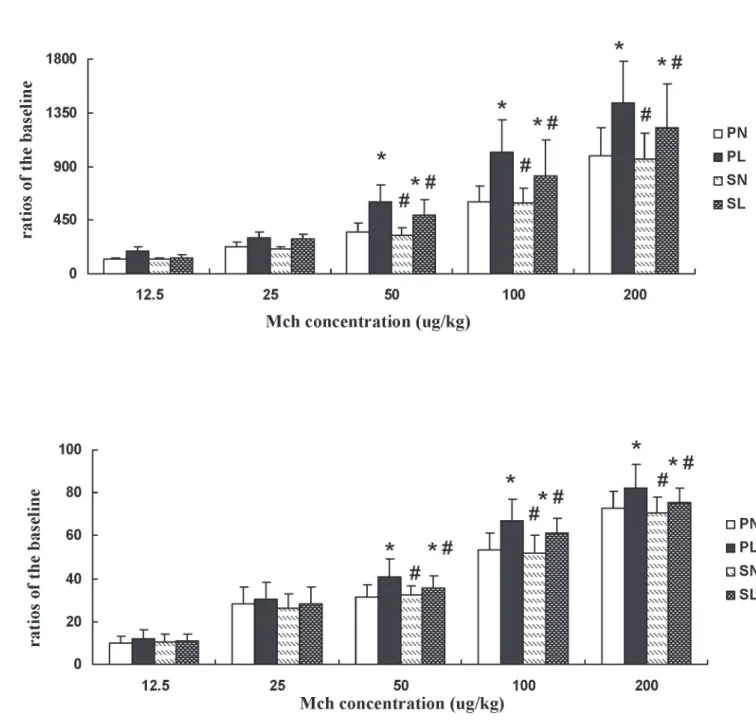
Airway responsiveness was calculated as the ratio of expiratory resistance (Re) and dynamic lung compliance (Cldyn) following exposure to different Mch doses. Re was significantly in-creased and Cldyn significantly dein-creased in mice pretreated with LPS at each challenge dose of Mch, whether in the absence or presence of sevoflurane (Fig 2A,P<0.05). The changes in
Re and Cldyn in mice with LPS exposure were significantly higher than the changes in those anesthetized with sevoflurane when challenged with doses of 50μg/kg, 100μg/kg, and 200μg/kg (P<0.05). There were no significant differences among the other groups (Fig 2B,P>0.05).
Additionally, exposure to LPS increased the protein content and neutrophil recruitment in BALF compared to that observed in controls. Sevoflurane inhaled after LPS sensitization de-creased cellular and neutrophil recruitment into BALF and the protein content increase (Fig 3B,P<0.05).
Fig 2. Effects of sevoflurane on expiratory resistance (Re) and dynamic lung compliance (Cldyn) in mice after different concentrations of methacholine (Mch) after LPS challenge.(A) Ratios of Re changed with different concentrations of Mch.(B) Ratios of Cldyn changed with different concentrations of Mch. Data are presented as means±SEM (n = 8 per group).*P<0.05 versus PN; #P<0.05, versus PL.
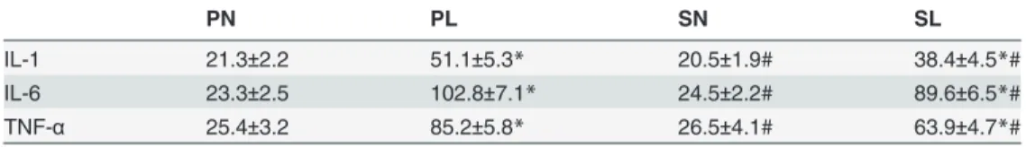
Cytokine measurement in BALF
Proinflammatory factors such as IL-1, IL-6, and TNF-αhave been implicated as important me-diators of inflammatory events. LPS challenge increased levels of IL-1, IL-6, and TNF-α Fig 3. Effects of sevoflurane on histopathologic outcome and cell counts in bronchoalveolar lavage fluid (BALF) after LPS challengein vivo.(A) Representative photomicrographs of lung sections stained with hematoxylin-and-eosin from mice anesthetized with phenobarbital or sevoflurane in the absence or presence of LPS challenge. The mice in group PN and SN showed much less inflammatory cell infiltration, alveolar septal thickening, and pulmonary edema in the lung compared with the mice exposed to LPS (NL+SL). Sevoflurane administered after intraperitoneal sensitization could mitigate lung inflammation upon re-challenge with LPS. Original magnification 100×. (B) Total cell and neutrophil counts in BALF. Data are presented as
means±SEM (n = 8 per group).*P<0.05 versus PN; #P<0.05, versus PL.
compared with the controls, regardless of anesthetic techniques. Treatment with sevoflurane after intraperitoneal sensitization halted the increase of the above factors after LPS aerosol challenge (Table 1,P<0.05).
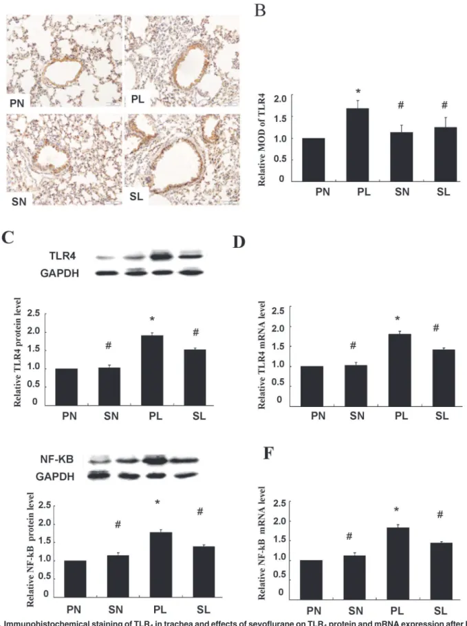
Measurement of TLR
4in lung tissue
Immunoreactivity of TLR4was observed only in the cell membrane and cytoplasm of the smooth muscle cell layer (Fig 4A). Quantitative analysis showed that sections taken from the mice exposed to LPS were stained more extensively and deeper than those in control groups. Immunoreactivity of TLR4in response to LPS was decreased after pretreatment with sevoflur-ane (Fig 4B,P<0.05). Activation of TLR4in the smooth muscle layer was closely related to
LPS-induced lung injury, and could be attenuated by sevoflurane inhalation. These findings were further confirmed and quantified by western blotting and real-time PCR (Fig 4C and 4D,
P<0.05).
Measurement of NF-
κ
B in lung tissue
The TLR signaling pathway culminates in the activation of the transcription factor NF-κB. We examined whether up-regulation of TLR4would lead to NF-κB activation and whether treat-ment with sevoflurane affected such a change. Western blot analysis indicated that markedly higher levels of NF-κB protein were expressed in mice exposed to LPS than in the controls, and this closely accompanied the up-regulation of TLR4(Fig 4D,P<0.05). Additionally, these
changes were ameliorated by sevoflurane preconditioning between sensitization and re-chal-lenge (Fig 4D,P<0.05).
Similar results were found with real-time PCR (Fig 4E,P<0.05). These results suggest that
TLR4and NF-κB are likely involved in the same signaling pathway, and the inhibition of NF-κB translocation mostly contributed to the protective effects of sevoflurane. However, it was
in-teresting to find that sevoflurane alone, in the absence of LPS challenge, did not affect either TLR4or NF-κB expression.
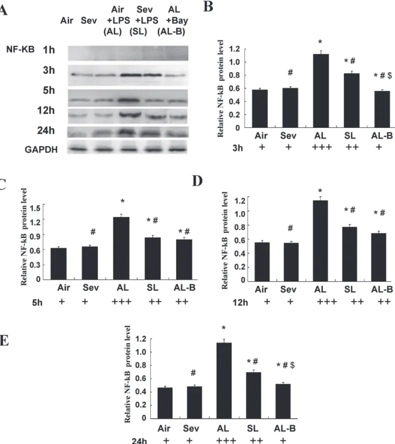
Effects of sevoflurane on protein expression of TLR
4and NF-
κ
B in
isolated ASMCs
We performed parallel experiments in cultured ASMCs. As shown in Figs5and6, significantly increased protein expression of TLR4and NF-κB was measured after LPS pre-sensitization of ASMCs and continuous LPS exposure (1 ng/ml) for 3, 5, 12, and 24 h (Group Air vs. Group AL,P<0.05). Sevoflurane prevented these increases in TLR4and NF-κB (Group AL vs. Group SL,P<0.05). There were no marked changes of TLR4and NF-κB in cultured ASMCs at the
Table 1. Effects of sevoflurane on concentrations of cytokines in BALF of mice exposed to LPS (ng/L).
PN PL SN SL
IL-1 21.3±2.2 51.1±5.3* 20.5±1.9# 38.4±4.5*#
IL-6 23.3±2.5 102.8±7.1* 24.5±2.2# 89.6±6.5*#
TNF-α 25.4±3.2 85.2±5.8* 26.5±4.1# 63.9±4.7*#
Data are presented as mean±SD (n = 8 per group).
*P<0.05 compared with PN; # P<0.05 compared with PL.
Fig 4. Immunohistochemical staining of TLR4in trachea and effects of sevoflurane on TLR4protein and mRNA expression after LPS challengein
above time points after treatment with sevoflurane or in controls without LPS challenge (Group Air vs. Group Sev,P>0.05).
We performed experiments with Bay 11–7082 (10μM), a specific inhibitor of NF-κB, to fur-ther confirm the signal mediating activation of TLR4. The results showed synergistically pro-tective actions to decrease the expression of TLR4and NF-κB throughout the 24 h observation period (Figs5and6).
ASM constrictor and relaxant responsiveness
in vitro
As a final assessment of the hypothesized interactions between NF-κB and sevoflurane, we compared the constrictor and relaxation responses in LPS-exposed isolated ASM segments in the absence and presence of sevoflurane and NF-κB inhibitors. Relative to control ASM, the maximal isometric contractile force responses to ACh were significantly increased in LPS-ex-posed rings (Tmax= 112.2 ± 8.4 g/g vs. 85.3 ± 8.6 g/g in control tissues,P<0.05). The enhanced responsiveness to ACh was mitigated in rings pretreated with sevoflurane (Tmax = 91.9 ± 9.9 g/g,
P<0.05). Similar protective effects were observed after pretreatment with Bay 11–7082 (96.9 ±
7.3 g/g,P>0.05).
Under the same treatment conditions, the relaxation responses to isoproterenol were signifi-cantly attenuated in LPS-exposed ASM, in which the mean Rmax responses amounted to 34.8 ± 4.7% vs. 59.1 ± 3.8% obtained in the control tissues (P<0.05). The decreased ratios of
Rmax were significantly mitigated in SL after LPS-challenge to 42.3± 3.6% (P<0.05), and
treatment with Bay 11–7082 provided similar Rmax values as the SL group (44.2± 4.0%,
P>0.05). Additionally, there were no marked changes in Tmax and Rmax in ASM of controls,
whether or not they were treated with sevoflurane (P>0.05) in the absence of LPS-challenge.
Discussion
It has been noted that surgical patients are more prone to airway hyperreactivity (AHR) during the perioperative period if they have previously had a respiratory infection. A better knowledge of the role of infection in AHR exacerbations might reveal new therapeutic interventions. TLR4 is ubiquitous in cells and is specifically activated by LPS. LPS is a major initiator of host im-mune responses and triggers expression of TLR4[24] and precipitates lung injury [7,10]. Therefore, intratracheal LPS instillation provides a useful experimental system for investigating the mechanisms of AHR in anesthetized and ventilated rats. Our study is among the first to demonstrate the effectiveness of sevoflurane inhalation in modifying AHR by interfering with the activation of NF-κB via TLR4on ASM after LPS-sensitization.
BALB/c mice were first treated with 50μg LPS i.p. to initiate local inflammatory reactions. The dosage of LPS and length of exposure were determined from preliminary experiments. There were no significant differences among the groups if the dose was too small or the expo-sure time was too short, whereas at a high dose or for a long time, ALI occurred during the sen-sitization period instead of during airway exposure. As previously reported, pulmonary
The presence of TLR4was observed only in the smooth muscle cell layer, whose cell membrane and cytoplasm stained tan as detected by horseradish peroxidase–labeled antibodies. Scale bars = 50μm. (B) Histogram for quantification of TLR4in smooth muscle cell layer. The mean optical density values (MOD) were calculated after normalizing against PN. (C) Representative western blot and quantitative analysis of TLR4isolated from smooth muscle cell layer after LPS challenge. (D) Quantitative real-time PCR of TLR4mRNA expressions in isolated smooth muscle cell layer. (E) Representative western blot and quantitative protein analysis of NF-κB in nuclear extracts from smooth muscle cell layer after LPS challenge. (F) Quantitative real-time PCR of NF-κB mRNA expressions in smooth muscle cell layer after LPS challenge. Sevoflurane prevented significant increases in protein and mRNA expressions of TLR4 and NF-κB in isolated smooth muscle cell layer from mice after LPS challenge. The relative integral density values (IDVs) were calculated after normalizing against GAPDH in each sample and presented as relative protein expression units. Data are presented as means±SEM (n = 8 per group).*P<0.05 versus PN; #P<0.05, versus PL.
Fig 5. Effects of sevoflurane on TLR4protein expression in airway smooth muscle cells (ASMCs) after continuous LPS exposure for 1, 3, 5, 12, and
function was obviously altered at 4 h after endotoxin administration in a mouse model of endo-toxin-induced lung injury [33]. This is the time period we chose for sensitization. In our model, we observed lung function alteration by airway responsiveness to methacholine after LPS exposure in the presence or absence of inhaled sevoflurane. The results of the present study showed that prolonged stimulation of ASM with LPS resulted in a marked increase in air-way responses to methacholine.
TLR4has been demonstrated to be a specific receptor for LPS. To further identify the role of TLR4after LPS exposure, the presence of TLR4was revealed in the smooth muscle cell layers by immunohistochemistry. Histological examination of lung tissues showed significant deteri-oration in mice with up-regulated TLR4expression compared to controls after LPS exposure, which is consistent with the role of TLR4in the regulation of immunity, as has been found in previous studies. Some studies showed a positive correlation with the level of TLR4expression and the extent of LPS-induced inflammatory cell recruitment in the airways [16,30]. The se-verity of AHR and pulmonary fibrosis was decreased and the prognosis improved by inhibiting TLR4expression [6,8,29,34]. All of these data indicate that the activation of TLR4on ASMs is important in the pathogenesis of lung inflammation after LPS exposure. The severity of lung inflammation is closely related to AHR (Fig 3andTable 1).
Sevoflurane is one of the most commonly used volatile anesthetics. Additionally, sevoflur-ane produces anti-inflammatory and bronchiodilatory effects by unknown mechanisms [21–
23]. Some studies showed that sevoflurane inhibited neutrophil function and the production of reactive oxygen species (ROS) in ischemic reperfusion injuries [25,35]. Sevoflurane was also found to suppress pro-inflammatory cytokine production and inducible NO synthase/NO bio-synthesis in LPS-activated macrophages [17,36]. With this in mind, we conducted this study of sevoflurane aimed to investigate the effect of sevoflurane on the expression of TLR4in lung tissue, since elevated expression levels of TLR4were closely associated with the extent of the acute pulmonary response to inhaled endotoxin. Our investigation strongly supported the anti-inflammatory effects of sevoflurane in ALI: sevoflurane improved LPS-induced ALIin vivoby improving lung histological alteration, decreasing inflammatory cytokine levels in
BALF, and inhibiting TLR4gene and protein expressions in lung tissue. Similar changes of NF-κB were detected by western blotting and real-time PCR. Volatile anesthetics have been
re-ported to reduce the LPS-induced inflammatory responses in airway smooth muscle by oppos-ing the actions of ERK1/2and p38 MAPK signaling [3,34]. Thus, we hypothesized that TLR4 was essential in the development of airway inflammation and that the critical role of TLR4was most likely to be mediated via NF-κB signaling pathway and reversed by
sevoflurane inhalation.
Epithelial cells and smooth muscle cells are the first to encounter invading microbes. There is increasing evidence demonstrating that ASMCs are not simple contractile elements, but also modulate immunity by releasing different cytokines and chemokines. Such a response could also be mediated via the release of inflammatory mediators from the bronchial epithelium and/ or inflammatory airway cells known to express a variety of TLR4, such as macrophages and neutrophils [30,34,37]. In our model, sevoflurane exerted anti-inflammatory effects in lung tissue and bronchial relaxation in isolated bronchial smooth muscle by attenuation of TLR4 ac-tivation, as illustrated by the changes of cell counts, IL-1, IL-6, and TNF-αlevels in BALF and tissues (Fig 3). The above findings were further confirmed and quantified by western blotting and quantitative real-time PCR of TLR4(Fig 4).
as relative protein expression units. Sevoflurane prevented TLR4increases in ASMCs at 3, 5, 12, and 24 h after continuous LPS exposure. Data are presented as means±SEM.*P<0.05 versus Group Air; #P<0.05 versus AL.
Based on this assumption and the resultsin vivo, the second part of our study was designed with
protocolsin vitro. Cultured human airway smooth muscle cells were found to express several TLRs,
with a pronounced expression of TLR2and TLR4,as had been demonstrated in mouse lung by real-time PCR analysis previously [10]. These findings validated thein vitrouse of ASMCs of mouse
tra-chea to further confirm the hypothesis proposed above. The ASMCs were first co-incubated with a small dose of LPS designed to imitate the circumstances of potential inflammatory stimuli. These observations were consistent with our previousin vivostudy, and showed that both gene and
pro-tein expressions of TLR4 and NF-κB in cultured ASMCs were significantly increased at 3, 5, 12 and 24 h after LPS exposure. The protective action of sevoflurane was indicated by its marked inhibition of TLR4and NF-κB mRNA expression and protein expressionin vitro. Furthermore, we performed additional experiments with a specific inhibitor of NF-κB (Bay 11–7082) to determine whether sevoflurane specifically inhibited NF-κB activation. The results showed that administration of the NF-κB inhibitor presented similar protective effects as sevoflurane with respect to the decreases in TLR4and NF-κB expressions at 3, 5, 12, and 24 h (Figs5and6) and improved the constrictor and relaxant responsiveness of ASM to ACh and isoproterenol at 24 h after exposure.
Studies have found that the mechanisms regulating TLR4expression in response to LPS var-ies in different tissues and cell types, partly contributing to different microenvironments or ex-tracellular matrices [30,34,37]. Therefore, cultured tracheal segments were used in the last part of our study, with the advantage not only in reducing phenotypic alternations of cultured cells and the potentially complex interactionsin vivo, but also of getting closer to the internal
environment that is in contact with indigenous structures of airways. Results from this work showed that neither LPS exposure nor activation of TLR4alone induced a direct contraction of the ASM. Instead, the activation of TLR4induced by LPS went on to play a role in the regula-tion of alternative splicing of nuclear NF-κB in lung after injury. The protective effects of sevo-flurane were shown with specific NF-κB inhibitor, too.
The results showing protective effects of sevoflurane on ALI are of clinical relevance. First, the protection by volatile anesthetics (such as isoflurane) on lung tissue could be observed after expo-sure to LPS [7,10], and the expression of TLR4was found in cultured human airway smooth cells [4,12]. Second, the experimental protocolsin vivoandin vitrowere closely related to clinical
practice. For example, the mice sensitized with LPS intraperitoneally ahead of sevoflurane treat-ment simulate patients with potential airway inflammation before undergoing general anesthesia. Third, the concentrations of sevoflurane used in thein vivoexperiments were comparable to
those in the plasma of people during general anesthesia in clinical practice. In addition, in thein vitroexperiments, the mean concentrations of sevoflurane in the solution (1.0%, 2.0%, and 3.0%
in the gas phase) were 0.17, 0.33, and 0.56 mM, respectively [38]. Each concentration of the anes-thetic had a close linear correlation with each concentration of the anesanes-thetic in the gas phase. The concentrations of sevoflurane obtained in the culture medium of ASMCs and tracheal seg-ments were 0.41 mM and 0.51 mM, with an equal amount in the gas phase.
Conclusion
The present study strongly demonstrate the importance of the TLR4/NF-κB-dependent signal-ing cascade for the pathogenesis of airway hyperresponsiveness and inflammatory injury. Sevo-flurane exerts direct relaxant and anti-inflammatory effects by decreased histological
alterations, decreased inflammatory cytokine release, and decreased responsiveness to Mch and ACh invivoandin vitroby inhibiting TLR4/NF-κB pathway.
in each sample and presented as relative protein expression units. Sevoflurane prevented NF-κB increases in ASMCs at 3, 5, 12, and 24h after continuous LPS exposure. Data are presented as means±SEM.*P<0.05 versus Group Air; #P<0.05 versus AL; $P<0.05 versus SL.
Author Contributions
Conceived and designed the experiments: XQL XLW. Performed the experiments: XJS XQL WFT. Analyzed the data: JKW. Contributed reagents/materials/analysis tools: JKW XJS XQL. Wrote the paper: JKW.
References
1. Howarth PH, Knox AJ, Amrani Y, Tliba O, Panettieri RA, Johnson M. Synthetic responses in airway smooth muscle. J Allergy Clin Immunol.2004; 114:S32–S50. PMID:15309017
2. Sukkar MB, Xie S, Khorasani NM, Kon OM, Stanbridge R, Issa R, et al. Toll-like receptor 2, 3, and 4 ex-pression and function in human airway smooth muscle.J Allergy Clin Immunol.2006; 118:641–648. PMID:16950283
3. Shan X, Hu A, Veler H, Fatma S, Grunstein JS, Chuang S, et al. (2006) Regulation of Toll-like receptor 4-induced proasthmatic changes in airway smooth muscle function by opposing actions of ERK1/2 and p38 MAPK signaling. Am J Physiol Lung Cell Mol Physiol. 2006; 291: 324–333 PMID:16581829
4. Månsson Kvarnhammar A, Tengroth L, Adner M, Cardell LO. Innate immune receptors in human airway smooth muscle cells: activation by TLR1/2, TLR3, TLR4, TLR7 and NOD1 agonists. PLoS One. 2013; 8:e68701. doi:10.1371/journal.pone.0068701PMID:23861935
5. Lavieri R, Piccioli P, Carta S, Delfino L, Castellani P, Rubartelli A. TLR Costimulation Causes Oxidative Stress with Unbalance of Proinflammatory and Anti-Inflammatory Cytokine Production. J Immuno. 2014; l192:5373–5381. doi:10.4049/jimmunol.1303480PMID:24771848
6. Li XQ, Lv HW, Tan WF, Fang B, Wang H, Ma H. Role of the TLR4 pathway in blood-spinal cord barrier dysfunction during the bimodal stage after ischemia/reperfusion injury in rats. J Neuroinflammation. 2014; 11:62. doi:10.1186/1742-2094-11-62PMID:24678770
7. Durán A, Alvarez-Mon M, Valero N. Role of toll-like receptors (TLRs) and nucleotide-binding oligomeri-zation domain receptors (NLRs) in viral infections. Invest Clin. 2014; 55:61–81. PMID:24758103
8. Li XQ, Wang J, Fang B, Tan WF, Ma H. Intrathecal antagonism of microglial TLR4reduces inflammatory damage to blood-spinal cord barrier following ischemia/reperfusion injury in rats. Mol Brain. 2014; 7:28. doi:10.1186/1756-6606-7-28PMID:24751148
9. Wei D, Huang Z. Anti-inflammatory Effects of Triptolide in LPS-Induced Acute Lung Injury in Mice. In-flammation. 2014; 37:1307–1316. doi:10.1007/s10753-014-9858-5PMID:24706025
10. Go H, Koh J, Kim HS, Jeon YK, Chung DH. Expression of toll-like receptor 2 and 4 is increased in the respiratory epithelial cells of chronic idiopathic interstitial pneumonia patients. Respir Med. 2104; 108:783–792.
11. Ni JQ, Ouyang Q, Lin L, Huang Z, Lu H, Chen X, et al. Role of toll-like receptor 4 on lupus lung injury and atherosclerosis in LPS-challenge ApoE-/-mice. Clin Dev Immunol. 2013; 2013:476856. doi:10. 1155/2013/476856PMID:24324506
12. Jiao H, Zhang Y, Yan Z, Wang ZG, Liu G, Minshall RD, et al.Caveolin-1 Tyr14 phosphorylation induces interaction with TLR4 in endothelial cells and mediates MyD88-dependent signaling and sepsis-in-duced lung inflammation. J Immunol. 2013; 191:6191–6199. doi:10.4049/jimmunol.1300873PMID: 24244013
13. Lajoie-Kadoch S, Joubert P, Letuve S, Halayko AJ, Martin JG, Soussi-Gounni A, et al. TNF-a and IFN-g inversely modulate expression of the IL-17E receptorin airway smooth muscle cells. Am J Physiol Lung Cell Mol Physiol. 2006; 290:1238–1246. PMID:16428271
14. Dragon S, Rahman MS, Yang J, Unruh H, Halayko AJ, Gounni AS. IL-17 enhances IL-1beta-mediated CXCL-8 release from human airway smooth muscle cells. Am J Physiol Lung Cell Mol Physiol. 2007; 292:1023–1029. PMID:17189320
15. Rodgers A, Walker N, Schug S, McKee A, Kehlet H, van Zundert A, et al.Reduction of postoperative mortality and morbidity with epidural or spinal anaesthesia: Results from overview of randomised trials. BMJ. 2000; 321:1–12. PMID:10875807
16. Chung IS, Kim JA, Kim JA, Choi HS, Lee JJ, Yang M, et al. Reactive oxygen species by isoflurane me-diates inhibition of nuclear factorκB activation in lipopolysaccharide-induced acute inflammation of the lung. Anesth Analg. 2013; 116:327–335. doi:10.1213/ANE.0b013e31827aec06PMID:23302986
17. Pang YL, Chen BS, Li SP, Huang CC, Chang SW, Lam CF, et al. The preconditioning pulmonary pro-tective effect of volatile isoflurane in acute lung injury is mediated by activation of endogenous iNOS. J Anesth. 2012; 26:822–828. doi:10.1007/s00540-012-1456-9PMID:22864653
19. Li QF, Zhu YS, Jiang H, Xu H, Sun Y. Isoflurane preconditioning ameliorates endotoxin-induced acute lung injury and mortality in rats. Anesth Analg. 2009; 109:1591–1597. doi:10.1213/ANE.
0b013e3181baf506PMID:19843795
20. Xu X, Feng J, Zuo Z. Isoflurane preconditioning reduces the rat NR8383 macrophage injury induced by lipopolysaccharide and interferon gamma. Anesthesiology.2008; 108:643–650. doi:10.1097/ALN. 0b013e318167aeb4PMID:18362596
21. Myers CF, Fontao F, Jánosi TZ, Boda K, Peták F, Habre W. Sevoflurane and desflurane protect cholin-ergic-induced bronchoconstriction of hyperreactive airways in rabbits. Can J Anaesth. 2011; 58:1007– 1015. doi:10.1007/s12630-011-9578-3PMID:21887602
22. Zhao S, Wu J, Zhang L, Ai Y. Post-conditioning with sevoflurane induces heme oxygenase-1 expres-sion via the PI3K/Akt pathway in lipopolysaccharide-induced acute lung injury. Mol Med Rep.2014; 9:2435–2440. doi:10.3892/mmr.2014.2094PMID:24691522
23. Kalimeris K, Zerva A, Matsota P, Nomikos T, Fragopoulou E, Politi AN, et al Pre-treatment with sevo-flurane attenuates direct lung injury. Minerva Anestesiol. 2013; 80:635–644. PMID:24299917
24. Kojima A, Kitagawa H, Omatsu-Kanbe M, Matsuura H, Nosaka S. Sevoflurane protects ventricular myocytes against oxidative stress-induced cellular Ca2+ overload and hypercontracture. Anesthesiolo-gy. 2013; 119:606–620. doi:10.1097/ALN.0b013e318292ee52PMID:23571639
25. Yang Q, Dong H, Deng J, Wang Q, Ye R, Li X, et al. Sevoflurane preconditioning induces neuroprotec-tion through reactive oxygen species-mediated up-regulaneuroprotec-tion of antioxidant enzymes in rats. Anesth Analg. 2011; 112:931–937. doi:10.1213/ANE.0b013e31820bcfa4PMID:21385986
26. Qiao S, Xie H, Wang C, Wu X, Liu H, Liu C. Delayed anesthetic preconditioning protects against myo-cardial infarction via activation of nuclear factor-κB and upregulation of autophagy. J Anesth.2013; 27:251–260. doi:10.1007/s00540-012-1494-3PMID:23143013
27. Wang H, Lu S, Yu Q, Liang W, Gao H, Liu P, et al. Sevoflurane preconditioning confers neuroprotection via anti-inflammatory effects. Front Biosci (Elite Ed). 2011; 3:604–615 PMID:21196338
28. Hollingsworth JW, Whitehead GS, Lisa K, CookLin DN, Nakano H, Schwartz DA, et al. TLR4Signaling Attenuates Ongoing Allergic Inflammation. J Immunol.2006; 176:5856–5862. PMID:16670292
29. Hamida H, Chieppa M, Perros F, Willart MA, Germain RN, Lambrecht BN. House dust mite allergen in-duces asthma via Toll-Like receptor 4 triggering of airway structural cells. Nature medicine. 2009; 15:410–416. doi:10.1038/nm.1946PMID:19330007
30. Dong L, Li H, Wang S, Li Y. Different doses of lipopolysaccharides regulates the lung inflammation of asthmatic mice via TLR4 pathway in alveolar macrophages. Journal of Asthma, 2009; 46:229–233. doi:10.1080/02770900802610050PMID:19373628
31. Hakonarson H, Maskeri N, Carter C, Hodinka RL, Campbell D Grunstein MM. Mechanism of rhinovirus-induced changes in airway smooth muscle responsiveness. J Clin Invest.1998; 102: 1732–1741. PMID:9802887
32. Chiba Y, Ueno A, Shinozaki K, Takeyama H, Nakazawa S, Sakai H, et al. Involvement of RhoA-mediat-ed Ca2+ sensitization in antigen-inducRhoA-mediat-ed bronchial smooth muscle hyperresponsiveness in mice. Respir Res. 2005; 6:4. PMID:15638941
33. Rojas M, Woods CR, Mora AL, Xu J, Brigham KL. Endotoxin-induced lung injury in mice: structural, functional, and biochemical responses. Am J Physiol Lung Cell Mol Physiol. 2005; 288: 333–341.
34. Joh EH, Gu W, Kim DH. Echinocystic acid ameliorates lung inflammation in mice and alveolar macro-phages by inhibiting the binding of LPS to TLR4 in NF-κB and MAPK pathways. Biochem Pharma-col.2012; 84(3):331–340. doi:10.1016/j.bcp.2012.04.020PMID:22564908
35. Xia Z, Irwin MG. Esmolol may abolish volatile anesthetic-induced postconditioning by scavenging reac-tive oxygen species. Anesthesiology. 2009; 111(4):924–925. doi:10.1097/ALN.0b013e3181b64c38 PMID:20029261
36. Mansson A, Cardell LO. Role of atopic status in Toll-like receptor (TLR) 7- and TLR9-mediated activa-tion of human eosinophils. J Leukoc Biol. 2009; 85:719–727. doi:10.1189/jlb.0808494PMID: 19129482
37. Kuzemtseva L, de la Torre E, Martín G, Soldevila F, Ait-Ali T, Mateu E, et al. Regulation of toll-like re-ceptors 3, 7 and 9 in porcine alveolar macrophages by different genotype 1 strains of porcine reproduc-tive and respiratory syndrome virus. Vet Immunol Immunopathol. 2014; 158:189–198. doi:10.1016/j. vetimm.2014.01.009PMID:24534144