• Nenhum resultado encontrado

Rev. dor vol.16 número1

N/A
N/A
Protected

Academic year: 2018

Share "Rev. dor vol.16 número1"

Copied!
6
0
0

Texto

(1)

ABSTRACT

BACKGROUND AND OBJECTIVES: Transcranial Direct Current Stimulation is a neuromodulation method to treat neuropsychiatric and other diseases. his study aimed at eva-luating the efect of this technique on pain and quality of life of ibromyalgia patients, in addition to studying brain images by Brain Perfusion Scintigraphy before and after procedures. METHODS: Prospective, double-blind, randomized, control-led study with parallel arms evaluated clinically and by CT scan with single photon emission (Brain Spect). Sample was made up of 20 female ibromyalgia patients divided in two groups of 10. Parameters evaluated were quality of life by the pain Visual Analog Scale, Quality of Life Questionnaire SF-36, Fi-bromyalgia Impact Questionnaire and pain level measurement on tender points with Fischer’s Algiometer. he efective group has received 1.0mA anodal stimulation for 20 minutes in M1 to the left, cathode positioned on the right supraorbital region once a week in a total of 10 consecutive weeks. he Sham group received no electric impulses.

RESULTS: here has been decrease in the Fibromyalgia Im-pact Questionnaire (55.36 to 35.42; p=0.0059) and the Visual Analog Scale (6.05 to 3.60; p=0.0316) scores. here has been statistically signiicant diference (p<0.05) in trigger points me-asurement in the efective group. For the efective group,

the-Transcranial direct current stimulation in fibromyalgia: effects on pain and

quality of life evaluated clinically and by brain perfusion scintigraphy*

Estimulação elétrica transcraniana por corrente contínua em fibromialgia: efeitos sobre a dor e

a qualidade de vida, avaliados clinicamente e por cintilografia de perfusão cerebral

Levi Higino Jales Junior1, Maria do Desterro Leiros Costa2, Levi Higino Jales Neto3, João Paulo Moraes Ribeiro4, Waleska

Jessiane Silva do Nascimento Freitas4, Manoel Jacobsen Teixeira5

*Received from Teaching Hospital Onofre Lopes, Federal University of Rio Grande do Norte, School of Medicine, University of São Paulo, São Paulo, SP, Brazil.

1. Federal University of Rio Grande do Norte, Teaching Hospital Onofre Lopes, Natal, RN, Brazil.

2. Federal University of Paraiba, Department of Morphology, João Pessoa, PB, Brazil. 3. Hospital São Camilo, Department of Rheumatology, São Paulo, SP, Brazil.

4. Clinical Pain Center, Norte Riograndense Society for the Study of Pain, Natal, RN, Brazil.

5. University of São Paulo, School of Medicine, Clinicas Hospital, Department of Neurol-ogy, São Paulo, SP, Brazil.

Submitted in October 15, 2014.

Accepted for publication in February 09, 2015. Conlict of interests: none – Sponsoring sources: none.

Correspondence to: Levi Higino Jales Junior

Avenida Hermes da Fonseca, 1078 – Petrópolis 59034-000 Natal, RN, Brasil.

E-mail: ipens@supercabo.com.br

© Sociedade Brasileira para o Estudo da Dor

re has been also evidence of statistically signiicant diference in the evaluation of SF-36 (p<0.05) in Functional Capacity, Physical and Emotional Aspects domains, where patients have increased respective indices after treatment. In Brain Spect, for 3 efective group patients, there have been signiicant changes in images with decreased biparietal hypoperfusion after proce-dures. No signiicant Brain Spect image changes were observed in the Sham group.

CONCLUSION: Transcranial Direct Current Stimulation was efective for therapeutic pain control and has improved quality of life of ibromyalgia patients. Brain Spect has contributed for complementary ibromyalgia evaluation.

Keywords: Chronic pain, Electric stimulation, Fibromyalgia, Neuromodulation, Scintigraphy.

RESUMO

JUSTIFICATIVA E OBJETIVOS: Estimulação Elétrica Transcraniana por Corrente Contínua é um método de neu-romodulação no tratamento de doenças neuropsiquiátricas e outras. O objetivo deste estudo foi avaliar o efeito dessa técnica sobre a dor e qualidade de vida em pacientes ibromiálgicos, além de estudar imagens cerebrais por Cintilograia de Perfusão Cerebral, antes e após procedimentos.

MÉTODOS: Estudo prospectivo, duplamente encoberto, ale-atório, controlado, braços paralelos, avaliados clinicamente e por tomograia computadorizada de emissão de fóton único (Spect Cerebral). Amostra constituída por 20 pacientes do gê-nero feminino ibromiálgicos, dois grupos de 10, sobre dor e qualidade de vida pela Escala Analógica Visual da dor, Ques-tionário de Qualidade de Vida-SF-36, QuesQues-tionário sobre Im-pacto da Fibromialgia e mensuração do nível de dor em pontos sensíveis, através do Algiômetro de Fischer. O grupo efetivo recebeu estimulação anódica 1,0mA, 20 minutos, em M1 à es-querda, catodo posicionado na região supraorbital direita, uma vez por semana perfazendo 10 semanas consecutivas. O grupo Sham não recebeu impulsos elétricos.

RESULTADOS: No grupo efetivo observou-se redução dos escores do Questionário de Impacto da Fibromialgia (55,36 para 35,42; p=0,0059) e da Escala Analógica Visual (6,05 para 3,60;p=0,0316). Houve diferença estatisticamente

(2)

icativa (p<0,05) em relação aos valores mensurados dos pon-tos-gatilho no grupo efetivo. No grupo efetivo observou-se evidência de diferença estatisticamente signiicativa na avalia-ção do SF-36 (p<0,05), quanto aos domínios de Capacidade Funcional, Aspectos Físicos e Emocionais, onde os pacientes após o tratamento aumentaram os respectivos índices estu-dados. No Spect Cerebral foram observadas em 3 pacientes do grupo efetivo, modiicações signiicativas nas imagens com redução da hipoperfusão biparietal observada antes do tDCS e aparecimento de novas áreas com hiperperfusão em núcle-os da base, após procedimentnúcle-os. No grupo Sham não foram observadas alterações signiicativas nas imagens dos Spects Cerebrais.

CONCLUSÃO: A Estimulação Elétrica Transcraniana por Corrente Contínua foi eficaz no controle terapêutico da dor e promoveu melhora na qualidade de vida dos pacien-tes fibromiálgicos. O Spect Cerebral contribuiu na avalia-ção complementar da Fibromialgia.

Descritores: Cintilografia, Dor crônica, Estimulação elé-trica, Fibromialgia, Neuromodulação.

INTRODUCTION

Fibromyalgia (FM) is a syndrome where patients refer chronic widespread pain throughout the body, associated to fatigue, sleep disorders (frequently insomnia), mood disorders (especially depression) and other clinical mani-festations1.

Pain is predominantly musculoskeletal, reported as heavy and may also present as burning, jumping, “tiring” and others, in general with higher intensity at night and in the morning at awakening2. Chronic fatigue is described as lack of energy, unwillingness to work, muscle weakness and mental tiredness2. At clinical evaluation, patients are in good general status, with the presence of tender points, perceived on different parts of the body2

. In 2010, new FM diagnostic criteria were established by the American Colle-ge of Rheumatology (ACR)3, but previous criteria of 1990 were not invalidated. New criteria shall meet the three following conditions:

1. Widespread Pain Index (WPI) ≥7 and severity symptoms (SS) in a scale of ≥ 5 or WPI between 3 and 6 and SS with score of ≥9;

2. Symptoms are present at similar level for at least three months;

3. Patients have no other disease which could justify the sensation of widespread pain4.

According to Brazilian Society of Rheumatology guidelines on Fibromyalgia (2004), treatment aims at: relieving pain, improving sleep quality and physical fitness, and decrea-sing fatigue, maintaining or reestablishing emotional ba-lance and specific clinical control of associated disorders4. Notwithstanding many advances in FM understanding, its treatment is still a challenge with a constant search for evidences of effective pharmacological and non-phar-macological interventions. Low-level electric current has

been suggested as a useful tool to treat neurological di-seases and for the rehabilitation of chronic didi-seases, such as: Parkinson’s disease, epilepsy, cerebrovascular diseases, stroke and several syndromes, which evolve with chronic pain5. A study on transcranial direct current stimulation in FM patients has shown significant pain decrease6.

Aiming at bringing new knowledge or at confirming re-sults of similar experiments, this study has evaluated the use of Transcranial Direct Current Stimulation (tDCS) in FM patients to clinically measure its effects on pain. In parallel, we have evaluated quality of life (QL) as well as we have studied brain images of those patients by brain per-fusion scintigraphy (Brain Spect), before and after tDCS procedures.

METHODS

This was a prospective, double-blind, randomized and placebo-controlled study on the effects of tDCS on FM, clinically evaluated by Brain Spect. SPECT is an abbrevia-tion for Single Photon Emission Computer Tomography, which is a specialized Nuclear Medicine exam to evaluate perfusion directly on brain blood flow, providing a pattern of human brain tissue images and which indirectly studies neuronal activities in the region7.

Data collection and applications were performed in ou-tpatient setting by an academic and multiprofessional team trained on the use of the tDCS device, in the Norte Riograndense Institute of Health Research and Teaching (IPENS), Natal/RN.

Inclusion criteria were patients with FM, aged between 25 and 65 years, of both genders and living in the city of Natal/RN. Patients were informed about the nature of the project and have agreed in participating, allowing the re-cording of their activities by signing the Free and Informed Consent Term (FICT). During the whole project, in both groups, there has been no interference with the conven-tional treatment to which individuals were submitted, so they continued using their pharmacological and non-phar-macological therapies, according to individual situations. Exclusion criteria were patients with severe cognitive de-ficits; illiterate; patients with previous and/or family his-tory of seizures; patients with arrhythmias and pacemaker; pregnant and breastfeeding females.

Patients were clinically evaluated with regard to pain cha-racteristics and QL by the following pain evaluation me-thods: Visual Analog Scale (VAS); Quality of Life Ques-tionnaire (SF-36); Fibromyalgia Impact QuesQues-tionnaire (FIQ), tender point pain level measurement by Force Dial Algiometer, also known as Fischer Algiometer. All patients had their clinical history and physical evaluation recorded and attached to individual brochures. All patients included in the project were also submitted to Brain Spect before and after procedures.

(3)

rest. Liposoluble radiotracers are injected in a forearm vein, which have the ability of crossing blood-brain barrier and concentrate inside brain cells, proportionally to blood supply, including functional demand (brain metabolism)8. This study has used the radiotracer HMPAO-99mTc (he-xamethyl propylene – animaoxine marked with technetium radioisotope)9,10. Brain images interpretation brings valu-able information on blood flow in different brain regions, correlating it to the possible metabolic function of these areas, with evidences of more or less activity7. Once the radiotracer is injected, ideal brain concentration time to obtain images is 15 to 40 minutes. After this period, the tomographic study was carried out. Brain spect is contrain-dicated for pregnant and breastfeeding females.

In this study, Brain Spect images were analyzed and com-pared with the Oasis international normative database, of the Segami program11.

With regard to tDCS procedures, two electrodes were ade-quately positioned on the scalp of patients without causing discomfort. The design has involved 20 female partici-pants, divided in two groups of 10 patients. One group, called effective, has received 1.0mA electric impulse, su-pplied by an electronic unit with direct current control, from the Cerebral Electronic Stimulator equipment (CES). The other group, called Sham, has not received electric impulses. During procedures, patients remained comforta-bly lying down in adequate beds, with the anodal electro-de positioned on the scalp, on the superior-lateral face of the skull, region corresponding to the left precentral gyrus (M1 or Brodman’s area 4) on its medial third.

The cathodic electrode was positioned on the contralateral supraorbital region. Between scalp and electrode a rubber sponge was placed measuring 3x5cm, previously moistened with 0.9% saline. Direct 1.0mA current was applied du-ring 20 minutes. For the Sham group (control), the same procedures were adopted, once a week for 20 minutes, for 10 consecutive weeks, with the fundamental difference that the tDCS device was not turned on. Patients, as well as investigator in charge and evaluators, were blind to the nature of applied stimulation; only 2 professionals knew the group to which patients belonged and the nature of applied stimulation. They recorded and filed data obtained for follow-up and analysis at the end of the sessions. Data were recorded and filed for follow-up and scientific analysis. Pain intensity by VAS, Questionnaires (FIQ and SF-36) and tender point pain level measurement through Fischer algiometer and patients’ Brain Spects, before and after treatment, were correlated.

Database was developed in XLSX format, and Excel 2010 was used to build tables, while for figures and statistical tests the free statistical software R, version 3.0.0 was used. Wilcoxon non-parametric statistical test was used to com-pare two paired samples12

.

The project was previously filed before the Plataforma Bra-sil and submitted to the appreciation of the Ethics and Re-search Committee, Teaching Hospital Onofre Lopes (CEP/

HUOL/UFRN), in compliance with Resolution 196/96 of the National Health Council and was approved by CEP/ HUOL 128183 of 10/15/2012.

Tender points pain level measured with Fischer Algiometer was carried out on classic sites, according to ACR recom-mendation as shown in figure 1.

Patient number: ��������

Total: ��������

Figure 1. Fischer Algiometer measurement points (18 tender points)

RESULTS

Participated in the study 20 female patients, with mean age of 46.40 years with standard deviation of 10.615 years. Half of them were aged up to 48 years.

he efective group has shown statistically signiicant FIQ di-ference (p>0.05) before and after treatment with decrease in the studied variable (Table 1).

Table 1. Fibromyalgia Impact Questionnaire results before and after treatment for both groups

Descriptive statistics

Minimum Maximum Median Mean Standard deviation Sham group

Pre-treatment 10.67 84.34 61.93 61.93 24.974 Post-treatment 12.67 85.20 47.65 42.42 28.241

p=0.192 Effective group

Pre-treatment 31.19 76.49 55.53 55.36 14.018 Post-treatment 17.53 60.43 32.26 35.42 12.841 p=0.005

(4)

Table 2. Values of the sum of trigger-points by Fischer Algiometer before and after treatment for both groups

Descriptive statistics

Minimum Maximum Median Mean Standard deviation Sham group

Pre-treatment 30.60 78.10 48.85 49.92 14.093 Post-treatment 34.30 80.40 49.90 52.38 14.821 p=0.556

Effective group

Pre-treatment 28.00 59.60 48.30 46.24 10.733 Post-treatment 31.30 74.00 54.45 53.14 14.008 p=0.037

here has been statistically signiicant diference for the efec-tive group (p<0.05) with regard to pain measured by VAS (Table 3).

Table 3. Pain intensity by the visual analog scale before and after treatment for both groups

Descriptive statistics

Minimum Maximum Median Mean Standard deviation Sham group

Pre-treatment 3.00 10.0 7.50 6.70 2.111 Post-treatment 2.00 9.00 6.00 5.60 2.503 p=0.277

Effective group

Pre-treatment 2.50 9.00 6.00 6.05 2.061 Post-treatment 0.00 6.00 3.50 3.60 1.838 p=0.031

here has been statistically signiicant diference in SF-36 do-mains (p<0.05) for the Sham group (control), only on emo-tional aspects, with increased scores (Table 4).

Table 4. Scores of quality of life questionnaire domains before and after procedures for the Sham group

Descriptive statistics

Minimum Maximum Median Mean Standard deviation Functional capacity

Pre-treatment 10.00 75.00 20.00 31.00 23.070 Post-treatment 10.00 95.00 32.50 38.00 26.895 p=0.120

Physical aspects

Pre-treatment 0.00 100.00 0.00 22.50 36.228 Post-treatment 0.00 100.00 25.00 47.50 41.583 p=0.123

Pain

Pre-treatment 40.00 90.00 60.00 62.00 13.166 Post-treatment 20.00 80.00 45.00 50.00 23.570 p=0.064

General health status

Pre-treatment 30.00 80.00 62.50 59.00 15.420 Post-treatment 50.00 85.00 62.50 63.50 9.443 p = 0.307

Vitality

Pre-treatment 40.00 65.00 55.00 54.50 6.852 Post-treatment 45.00 80.00 55.00 58.00 10.055 p=0.142

Table 4. Scores of quality of life questionnaire domains before and after procedures for the Sham group – continuation

Descriptive statistics

Minimum Maximum Median Mean Standard deviation General aspects

Pre-treatment 25.00 75.00 50.00 50.00 15.590 Post-treatment 25.00 75.00 50.00 50.00 16.667 p=1

Emotional aspects

Pre-treatment 0.00 33.33 16.67 16.67 17.566 Post-treatment 0.00 100.00 66.67 60.00 43.886 p=0.033

Mental health

Pre-treatment 36.00 68.00 52.00 51.60 10.741 Post-treatment 36.00 68.00 54.00 54.00 11.963 p=0.399

here has been evidence of statistically signiicant diference in SF-36 evaluation for the efective group (p<0.05) with re-gard to functional capacity, physical and emotional aspects, where patients after treatment have increased the respective indices (Table 5).

Table 5. Scores of quality of life questionnaire domains before and after procedures for the effective group

Descriptive statistics

Minimum Maximum Median Mean Standard deviation Functional capacity

Pre-treatment 20.00 65.00 50.00 48.00 16.364 Post-treatment 50.00 85.00 70.00 68.50 11.068 p=0.014

Physical aspects

Pre-treatment 0.00 75.00 12.50 17.50 23.717 Post-treatment 0.00 100.00 75.00 75.00 31.180 p=0.008

Pain

Pre-treatment 10.00 90.00 55.00 55.00 22.730 Post-treatment 10.00 70.00 40.00 43.00 18.288 p=0.289

General health status

Pre-treatment 50.00 80.00 60.00 63.50 10.554 Post-treatment 40.00 75.00 55.00 58.00 11.106 p=0.523

Vitality

Pre-treatment 30.00 60.00 45.00 46.50 10.014 Post-treatment 45.00 70.00 52.50 53.50 9.144 p=0.098

General aspects

Pre-treatment 25.00 75.00 50.00 51.25 17.129 Post-treatment 12.50 62.50 50.00 47.50 15.366 p=0.429

Emotional aspects

Pre-treatment 0.00 66.67 16.67 26.67 30.633 Post-treatment 0.00 100.00 100.00 80.00 35.833 p=0.013

Mental health

Pre-treatment 44.00 68.00 54.00 53.60 8.044 Post-treatment 32.00 76.00 60.00 58.40 11.345 p=0.124

(5)

hree efective group patients (Figure 2) had signiicant Brain Spect images changes with decreased biparietal hypoperfusion observed before tDCS and appearance of new hyperperfusion areas in basal ganglia after procedures. here have been no Brain Spect images changes for the Sham group.

No change 7

Perfusion change 3 10

8

6

4

2

0

Figure 2. Signiicant Brain Spect images changes for the effective group after tDCS in 3 patients (30%).

DISCUSSION

This scientific preliminary study with 20 female patients, with mean age of 46.40 years has shown statistically signifi-cant difference for the effective group in FIQ, VAS, Tender Points and SF-36 domains: Functional Capacity, Physical and Emotional Aspects, with improvement of such indica-tors as compared to the Sham group.

TDCS is a painless procedure causing just an occasional and transient itching sensation on skin below the electrodes. The application for 10-20 minutes of electric stimulation with intensity of 1 to 2mA does not injure neural tissue13. Functional and neuro-image studies have shown that anodic tDCS is related to local blood flow change and does not cause edema or blood-brain barrier disorders which, in part, assures procedure safety6. Anodic stimulation induces sig-nificant increase in oxy-hemoglobin concentration while a negligible effect is observed on the cathode, confirming that electric stimulation reaches brain cortex in different ways14. tDCS is easy to apply and may be used as complementary strategy to control pain15. There is also a significant correla-tion of improvement in QL, evaluated by FIQ changes. The highest the score, the highest the impact on QL of indivi-duals16. tDCS in this study, as compared to FIQ, has shown significant effectiveness in decreasing the negative disease impact on patients submitted to the treatment, as compared to the placebo group. There has been decrease of mean sco-res in the effective group from 55.36 to 35.42, with expsco-res- expres-sive change in daily activities performance.

With regard to tender points, the sum of sensitivity scores found on Tender Points in this study has significantly incre-ased for the effective group, with mean increase from 49.92 to 52.38. tDCS has significantly decreased hypersensitivity in these regions as compared to the placebo group. This sa-tisfactory result was also found by other research centers. Randomized double-blind and controlled studies17,18 using

transcranial stimulation have also shown significant pain in-tensity decrease in FM patients. A study19 has carried out a systematic review in journals such as Medline, PshychUN-FO, CINAHL and Cochrane Library on the use of rTMS and tDCS to treat FM patients pain, and has found pain relief in 100% of studies with adequate scientific methodo-logy using tDCS and in 80% of studies using rTMS. VAS was representative to help checking pain intensity of patients of this study. tDCS in the effective group has con-siderably decreased pain intensity scores, with mean score decrease from 6.05 to 3.60 for patients submitted to stimu-lation, showing therapeutic benefits. According to Fregni et al.6, patients submitted to tDCS with active stimulation on M1 have also shown significant pain decrease by VAS6. The SF-36 questionnaire is an extraordinary tool to evaluate QL20. tDCS with regard to SF-36 of this study has influen-ced factors related to functional, physical and emotional ca-pacity, has promoted performance and QL improvement for the effective group, with mean score increase from 48.00 to 68.50 in functional capacity domain. In physical aspects there has been strengthening with mean score increase from 17.50 to 75.00. In emotional aspects mean increase was from 26.67 to 80.00. These data show QL improvement for patients submitted to tDCS. Similar results, however with 2mA stimulation were also found by other research centers using EDCS in FM patients. Riberto et al.20, using simi-lar procedures, have shown pain decrease and QL improve-ment. Fagerlund et al.21 have recently published that tDCS was effective to treat FM and the results of this study suggest that the therapeutic method has strong potential of indu-cing pain relief in FM patients.

FM patients may present changes in brain perfusion pat-tern8,9. Brain Spect has been used in scientific research to study correlations of brain images in FM patients22. Eric Guedj of the Central Service of Biophysique et de Médecine Nucléaire in Assistance Publique dês Hôpitaux de Marseille and colleagues have investigated specific clinical correla-tions of functional brain abnormalities using SPECT. Twen-ty FM patients have presented frontal lobe, cingulated, tem-poral and cerebellar cortex hypoperfusion23. In our study, brain Spect evaluation of the effective group has suffered significant changes in brain perfusion in 3 patients, showing decreased biparietal hypoperfusion, observed before proce-dures, and appearance of new images with areas of hyper-perfusion in basal ganglia after tDCS. Remaining results were inconclusive. The Sham group had no significant brain images changes.

CONCLUSION

(6)

REFERENCES

1. Santos AM, Assumpção A, Matsutani LA, Pereira CA, Lage LV, Marques AP. De-pressão e qualidade de vida em pacientes com ibromialgia. Rev Bras Fisioter. 2006;10(3):317-24.

2. Weidebach WF. [Fibromyalgia: evidence of a neurophysiologic substrate]. Rev Assoc Med Bras. 2002;48(4):291. Portuguese.

3. Wolfe F, Clauw DJ, Fitzcharles MA, Goldenberg DL, Katz RS, Mease P, et al. he American College of Rheumatology preliminary diagnostic criteria for ibromyalgia and measurement of symptom severity. Arthritis Care Res. 2010;62(5):600-10. 4. Heymann RE, Paiva Edos S, Helfenstein M Jr, Pollak DF, Martinez JE, Provenza JR,

et al. [Brazilian consensus on the treatment of ibromyalgia]. Rev Bras Reumatol. 2004;50(1):56-66. English, Portuguese.

5. Talelli P, Rothwell J. Does brain stimulation after stroke have a future? Curr Opin Neurol. 2006;19(56):543-50.

6. Fregni F, Gimenes R, Valle AC, Ferreira MJ, Rocha RR, Natalle L, et al. A ran-domized, sham-controlled, proof of principle study of transcranial direct cur-rent stimulation for the treatment of pain in ibromyalgia. Arthritis Rheum. 2006;54(12):3088-98.

7. Sokolof L. Circulation and energy metabolism of the brain. In: Siegel GJ, Albert RW, et al. (editors): Basic Neurochemistry, 2nd ed. Boston: Little Brown; 1972. 388-413p.

8. Mena GI. Neurospect: imagenología funcional em psiquiatría. Rev Med Nucl Alasbi-mn J. 2009;11(45):AJ45-1.

9. Mena I, Dörr A, Viani S, Neubauer S, Gorostegui ME, Dörr MP et al. Efectos del consu-mo de marihuana en escolares sobre funciones cerebrales deconsu-mostrados mediante pruebas neuropsicológicas e imágenes de neuro-SPECT. Salud Ment. 2013;36(5):367-74. 10. Mena FJ, Mena I, Alamos F, Solé C, Neubauer S, Barra F, Garcia R. Children normal

HMPAO brain SPECT. Alasbimn J. 1998;1(1):AJ01-3.

11. Withofs N, Bernard C, Van der Rest C, Martinive P, Hatt M, Jodogne S, et al. FDG PET/CT for rectal carcinoma radiotherapy treatment planning: comparison of func-tional volume delineation algorithms and clinical challenges. J Appl Clin Med Phys. 2014;15(5):4696.

12. Callegari SMJ. Bioestatística: princípios e aplicações. Porto Alegre: Artmed; 2003. 13. Roizenblatt S, Fregni F, Gimenez R, Wetzel T, Rigonatti SP, Tuik S, et al. Site-speciic

efects of transcranial direct current stimulation on sleep and pain in ibromyalgia: a randomized, sham-controlled study. Pain Pract. 2007;7(4):297-306.

14. Merzagora AC, Fofani G, Panyavin I, Mordillo-Mateos L, Aguilar J, Onaral B, et al. Prefrontal hemodynamic changes produced by anodal direct current stimulation. Neuroimage. 2010;49(3):2304-10.

15. Groppa S, Bergmann TO, Siems C, Möle M, Marshall L, Siebner HR. Slow-oscilla-tory transcranial direct current stimulation can induce bidirectional shifts in motor cortical excitability in awake humans. Neuroscience. 2010;166(4):1219-25. 16. Marques AP, Santos AM, Assumpção A, Matsutani LA, Lage LV, Pereira CA.

Vali-dação da Versão Brasileira do Fibromyalgia Impact Questionnaire (FIQ). Rev Bras Reumatol. 2006;46(1):24-31.

17. Mishkin MM. Contrast media safety: what do weknow and how do we know it? Am J Cardiol.1990;66(14):34F-36F.

18. Webb JA, Stacul F, homsen HS, Morcos SK. Late adverse reactions to intravascular iomedia. Eur Radiol. 2003;13(1):181-4.

19. Short EB, Borckardt JJ, Anderson BS, Frohman H, Beam W, Reeves ST, George MS. Ten sessions of adjuvantive left prefrontal TMS signiicantly reduces ibromyalgia pain: a randomized, controlled pilot study. Pain.2011;152(11):2477-84.

20. Riberto M, Marcon Alieri F, Monteiro de Benedetto Pacheco K, Dini Leite V, Nemo-to Kaihami H, Fregni F, et al. Eicacy of transcranial direct current stimulation cou-pled with a multidisciplinary rehabilitation program for the treatment of ibromyalgia. Open Rheumatol J. 2011;5:45-50.

21. Fagerlund AJ, Hansen OA, Aslaksen PM. Transcranial direct current stimulation as a treatment for patients with ibromyalgia: a randomized controlled trial. Pain. 2015;156(1):62-71

22. Marlow NM, Bonilha HS, Short EB. Eicacy of transcranial direct current stimula-tion and repetitive transcranial magnetic stimulastimula-tion for treating ibromyalgia syndro-me: a systematic review. Pain Pract. 2013;13(2)131-45

Imagem

Table 1. Fibromyalgia Impact Questionnaire results before and after  treatment for both groups
Table 2. Values of the sum of trigger-points by Fischer Algiometer  before and after treatment for both groups
Figure 2. Signiicant Brain Spect images changes for the effective  group after tDCS in 3 patients (30%).

Referências

Documentos relacionados

ADAMTS13 (a desintegrin-like and metalloprotease with thrombospondin type I repeats); complement regulation factors; malignant hypertension; microan- giopathic haemolytic

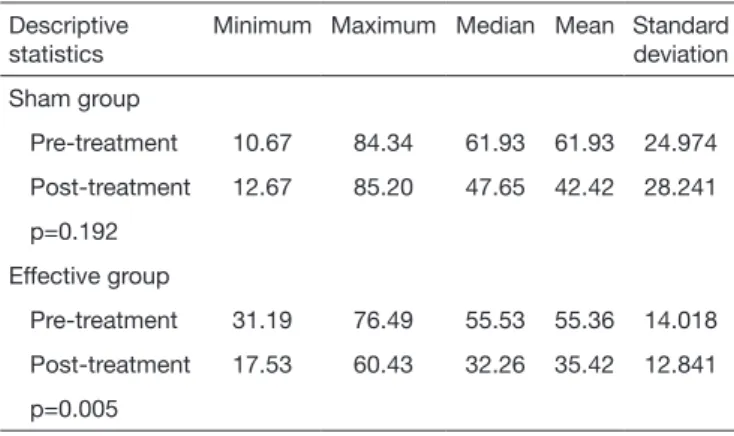
Os resultados sugerem que a ETCC simultânea (anódica e catódica) é um método que pode auxiliar a reabilitação de pacientes com afasia do tipo anômica e de Broca, principalmente

Esta linha de pensamento revela a concretização dos objetivos que se pretendiam alcançar com a estratégia do portefólio: promover a prática de uma avaliação para as aprendizagens

2.2 O novo planejamento e controle da produção na empresa O processo de planejamento e controle da produção facilita a implementação dos princípios da Construção Enxuta, na medida

Keywords: transcranial direct current stimulation (tDCS), chronic pain, pain neuromatrix, transcranial magnetic stimulation (TMS), motor cortex

Por detrás da carta e na base da mesma estavam, no entanto, muito mais do que simples considerandos oportunistas sobre os melhores mecanismos para casar reis e povos. E o cartismo

Além do custo por atividade, apuramos a margem de contribuição por saca e por hectare colhida, fornecendo assim ao agricultor todas as informações que lhe são necessárias para

Durante as entrevistas, os sujeitos foram questionados acerca de maneiras como os elementos sensoriais relacionados a cada um dos sentidos poderiam ser trabalhados