Mechanism of Action of Salivaricin 9 Lantibiotic
Produced by
Streptococcus salivarius
NU10
Abdelahhad Barbour
1, Koshy Philip
1*, Sekaran Muniandy
21 Institute of Biological Sciences, Microbiology Division, Faculty of Science, University of Malaya, Kuala Lumpur, Malaysia, 2 Department of Molecular Medicine, Faculty of Medicine, University of Malaya, Kuala Lumpur, Malaysia
Abstract
Background: Lantibiotics are small lanthionine-containing bacteriocins produced by lactic acid bacteria. Salivaricin 9 is a newly discovered lantibiotic produced by Streptococcus salivarius. In this study we present the mechanism of action of salivaricin 9 and some of its properties. Also we developed new methods to produce and purify the lantibiotic from strain NU10.
Methodology / Principal Findings: Salivaricin 9 was found to be auto-regulated when an induction assay was applied and this finding was used to develop a successful salivaricin 9 production system in liquid medium. A combination of XAD-16 and cation exchange chromatography was used to purify the secondary metabolite which was shown to have a molecular weight of approximately 3000 Da by SDS-PAGE. MALDI-TOF MS analysis indicated the presence of salivaricin 9, a 2560 Da lantibiotic. Salivaricin 9 is a bactericidal molecule targeting the cytoplasmic membrane of sensitive cells. The membrane permeabilization assay showed that salivaricin 9 penetrated the cytoplasmic membrane and induced pore formation which resulted in cell death. The morphological changes of test bacterial strains incubated with salivaricin 9 were visualized using Scanning Electron Microscopy which confirmed a pore forming mechanism of inhibition. Salivaricin 9 retained biological stability when exposed to high temperature (90-100°C) and stayed bioactive at pH ranging 2 to 10. When treated with proteinase K or peptidase, salivaricin 9 lost all antimicrobial activity, while it remained active when treated with lyticase, catalase and certain detergents.
Conclusion: The mechanism of antimicrobial action of a newly discovered lantibiotic salivaricin 9 was elucidated in this study. Salivaricin 9 penetrated the cytoplasmic membrane of its targeted cells and induced pore formation. This project has given new insights on lantibiotic peptides produced by S. salivarius isolated from the oral cavities of Malaysian subjects.
Citation: Barbour A, Philip K, Muniandy S (2013) Enhanced Production, Purification, Characterization and Mechanism of Action of Salivaricin 9 Lantibiotic Produced by Streptococcus salivarius NU10. PLoS ONE 8(10): e77751. doi:10.1371/journal.pone.0077751
Editor: Eric Cascales, Centre National de la Recherche Scientifique, Aix-Marseille Université, France Received June 13, 2013; Accepted September 6, 2013; Published October 16, 2013
Copyright: © 2013 Barbour et al. This is an open-access article distributed under the terms of the Creative Commons Attribution License, which permits unrestricted use, distribution, and reproduction in any medium, provided the original author and source are credited.
Funding: The authors wish to acknowledge the support by way of facilities from University of Malaya and the High Impact Research – Malaysian Ministry of Higher Education grant designated as UM.C/625/1/HIR/MOHE/SC/08 with account F000008-21001 under the Principal Investigator Koshy Philip for the study. The funders had no role in study design, data collection and analysis, decision to publish, or preparation of the manuscript.
Competing interests: The authors have declared that no competing interests exist. * E-mail: kphil@um.edu.my
Introduction
Different genera of lactic acid bacteria (LAB) can produce different kinds of antimicrobial peptides and bacteriocins such as plantaricin produced by Lactobacillus plantarum [1], enterococcin produced by Enterococcus faecium [2], leucocin produced by Leuconostoc carnosum [3], pediocin produced by
Pediococcus acidilactici [4] and others. Interest in bacteriocins has increased recently due to their antimicrobial activity towards Gram-positive pathogens [5-7]. Bacteriocins produced by oral cavity microorganisms have been reported previously [8-12]. Most of the bacteriocins produced by human oral
Many bacteriocin-producing strains have already been used as probiotics. S. salivarius K12 is an oral probiotic producing two kinds of antimicrobial peptides referred as salivaricin A2 and salivaricin B [9]. Both bacteriocins can be recovered by a freeze-thaw extraction method after the producer is grown on solid medium. Pore formation is a common mode of action of lantibiotics [24,25]. The permeabilization of the cytoplasmic membrane of targeted cells has been studied to investigate whether bioactive lantibiotics can penetrate the cell membrane of certain potential pathogens [26,27]. In this study we developed a new induction assay to produce salivaricin 9 in liquid medium for the first time using S. salivarius strain NU10 isolated from a Malaysian subject. The purification method used to recover salivaricin 9 was XAD-16 chromatography followed by cation exchange chromatography. Tris-Tricine SDS PAGE indicated that the peptide has a molecular weight of approximately 3,000 Da. Matrix assisted laser desorption ionization time of flight mass spectrometry MALDI-TOF (MS) analysis indicated a molecular weight of 2560 Da. We also studied the mechanism of action of the pure salivaricin 9 using SYTOX® Green. Flow cytometry analysis was also used to demonstrate membrane disruption using propidium iodide to probe cells with compromised membranes. Scanning electron microscopy was used to detect the morphological changes of the targeted indicator microorganisms after treating with salivaricin 9. Investigating the mechanism of action of lantibiotics produced by oral streptococci can assist with the development of new antimicrobials and probiotics that can be used to enhance the health of the human oral cavity and upper respiratory tract.
Results
Simultaneous antagonism test
Strain NU10 isolated from a Malaysian subject showed significant inhibitory activity when tested in the simultaneous antagonism test. When both producer and indicator were grown at the same time on blood agar, the producer strain NU10 inhibited the indicator growth. Figure 1 shows a comparison of the inhibition zones caused by strains NU10 and K12 (a commercial probiotic).
Distribution of salA and sivA structural genes in strains NU10 and YU10
Both strains NU10 and YU10 were shown to harbour the structural genes sivA and salA encoding the production of salivaricin 9 and A respectively. sivA from strain NU10 was sequenced and translated to protein using in silico analysis [28] (Figure 2). The sivA sequence of strain NU10 showed 100% homology with sivA of strain 9 (accession number: DQ889747.1).
Auto-inducing and cross inducing activities of salivaricins 9 and A
Bacteriocins produced by S. salivarius are often not expressed in liquid media. In this study, we tried to enhance bacteriocin production by using specific induction. Table 1
shows that salivaricin 9 production appeared to be auto-regulated. When salivaricin 9 was added to NU10 cultures it induced the production of antimicrobial activity. This auto-induction capability was used to enhance salivaricin 9 production in liquid medium. Crude bacteriocin preparations from all salivaricin producers were designated as BLIS (BLIS-NU10, BLIS-YU10 and BLIS-K12) and each of these preparations contains more than one kind of bacteriocin molecule that was tested as an inducer in this study. BLIS-NU10 extracted from strain BLIS-NU10 cells contained salivaricins A and 9 and it induced bacteriocin production in strains YU10 and K12 since all S. salivarius strains tested in this study harbour
salA structural gene encoding the production of salivaricin A. The pure FPLC-fraction of salivaricin 9 was also used as an inducer and was shown to induce bacteriocin production only in strains NU10 and YU10 as both harbour the sivA gene encoding salivaricin 9 production. However, pure salivaricin 9 had no induction activity when incubated with strain K12, which is PCR-negative for the structural gene of salivaricin 9. Nisin did not show any induction activity when incubated with any of the S. salivarius strains but it induced the production of the inhibitory activity when incubated with nisin producer
Lactococcus lactis strain ATCC11454 (Table 1).
Production of salivaricin 9 in liquid medium
Induced cultures of S. salivarius NU10 showed detectable inhibitory activity. After 8 hours the level of salivaricin 9 started to increase gradually until it reached more than 1200 arbitrary units per millilitre (AU/ml) at 16 hours. Once the growth kinetics of strain NU10 reached the stationary phase, the levels of the inhibitory activity remained stable (Figure 3). The arbitrary units representing bacteriocin titre are expressed as the reciprocal of the highest dilution that showed inhibitory activity against indicator strain, Micrococcus luteus.
Salivaricin 9 purification and minimum inhibitory concentration (MIC)
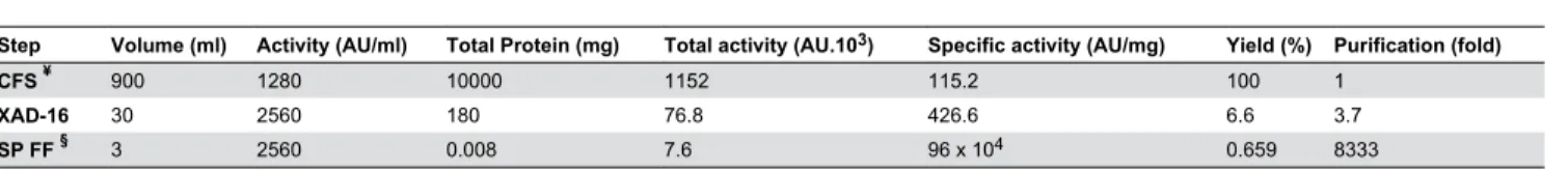
XAD-16 and cation exchange chromatography were used to purify salivaricin 9 from liquid cultures. The inhibitory activity bound to the strong cation exchanger SP FF column very efficiently. Salivaricin 9 could not be detected at a UV wavelength of 280 nm (Figure 4) indicating a relative absence of aromatic amino acid residues in the peptide. Therefore, two additional wave lengths of 207 nm and 214 nm were used. The inhibitory activity started to elute from the column at 23% NaCl. Purification steps are described in Table 2. The three 1ml active fractions were tested on Tris-Tricine SDS PAGE to estimate the molecular weight and check for the purity of the final product. Each of the three active fractions showed single protein bands with a molecular weight of approximately 3,000 Da compared to the protein standard (Figure 5). Micrococcus luteus and Corynebacterium spp showed to be the most sensitive indicators to the salivaricin 9 peptide and the MIC values of these strains were low (4-8 µg.mL-1). Streptococcus
pyogenes strain ATCC12344 was inhibited by a higher concentration of salivaricin 9 (MIC 32 µg.mL-1). MIC values of
Salivaricin 9 identification
Pure lantibiotic was subjected to MALDI-TOF (MS) analysis and showed the presence of a 2560 Da peptide representing salivaricin 9 (Figure 6).
Salivaricin 9 mode of action
When added to different growth phases of Micrococcus luteus culture, salivaricin 9 induced cell lysis. Figure 7 shows
the decreased OD600 reading when salivaricin 9 was added to
the culture, indicating that salivaricin 9 is a bactericidal and bacteriolytic peptide.
Permeabilization assay
The pure bioactive peptide was tested for inhibitory action against Corynebacterium spp and Streptococcus equisimilis
ATCC 12388. The ability of salivaricin 9 to penetrate the
Figure 1. Simultaneous Antagonism Assay. A: NU10 was used as a bacteriocin producer, B: K12 (producer of salivaricin A and salivaricin B) was used as a positive control. Micrococcus luteus was used as target indicator strain.
cytoplasmic membrane of the Corynebacterium spp was greater compared to the permeabilization activity against S. equisimilis. The detected signal at 520 nm was the result of binding between the inner nucleic acid and SYTOX® Green
dye. Unlike SYTO9® stain which can permeate both live and
dead cells, the SYTOX® Green stain is a high-affinity nucleic
acid stain that easily penetrates cells with compromised plasma membranes and yet will not cross the intact membranes of live cells [29]. In this study the fluorescence signal from membrane-compromised bacteria labelled with SYTOX® Green stain was detected by Real-Time PCR
thermocycler since the fluorescence spectra of SYTOX® Green
(504/523) are close to those of SYBR green (494/521). Immediately after adding salivaricin 9 to the indicator strains the fluorescence started to increase gradually, indicating membrane permeabilization activity. In this assay 70% ethanol which is known to attack bacterial cell membranes [30]was used as a positive control. When tetracycline was assessed in this assay no significant fluorescence was detected due to its different mechanism of action. Tetracycline binds reversibly to
the 30S ribosomal subunit to block the binding of the aminoacyl-tRNA to the acceptor site on the mRNA-ribosome complex and as a result protein synthesis is inhibited leading to bacteriostatic activity [31,32]. Figure 8 shows the membrane permeabilizing activity of salivaricin 9.
Flow Cytometry analysis of membrane disruption
In this study potential membrane disruption in Micrococcus luteus ATCC10240 by salivaricin 9 and nisin was probed using the fluorescent dye propidium iodide (PI) which can enter dead cells in the presence of the pore forming agent, but cannot penetrate live cells with intact membranes. As expected, nisin (a pore forming lantibiotic) produced a large increase in cell-associated geometric mean fluorescence intensity (MFI). However, when cells were treated with salivaricin 9, an increase in fluorescence intensity occurred consistent with pore formation and loss of membrane integrity (Figure 9).
Figure 2. Gene encoding salivaricin 9 production. A: salA structural gene encoding salivaricin A production in strains NU10 (1) and YU10 (2). B: sivA structural gene encoding salivaricin 9 production in strains NU10 (1) and YU10 (2). (M) 100 bp DNA leader. Gel electrophoresis was performed using 2% (w/v) agarose and stained using GelRedTM. C: Assembled sivA gene sequence. The
open reading frame ORF encoding the production of the leader and mature peptide is highlighted in red. D: Insilico DNA to protein translation, leader peptide (red) and mature salivaricin 9 (blue).
doi: 10.1371/journal.pone.0077751.g002
Table 1. Induction of inhibitor production by S. salivarius strains NU10, YU10, K12 and nisin-producing strain ATCC11454 using crude preparations, purified salivaricin 9 and nisin.
Inhibitor-positive preparation tested for inducing activity
Lantibiotic peptide(s) in
preparation Preparation induces inhibitor production in S. salivarius strains and nisin-producing strain
NU10 YU10 K12 ATCC11454 †
BLIS-NU10 α Sal A & 9 Yes Yes Yes No
Pure salivaricin 9 β Sal 9 Yes Yes No No
BLIS-YU10 α Sal A & 9 Yes Yes Yes No
BLIS-K12 α Sal A & B Yes Yes Yes No
Nisin (Sigma) Nisin No No No Yes
α. BLIS (bacteriocin-like inhibitory substances) representing the crude extract of each producer strain. β. FPLC-purified fraction of salivaricin 9 (sal9) produced by strain NU10.
Scanning Electron Microscopy
Salivaricin 9 caused major morphological changes in treated strains of Gram-positive bacteria. The pores formed after
incubation with salivaricin 9 showed differences for the three tested strains. After a few minutes exposure to salivaricin 9, pores developed in the cell envelopes of Micrococcus luteus
Figure 3. Growth kinetics of strain NU10 during salivaricin 9 production. Inhibitory activity of the cell free supernatant tested against Micrococcus luteus. Salivaricin 9 production was stable and consistent when strain NU10 reached the stationary phase of growth.
doi: 10.1371/journal.pone.0077751.g003
Figure 4. FPLC profile showing purification of salivaricin 9 using SP FF column. Salivaricin 9 was bound to the strong cation exchanger efficiently and eluted using linear gradient of increasing NaCl concentrations. Salivaricin 9 was detected only at wave lengths of 207 and 214 nm.
Table 2. Purification of salivaricin 9 using XAD-16 and cation exchange chromatography.
Step Volume (ml) Activity (AU/ml) Total Protein (mg) Total activity (AU.103) Specific activity (AU/mg) Yield (%) Purification (fold)
CFS ¥ 900 1280 10000 1152 115.2 100 1
XAD-16 30 2560 180 76.8 426.6 6.6 3.7
SP FF § 3 2560 0.008 7.6 96 x 104 0.659 8333
¥. Cell free supernatant from induced culture §. Strong cation exchanger column doi: 10.1371/journal.pone.0077751.t002
Figure 5. Tris-Tricine SDS page of the purified peptide. Lane 1: Dual Xtra protein marker (Bio Rad). Lanes: 2, 3 and 4: active fractions eluted from FPLC system.
and Corynebacterium spp. indicating significant changes in their cell morphology. On the other hand S. equisimilis
displayed less morphological damage following exposure to salivaricin 9. Nevertheless some pore formation was detected as the inner materials of the cells oozed through the pores resulting in cell death (Figure 10).
Salivaricin 9 stability
Salivaricin 9 retained biological stability when exposed to high temperature (90-100°C) for 30 minutes. The antimicrobial activity of salivaricin 9 was also retained after exposure to a wide pH range of 2 to 10. However salivaricin 9 appeared to be more stable in acidic and neutral conditions. Salivaricin 9 lost all antimicrobial activity when treated with proteinase K and peptidase (Table 4).
Table 3. Simultaneous Antagonism Test and Minimal Inhibitory Concentration (MIC).
Indicator microorganism Simultaneous antagonism test MIC (µg/ml) of salivaricin 9
Micrococcus luteus ATCC10240 ++++ 4
Micrococcus luteus GAB13 +++ 16
Haemophilus parainfluenza TONEJ11 -
-Actinomyces naeslundii TG2 -
-Streptococcus equisimilis ATCC12388 ++ 64
Staphylococcus aureus RF122 -
-Corynebacterium spp GH17 ++++ 8
Lactococcus lactis ATCC11454 - 128
Streptococcus mutans GEJ11 -
-Streptococcus pyogenes ATCC12344 - 32
Streptococcus pyogenes ATCC12384 - 32
Bacillus cereus ATCC14579 -
-++++ inhibition zone >12 mm, +++ inhibition zone = 10 mm, ++ inhibition zone < 5 mm, - No inhibition. doi: 10.1371/journal.pone.0077751.t003
Figure 6. MALDI-TOF MS analysis of salivaricin 9. Active peak indicating the molecular weight of salivaricin 9 at 2560 Daltons.
Discussion
It has been reported that some bacteriocins including salivaricin A, salivaricin B [9,13] and mutacin [8] are controlled by quorum sensing mechanisms and they are better expressed when the producer bacteria grow profusely on solid media. This production strategy results in greater bacteriocin expression in solid culture media compared to that obtained from low density bacterial growth in liquid medium [33,34]. Usually, to produce these cell density-dependent bacteriocins, a freeze thaw method is used following growth of the inhibitory bacteria on solid or semi-solid media containing agar or agarose. In this study, a new method was developed to produce this type of bacteriocin in liquid medium. When bacteriocin production was first evaluated in liquid media using S. salivarius NU10 as the producer, no inhibitory activity could be detected. Most lantibiotic biosynthesis can be auto regulated by a signal transduction system for example bovicin HJ50 production by
Streptococcus bovis [35], salivaricin A production by S. salivarius 20P3 [36] and nisin production by Lactococcus lactis
[37]. To investigate at what stage of the bacterial growth the optimum quantity of salivaricin 9 can be recovered, a liquid medium system was used to enable estimates to be made of both the cell count and inhibitor titre. The induction procedure showed that salivaricin 9 production by strain NU10 is auto-regulated. When the culture was grown in the presence of supplementary salivaricin 9 (added at inducing levels) the yield of salivaricin 9 was increased to 1200 AU/ml (Figure 3).
Feeding the induced culture with fresh medium helped to scale up the productivity while the culture itself worked as inoculum and inducer at the same time. Strains NU10 and YU10 are both PCR-positive for the salivaricin A structural gene, another auto-inducible lantibiotic shown to be produced by certain S. salivarius strains such as the prototype probiotic strain K12 (personal communication John Tagg). Crude peptide preparations from strains K12 and YU10 induced bacteriocin production in strain NU10. Strain K12 does not harbour sivA
(the gene encoding salivaricin 9 production) and the induction activity of strain K12 toward NU10 is due to the production of salivaricin A by both strains rather than salivaricin 9 production. However, pure salivaricin 9 induced the inhibitor production in strain NU10 and YU10 (harbouring sivA) but not in strain K12 (sivA-negative). The use of cation exchange chromatography to purify lantibiotics has been described previously [38,39]. Salivaricin B has been purified using XAD-2 chromatography and RPHPLC [9] and also to separate the lantibiotics salivaricin A2 and B produced by S. salivarius strain K12. The current study is the first report of salivaricin 9 purification using SP Sepharose, a method resulting in improved recovery of salivaricin 9. Use of an XAD-16 resin was preferred over XAD-2 due to its higher capacity to bind salivaricin 9. XAD-2 particles had been used previously to adsorb salivaricin A [13] while amberlite XAD-4 was used to adsorb nisin [40]. The use of XAD-16 hydrophobic resin was a critical step in the protocol to achieve clear and desalted crude peptide. XAD-16 amberlite was used previously to adsorb LtnA1 and LtnA2 that comprise
Figure 7. Bactericidal mode of action of salivaricin 9. Salivaricin 9 was added to different phases of bacterial growth. Salivaricin 9 induced bacterial lysis and decreased the indicator bacterial growth significantly. The sensitive bacteria Micrococcus luteus lost the ability to grow again after salivaricin 9 was added.
the two components of lacticin 3147 [41]. The FPLC system used in this study separated salivaricin 9 from the crude fraction prepared by XAD-16 chromatography. This purification protocol resulted in a single protein intensive band when Tris-Tricine SDS-PAGE was applied. Most of the impurities failed to bind to the SP sepharose resin and several peaks were detected after elution with a linear gradient of increasing salt concentration. The bioactivity test of all fractions showed that three active fractions corresponding to a single peak were eluted at a NaCl concentration of 23%. There is a lack of information about the mechanism of action of lantibiotics produced by oral streptococci. The permeabilization activity of some lantibiotics toward gram positive bacteria has been studied previously [26]. The depolarization of the cytoplasmic membrane of gram positive bacteria by salivaricin 9 was assessed by the de-quenching of SYTOX® Green fluorescence
in this study. Flow cytometry analysis was used to confirm the permeability activity of salivaricin 9 as a pore former. Both molecular probes SYTOX® Green and propidium iodide are
impermeable stains and yet cannot penetrate cells with intact membranes [42]. Propodium iodide has been used previously to check for pore formation by some bacteriocins, antibiotics and other antimicrobial agents [43-46]. Treating Micrococcus luteus ATCC10240 cells with salivaricin 9 allowed the (PI) dye to enter the cells through damaged cytoplasmic membranes. However, there is correlation between MFI and lantibiotic concentration. The MIC value of salivaricin 9 for this strain (4µg/ml) did not show significant increase in the MFI after 30 minutes of incubation. However, increases of 3-fold (12µg/ml) and 5-fold (20µg/ml) above the MIC value showed significant MFI increments. An overview of this data suggests that salivaricin 9 is able to form pores in bacterial membranes which
Figure 8. Membrane permeabilization assay of salivaricin 9. A: Salivaricin 9 permeabilization activity towards cytoplasmic membrane of S. equisimilis. B: Salivaricin 9 permeabilization activity towards cytoplasmic membrane of Corynebacterium spp. Negative controls comprise targeted bacteria without adding salivaricin 9. Positive control used 70% ethanol. Tetracycline did not show any permeability activity in this test.
Figure 9. Flow cytometry analysis of pore-forming activity of salivaricin 9. Like nisin, salivaricin 9 alters the membrane permeability of Micrococcus luteus ATCC10240 as measured by propidium iodide (PI) uptake. (A) Average MFI of triplicate measurements for nisin at a concentration of 20µg/ml and a range of salivaricin 9 concentrations of 3-fold and 5-fold above its MIC value. (B) Representative histogram of cell count versus PI fluorescence intensity at antibiotic concentrations shown in panel A.
Figure 10. morphological changes of sensitive bacterial cells incubated with salivaricin 9. A: Untreated Micrococcus luteus
used as a control. B: Morphological changes of Micrococcus luteus treated with salivaricin 9. C: Untreated S. equisimilis used as a control. D: Morphological changes of S. equisimilis treated with salivaricin 9. E: Untreated Corynebacteriumspp used as a control. F: Morphological changes of Corynebacteriumspp treated with salivaricin 9. White arrows indicate pores formed by salivaricin 9.
will lead to loss in membrane integrity of the cells. This conclusion may explain the morphological changes that salivaricin 9 can cause in the targeted cells. Type A lantibiotics (e.g. nisin and epidermin) are amphipathic and elongated molecules which mainly act by forming pores into the cytoplasmic membrane of the targeted bacterial cell [25,47,48]. Binding to lipid II is an initial factor inducing a transmembrane orientation of the pore forming lantibiotic [25]. At some stage lantibiotics such as nisin can form pores and can inhibit cell wall biosynthesis when they bind to the peptidoglycan precursor lipid II [49]. Salivaricin 9 showed similar characteristics in this study to other lantibiotics in bringing about membrane permeabilization by pore formation in the cytoplasmic membrane. Further work is now in progress to evaluate the mechanism of binding of salivaricin 9 to lipid II precursor by using fluorimetric spectroscopy. Mass production using cloning and expression techniques is also being investigated so that the lantibiotic can be commercialized as a biopharmaceutical.
Conclusions
Salivaricin 9 produced by strain S. salivarius NU10 isolated from a Malaysian subject was produced in liquid medium using an induction method. XAD-16 and cation exchange chromatography were used to purify the active agent. MALDI-TOF analysis showed that the pure peptide has a molecular weight of 2560 Da. Salivaricin 9 displayed bactericidal and
bacteriolytic activity against and induced cytoplasmic membrane permeability in susceptible bacterial cells. Pore formation activity was detected in sensitive strains incubated with salivaricin 9. This study has revealed additional information on an antimicrobial peptide produced by S. salivarius that can expedite the further development of specifically targeted novel antibiotics in this era of increasing antibiotic resistance.
Materials and Methods
Bacterial strains and media
S. salivarius NU10 and YU10 were isolated from two healthy Malaysian subjects and then deposited in NCBI Gene Bank under accession numbers KC796011 and KC796012 respectively. Strains NU10 and YU10 were isolated from the two subjects who were required to sign a consent form to isolate S. salivarius from the tongue surface using sterile cotton swabs. No approval for sampling from tongue surface is required from the IRB as a pro forma for written consent from the subject is approved by the IRB. The ethics committee IRB Reference No. is DF OP1304/0019 (P) for our Institution (University of Malaya). We have discussed this with the ethics committee (IRB) and the protocol complied with Good Laboratory Practices and no IRB approval is required prior to sampling in this cases. This work was entirely done in our own home country Malaysia within the University campus that
Table 4. Salivaricin 9 stability (thermal / pH/ proteinase K/ chemicals).
Stability Concentration Inhibition zone
Temperature (°C)
4, 20, 30, 37, 40, 50, 60, 70, 80 for 1 hour ++++
90, 100 for 30 min +++
121 for 20 min
-pH value
2- 7 ++++
8-10 +++
11-12
-Enzymes 1 mg.ml-1
Proteinase
-Proteinase K
-Peptidase
-Lyticase ++++
Catalase ++++
Detergents / Chemicals 1% (w/v)
Tween 80 ++++
Tween 20 ++++
Tritone X100 ++
SDS ++++
EDTA ++++
Urea ++++
NaCl ++++
(++++) : inhibition zone >20 mm; (+++) : inhibition zone =20 mm; (++): inhibition zone < 20 mm; (-): no bacterial inhibition. Micrococcus luteus was used as indicator. Salivaricin 9 titre: 800 AU/ml.
includes a university hospital. S. salivarius K12 a commercial probiotic (BLIS Technologies Ltd, New Zealand) was provided by John Tagg (University of Otago, New Zealand) for use as a positive control. Indicator strains used in this study are listed in Table 3. All the indicator strains are either purchased from American Type Culture Centre (ATCC) or were taken from the culture collection of Microbial Biotechnology Laboratory, Division of Microbiology, Institute of Biological Science, Faculty of Science, University of Malaya, Kuala Lumpur, Malaysia. Todd Hewitt broth (THB) was used to propagate all the bacterial strains in this study. Mitis Salivarius agar (MSA) was used to isolate pure colonies of S. salivarius strains. M17 broth supplemented with 2% yeast extract, 1% sucrose, 0.1% calcium carbonate (M17YESUCa) was used to produce salivaricin 9 in a liquid medium system. Columbia agar base supplemented with 5% whole human blood and 0.1% calcium carbonate (BACa) was used in simultaneous antagonism tests. All media was purchased from Difco, Becton Dickinson, Sparks, Md., USA.
Simultaneous antagonism assay
To check for antimicrobial activity, S.salivarius NU10 was stabbed into blood agar plate supplemented with 0.1% calcium carbonate previously seeded with a lawn of Micrococcus luteus. S. salivarius K12 (producer of salivaricin A2 and salivaricin B) was used as a positive control (Figure 1).
DNA isolation and detection of sivA structural gene encoding salivaricin 9 production
S. salivarius strain NU10 was grown aerobically in THB for 18 hours at 37°C. Cells were collected by centrifugation at 8000 x g for 3 minutes and washed twice with 0.85% NaCl before suspending in lysis buffer comprising of 20 mM Tris-HCl at pH 8.0, 2 mM sodium EDTA, 1.5 % Triton X-100 and 50 mg/ml lysozyme. DNA was isolated and purified using DNeasy®
Blood & Tissue kit (QIAGEN) following the manufacturer’s instructions. To detect sivA, primers sivF (5`-AAAAAGGCGCTTCTATATCCATGA-3`) and sivR (5`-ATCTTTACCTCAAACTTTTAAGTCCATT-3`) were used as described previously [12]. Primer pair SalAUS GTAGAAAATATTTACTACATACT-3`) and SalADS (5`-GTTAAAGTATTCGTAAAACTGATG-3`) were used to detect
salA encoding salivaricin A production in strains NU10 and YU10 as described previously [11]. sivA of strain NU10 was sequenced and assembled using DNA baser V.3 software and analysed using in silico DNA to protein translation tool [28] (Figure 2).
Salivaricin 9 induction assay
The bacteriocin-inducing activities present in crude extracts of S. salivarius strains NU10, YU10, K12 and in FPLC-purified salivaricin 9 peptide fractions having anti-Micrococcus luteus
activity was investigated in this study. The crude extracts of producer cells designated as BLIS-NU10, BLIS-YU10 and BLIS-K12 were isolated by acidic methanol extraction of the producer cells as described previously [11,12]. These crude BLIS extracts had the potential to contain at least two lantibiotics on the basis of PCR assessment for their content of
lantibiotic structural genes. Strains NU10 and YU10 had the structural genes for salivaricin 9 and salivaricin A and strain K12 had salivaricin A and salivaricin B structural genes. Salivaricin 9 was shown to be auto-regulated whereby a small amount of the active peptide can induce enhanced production in a large scale production system. Colonies from an 18 h culture of each S. salivarius strain NU10, YU10 and K12 grown anaerobically on BACa agar was used to inoculate 10 ml of M17YESUCa broth and incubated again under the same conditions. The cells were then collected by centrifugation at 18000 x g for 5 min and washed three times in saline buffer to reduce the levels of cell surface-associated lantibiotic before suspending in 10 ml of the same buffer. 1.5 ml Eppendorf tubes containing 0.9 ml of M17YESUCa were inoculated with 0.1 ml of the washed cells before 0.05 ml of the test sample (BLIS-extracts or pure salivaricin 9 of titre 4 AU/ml) was added and these tubes marked as “induced”. Tubes with no BLIS-extract or salivaricin 9 added were marked as “control” (uninduced). Both induced and control tubes were incubated anaerobically for 18 h before 0.05 ml of the test sample (BLIS-extract or salivaricin 9 of titre 4 AU/ml) was added to the control tube. 50µl aliquots of each sample (induced and control) were used to test for antimicrobial activity using the well diffusion assay. Induction of salivaricin 9 or salivaricin A production was recorded as inhibitory zones surrounding samples from the induced tubes but not the control tubes. Nisin producing
Lactococcus lactis strain ATCC11454 was also tested for bacteriocin-inducing activity as a positive control (Table 1).
Production of salivaricin 9 in liquid medium
Usually bacteriocins produced by S. salivarius can be isolated from cultures grown on solid medium using the freeze thaw method described previously [9]. Strain NU10 is also able to produce the bioactive peptide in solid phase medium and showed extremely weak expression of inhibitory activity in liquid media. To overcome this limitation, a new technique was used to enhance the production of salivaricin 9 lantibiotic in liquid media. Strain NU10 was grown on MSA for 18 h under anaerobic condition at 37°C before one colony (dome-shape in appearance) was used to inoculate 20 ml of M17YESUCa broth and incubated under the same conditions mentioned above on an orbital shaker at 150 rpm. The cells were collected by centrifugation at 6000 x g for 5 min at 4°C and then washed twice in 0.85% NaCl prior to dissolving in 20 ml of the same solution. The cell suspension was used to inoculate 50 ml of fresh M17YESUCa broth at 37°C in anaerobic condition for 20 h. The resulting culture was fed with fresh 50 ml of the same medium and 0.05 g/ml of pure salivaricin 9 was added as an inducer before the 100 ml culture was incubated for another 18 h. 80 ml of the final culture was used to inoculate 820 ml of M17YESUCa broth. The pH was adjusted to 6.5 using concentrated HCl. CFU and AU values during production were recorded (Figure 3).
Salivaricin 9 purification and determination of minimal inhibitory concentration (MIC)
was filtered through 0.2 µm sterilized cellulose membrane (Millipore) to ensure that all cell debris was removed. The filtered crude bacteriocin preparation was passed through 100 ml XAD-16 particles (Sigma) packed in a glass column. The column was washed with 400 ml of distilled water followed by 200 ml of 70% methanol before the active fraction was eluted using 200 ml of 95% methanol (adjusted to pH 2) at a flow rate of 15 ml/min. The methanol was removed using a rotary evaporator and the resulting peptide preparation was lyophilized and stored at -20°C. The lyophilized peptide pellets were dissolved in 20 mM sodium phosphate pH 5.8 (binding buffer). Then the sample was injected into a fast protein liquid chromatography (FPLC) system (ÄKTA Purifier™) using HiTrap SP FF 5ml strong cation exchanger column (GE Healthcare) equilibrated with the same buffer at a flow rate of 1 ml/min. Then the column was washed with 10X column volume of the binding buffer before salivaricin 9 was eluted using a linear gradient of 0 to 1M NaCl in 20 mM sodium phosphate buffer at pH 5.8 (Figure 4). All fractions were tested for inhibitory activity using Micrococcus luteus as the indicator. Purification steps, fold and yield are listed in Table 2. MIC values of pure salivaricin 9 against a set of bacterial indicator strains were determined by following the National Committee for Clinical Laboratory Standards (NCCLS) (Table 3).
Tris-Tricine SDS page
The active fractions obtained by FPLC were subjected to 16.5% sodium dodecyl sulphate (SDS) electrophoresis as described previously [50] using a Tris-Tricine buffer system. Mini-Protean Tetra Cell (Bio Rad) was used according to manufacturer’s instructions. Dual Xtra protein marker (Bio Rad) was used to estimate the molecular weight of the pure salivaricin 9. The gel run was at 125 V for 45 min and stained using SimplyBlue™ SafeStain (Life Technologies-Invitrogen). After de-staining, the gel was visualized and the molecular weight estimated (Figure 5).
Identification of the antimicrobial peptide
The FPLC purified peptide was subjected to matrix-assisted laser desorption-time of flight (MALDI-TOF) mass spectrometry (MS) at the Medical Biotechnology Laboratory, Faculty of Medicine, University of Malaya.
Membrane permeabilization assay
The membrane permeabilization activity of salivaricin 9 towards the cytoplasmic membrane of targeted bacterial cells was examined using SYTOX® Green dye. A log phase culture
of the targeted bacteria was centrifuged at 9000 x g for 5 minutes and then the cells were washed 3 times with 10 mM HEPES buffer before re-suspending in the same buffer. The CFU were counted to be 9 x 10⁵. 5µM of SYTOX® Green
(Invitrogen™) was added to the bacterial suspension after which 100µl of this mixture was transferred to a 96 well PCR plate before 100µg/ml of salivaricin 9 was added to the mixture (SYTOX® Green + bacterial cells). Wells containing only
bacterial cells and SYTOX® Green were designated as
negative control wells. 70% ethanol was used as the positive control. Tetracycline (1.5µg/ml) was also tested in this assay as
a known antimicrobial having a different mode of action. The fluorescence generated from the binding between SYTOX®
Green and nucleic acid of the dead bacterial cells with compromised membranes was detected at 520 nm and 37°C exposing for over a period of 60 and 80 minutes for S. equisimilis and Corynebacterium spp respectively (Figure 8). The experiment was performed in triplicate and raw data was analyzed using Microsoft Excel software.
Flow Cytometry analysis of membrane disruption
For membrane integrity assay using the dye propidium iodide (PI), cultures of Micrococcus luteus ATCC10240 were grown over night at 37°C in THB supplemented with 0.5% glucose (THBG) and then diluted with fresh THBG to an OD600 of 0.1.
The cells were centrifuged at 4000 x g for 5 minutes at 4°C and washed twice with washing buffer (2 mM HEPES + 2 mM glucose) before re-suspending in the same buffer and adjusting to OD600 0.1. The cells were exposed to salivaricin 9 (12, 20
µg/ml) or nisin (20 µg/ml) (Sigma) and PI (final concentration 25 µM) before incubating for 30 minutes at 37°C. Nuclease-Free Water (Promega, USA) was added instead of antibiotic and served as a negative control. Samples were analyzed using BD FACSCantoTM II flow cytometer using excitation at
488 nm and with the standard filter setup. Each sample was examined in triplicate and the geometric mean fluorescence intensity (MFI) of gated cell populations was calculated (Figure 9).
Scanning electron microscopy (SEM)
To study the morphological changes of the targeted bacterial cells after treating with pure salivaricin 9, indicator microorganisms were grown in THB to reach the exponential phase of growth at (OD600 0.7) and then the bacterial cultures
were diluted to OD600 0.2 before pure salivaricin 9 was added to
the cultures and incubated at 37°C for 260 minutes. After incubation, the bacterial pellets were centrifuged at 6000 x g for 10 minutes at 4°C and washed twice with phosphate buffer saline PBS at pH 7 before the samples were fixed with 8% glutaraldehyde in 1:1 (v/v) Sorensen’s phosphate buffer for one hour. 1:1 (v/v) of Sorensen’s phosphate buffer and water mixture was applied before the samples were fixed with 4% OsO4 mixed with 1:1 (v/v) H2O. After overnight incubation,
samples were washed with deionized water for 15 minutes and then dehydrated in increasing concentrations of ethanol. Dehydration with ethanol-acetone mixture followed by pure acetone was applied before critical point drying (CPD). After sample preparation JEOL JSM-7001F Scanning Electron Microscopy was used to visualize the morphological changes of bacterial cells treated with salivaricin 9 compared with non-treated cells (Figure 10).
Stability tests on salivaricin 9
wells of 6mm diameter each separated by a distance of 4 mm in CAB plates. One well was filled with 50µl of pure salivaricin 9 while the other was filled with 50µl of the tested enzyme. The plate was incubated at 50°C for 2 hours and then incubated at 37°C overnight before seeding the indicator strain onto the agar surface using a cotton swab. Then the plate was re-incubated at 37°C for 18 hours and the zones of inhibitions that appeared indicated residual activity of salivaricin 9 while absence of inhibition zones indicated the denaturation of the antimicrobial peptide by the applied enzymes namely proteinase K, peptidase, lyticase and catalase. The stability of salivaricin 9 to treatment with the chemicals EDTA, SDS, urea, NaCl and β-mercaptoethanol was examined by adding 1% of these chemicals to the bacteriocin followed by incubation for two hours at RT before the samples were centrifuged and the supernatant tested as mentioned above. If the chemical was a solvent such as β-mercaptoethanol, then the solvent was evaporated before testing for antimicrobial activity (Table 4).
Acknowledgements
The authors wish to acknowledge the laboratory facilities given by University of Malaya, Malaysia and BLIS Technologies Ltd, New Zealand for providing the probiotic control strain K12 for the study.
Author Contributions
Performed the experiments: AB KP. Analyzed the data: AB KP. Contributed reagents/materials/analysis tools: AB KP SM. Wrote the manuscript: AB KP. Carried out the laboratory work: AB. Planned, designed and guided the project: KP. Read the manuscript: SM. Proof reading the manuscript: AB KP SM.
References
1. Tiwari SK, Srivastava S (2008) Purification and characterization of plantaricin LR14: a novel bacteriocin produced by Lactobacillus plantarum LR/14. Appl Microbiol Biotechnol 79: 759-767. doi:10.1007/ s00253-008-1482-6. PubMed: 18496687.
2. Klocke M, Mundt K, Idler F, Jung S, Backhausen JE (2005) Heterologous expression of enterocin A, a bacteriocin from Enterococcus faecium, fused to a cellulose-binding domain in Escherichia coli results in a functional protein with inhibitory activity against Listeria. Appl Microbiol Biotechnol 67: 532-538. doi:10.1007/ s00253-004-1838-5. PubMed: 15660219.
3. Wan X, Li R, Saris PE, Takala TM (2012) Genetic characterisation and heterologous expression of leucocin C, a class IIa bacteriocin from Leuconostoccarnosum 4010. Appl Microbiol Biotechnol 97: 3509-3518. PubMed: 23053070.
4. Papagianni M, Anastasiadou S (2009) Pediocins: The bacteriocins of Pediococci. Sources, production, properties and applications. Microb Cell Factories 8: 1-16. doi:10.1186/1475-2859-8-1.
5. Abee T, Krockel L, Hill C (1995) Bacteriocins: modes of action and potentials in food preservation and control of food poisoning. Int J Food Microbiol 28: 169-185. doi:10.1016/0168-1605(95)00055-0. PubMed: 8750665.
6. Lee JH, Li X, O'Sullivan DJ (2011) Transcription analysis of a lantibiotic gene cluster from Bifidobacterium longum DJO10A. Appl Environ Microbiol 77: 5879-5887. doi:10.1128/AEM.00571-11. PubMed: 21742926.
7. Wilson-Stanford S, Smith L (2011) Commercial development and application of type A lantibiotics. Recent Pat Antiinfect Drugs Discov 6: 175-185. doi:10.2174/157489111796064632. PubMed: 21517737. 8. Nicolas GG, LaPointe G, Lavoie MC (2011) Production, purification,
sequencing and activity spectra of mutacins D-123.1 and F-59.1. BMC Microbiol 11: 1-10. doi:10.1186/1471-2180-11-1. PubMed: 21194490. 9. Hyink O, Wescombe PA, Upton M, Ragland N, Burton JP et al. (2007)
Salivaricin A2 and the novel lantibiotic salivaricin B are encoded at adjacent loci on a 190-kilobase transmissible megaplasmid in the oral probiotic strain Streptococcussalivarius K12. Appl Environ Microbiol 73: 1107-1113. doi:10.1128/AEM.02265-06. PubMed: 17194838. 10. Wescombe PA, Dyet KH, Dierksen KP, Power DA, Jack RW et al.
(2012) Salivaricin G32, a homolog of the prototype Streptococcus pyogenes nisin-Like Lantibiotic SA-FF22, produced by the commensal species Streptococcussalivarius. Int J Microbiol 2012: 1-10 PubMed: 22567013
11. Wescombe PA, Upton M, Dierksen KP, Ragland NL, Sivabalan S et al. (2006) Production of the lantibiotic salivaricin A and its variants by oral streptococci and use of a specific induction assay to detect their presence in human saliva. Appl Environ Microbiol 72: 1459-1466. doi: 10.1128/AEM.72.2.1459-1466.2006. PubMed: 16461700.
12. Wescombe PA, Upton M, Renault P, Wirawan RE, Power D et al. (2011) Salivaricin 9, a new lantibiotic produced by Streptococcus salivarius. Microbiology 157: 1290-1299. doi:10.1099/mic.0.044719-0. PubMed: 21310787.
13. Ross KF, Ronson CW, Tagg JR (1993) Isolation and characterization of the lantibiotic salivaricin A and its structural gene salA from Streptococcussalivarius 20P3. Appl Environ Microbiol 59: 2014-2021. PubMed: 8357242.
14. Al-Mahrous MM, Upton M (2011) Discovery and development of lantibiotics; antimicrobial agents that have significant potential for medical application. Expert Opin Drugs Discov 6: 155-170. doi: 10.1517/17460441.2011.545387. PubMed: 22647134.
15. Zhao M (2011) Lantibiotics as probes for phosphatidylethanolamine. Amino Acids 41: 1071-1079. doi:10.1007/s00726-009-0386-9. PubMed: 21573677.
16. Perin LM, Moraes PM, Silva A Jr., Nero LA (2012) Lantibiotics biosynthesis genes and bacteriocinogenic activity of Lactobacillus spp. isolated from raw milk and cheese. Folia Microbiol (Praha) 57: 183-190. doi:10.1007/s12223-012-0113-x. PubMed: 22447149.
17. González-Toledo SY, Domínguez-Domínguez J, García-Almendárez BE, Prado-Barragán LA, Regalado-González C (2010) Optimization of nisin production by Lactococcuslactis UQ2 using supplemented whey as alternative culture medium. J Food Sci 75: M347-M353. doi: 10.1111/j.1750-3841.2010.01670.x. PubMed: 20722935.
18. Flôres SH, Alegre RM (2001) Nisin production from Lactococcuslactis A.T.C.C. 7962 using supplemented whey permeate. Biotechnol Appl Biochem 34: 103-107. doi:10.1042/BA20010030. PubMed: 11592916. 19. Lv W, Zhang X, Cong W (2005) Modelling the production of nisin by
Lactococcuslactis in fed-batch culture. Appl Microbiol Biotechnol 68: 322-326. doi:10.1007/s00253-005-1892-7. PubMed: 15692804. 20. Cheigh CI, Park H, Choi HJ, Pyun YR (2005) Enhanced nisin
production by increasing genes involved in nisin Z biosynthesis in Lactococcuslactis subsp. lactis A164. Biotechnol Lett 27: 155-160. 21. Liu W, Zheng H, Wu Z, Wang Y (2010) Effects of pH profiles on nisin
fermentation coupling with foam separation. Appl Microbiol Biotechnol 85: 1401-1407. doi:10.1007/s00253-009-2217-z. PubMed: 19730846. 22. Pongtharangkul T, Demirci A (2006) Effects of fed-batch fermentation
and pH profiles on nisin production in suspended-cell and biofilm reactors. Appl Microbiol Biotechnol 73: 73-79. doi:10.1007/ s00253-006-0459-6. PubMed: 16733734.
23. Cotter PD, Hill C, Ross RP (2005) Bacterial lantibiotics: strategies to improve therapeutic potential. Curr Protein Pept Sci 6: 61-75. doi: 10.2174/1389203053027584. PubMed: 15638769.
24. Garcerá MJ, Elferink MG, Driessen AJ, Konings WN (1993) In vitro pore-forming activity of the lantibiotic nisin. Role of protonmotive force and lipid composition. Eur J Biochem 212: 417-422. doi:10.1111/j. 1432-1033.1993.tb17677.x. PubMed: 8444179.
25. van Heusden HE, de Kruijff B, Breukink E (2002) Lipid II induces a transmembrane orientation of the pore-forming peptide lantibiotic nisin. Biochemistry 41: 12171-12178. doi:10.1021/bi026090x. PubMed: 12356318.
27. Zendo T, Yoneyama F, Sonomoto K (2010) Lactococcal membrane-permeabilizing antimicrobial peptides. Appl Microbiol Biotechnol 88: 1-9. doi:10.1007/s00253-010-2764-3. PubMed: 20645082.
28. Bikandi J, San Millán R, Rementeria A, Garaizar J (2004) In silico analysis of complete bacterial genomes: PCR, AFLP-PCR and endonuclease restriction. Bioinformatics 20: 798-799. doi:10.1093/ bioinformatics/btg491. PubMed: 14752001.
29. Lebaron P, Catala P, Parthuisot N (1998) Effectiveness of SYTOX Green stain for bacterial viability assessment. Appl Environ Microbiol 64: 2697-2700. PubMed: 9647851.
30. Bourbon C, Bry C, Roggemans C, Soulard C, Thizon C et al. (2008) Use of a real-time polymerase chain reaction thermocycler to study bacterial cell permeabilization by antimicrobial peptides. Anal Biochem 381: 279-281. doi:10.1016/j.ab.2008.07.005. PubMed: 18656439. 31. Chopra I, Hawkey PM, Hinton M (1992) Tetracyclines, molecular and
clinical aspects. J Antimicrob Chemother 29: 245-277. doi:10.1093/jac/ 29.3.245. PubMed: 1592696.
32. Chopra I, Roberts M (2001) Tetracycline antibiotics: mode of action, applications, molecular biology, and epidemiology of bacterial resistance. Microbiol Mol Biol Rev 65: 232-260 ; second page, table of contents doi:10.1128/MMBR.65.2.232-260.2001. PubMed: 11381101. 33. Qi F, Chen P, Caufield PW (2000) Purification and biochemical
characterization of mutacin I from the group I strain of Streptococcus mutans, CH43, and genetic analysis of mutacin I biosynthesis genes. Appl Environ Microbiol 66: 3221-3229.
34. Kleerebezem M, Quadri LE, Kuipers OP, de Vos WM (1997) Quorum sensing by peptide pheromones and two-component signal-transduction systems in Gram-positive bacteria. Mol Microbiol 24: 895-904. doi:10.1046/j.1365-2958.1997.4251782.x. PubMed: 9219998. 35. Ni J, Teng K, Liu G, Qiao C, Huan L et al. (2011) Autoregulation of
lantibiotic bovicin HJ50 biosynthesis by the BovK-BovR two-component signal transduction system in Streptococcusbovis HJ50. Appl Environ Microbiol 77: 407-415. doi:10.1128/AEM.01278-10. PubMed: 21075878.
36. Upton M, Tagg JR, Wescombe P, Jenkinson HF (2001) Intra- and interspecies signaling between Streptococcus salivarius and Streptococcus pyogenes mediated by SalA and SalA1 lantibiotic peptides. J Bacteriol 183: 3931-3938. doi:10.1128/JB. 183.13.3931-3938.2001. PubMed: 11395456.
37. Kuipers OP, Beerthuyzen MM, de Ruyter PG, Luesink EJ, de Vos WM (1995) Autoregulation of nisin biosynthesis in Lactococcus lactis by signal transduction. J Biol Chem 270: 27299-27304. doi:10.1074/jbc. 270.45.27299. PubMed: 7592991.
38. Furmanek B, Kaczorowski T, Bugalski R, Bielawski K, Bohdanowicz J et al. (1999) Identification, characterization and purification of the
lantibiotic staphylococcin T, a natural gallidermin variant. J Appl Microbiol 87: 856-866. doi:10.1046/j.1365-2672.1999.00937.x. PubMed: 10664909.
39. Sahl HG (1994) Staphylococcin 1580 is identical to the lantibiotic epidermin: implications for the nature of bacteriocins from gram-positive bacteria. Appl Environ Microbiol 60: 752-755. PubMed: 8135526. 40. Tolonen M, Saris PE, Siika-Aho M (2004) Production of nisin with
continuous adsorption to Amberlite XAD-4 resin using Lactococcus lactis N8 and L. lactis LAC48. Appl Microbiol Biotechnol 63: 659-665. doi:10.1007/s00253-003-1413-5. PubMed: 12910326.
41. Morgan SM, O'Connor PM, Cotter PD, Ross RP, Hill C (2005) Sequential actions of the two component peptides of the lantibiotic lacticin 3147 explain its antimicrobial activity at nanomolar concentrations. Antimicrob Agents Chemother 49: 2606-2611. doi: 10.1128/AAC.49.7.2606-2611.2005. PubMed: 15980326.
42. Swe PM, Cook GM, Tagg JR, Jack RW (2009) Mode of action of dysgalacticin: a large heat-labile bacteriocin. J Antimicrob Chemother 63: 679-686. doi:10.1093/jac/dkn552. PubMed: 19213799.
43. Spyr CA, Käsermann F, Kempf C (1995) Identification of the pore forming element of Semliki Forest virus spikes. FEBS Lett 375: 134-136. doi:10.1016/0014-5793(95)01197-M. PubMed: 7498462. 44. Chehimi S, Sablé S, Hajlaoui MR, Limam F (2010) Mode of action of
thuricin S, a new class IId bacteriocin from Bacillusthuringiensis. Can J Microbiol 56: 162-167. doi:10.1139/W09-125. PubMed: 20237578. 45. Gut IM, Blanke SR, van der Donk WA (2011) Mechanism of Inhibition
of Bacillusanthracis spore outgrowth by the lantibiotic nisin. Acs. Chem Biol 6: 744-752.
46. Knerr PJ, Oman TJ, Garcia De Gonzalo CV, Lupoli TJ, Walker S et al. (2012) Non-proteinogenic amino acids in lacticin 481 analogues result in more potent inhibition of peptidoglycan transglycosylation. Acs. Chem Biol 7: 1791-1795.
47. Kordel M, Schüller F, Sahl HG (1989) Interaction of the pore forming-peptide antibiotics Pep 5, nisin and subtilin with non-energized liposomes. FEBS Lett 244: 99-102. doi: 10.1016/0014-5793(89)81171-8. PubMed: 2924913.
48. Moll GN, Roberts GC, Konings WN, Driessen AJ (1996) Mechanism of lantibiotic-induced pore-formation. Antonie Van Leeuwenhoek 69: 185-191. doi:10.1007/BF00399423. PubMed: 8775978.
49. Wiedemann I, Breukink E, van Kraaij C, Kuipers OP, Bierbaum G et al. (2001) Specific binding of nisin to the peptidoglycan precursor lipid II combines pore formation and inhibition of cell wall biosynthesis for potent antibiotic activity. J Biol Chem 276: 1772-1779. PubMed: 11038353.