ORIGINAL
ARTICLE
Molecular identification and typing of
Mycobacterium massiliense
isolated from
postsurgical infections in Brazil
Authors Fernanda Monego1
Rafael Silva Duarte2
Sueli Massumi Nakatani3
Wildo Navegantes Araújo4
Irina Nastassja Riediger5
Sonia Brockelt6
Verena Souza4
Jamyra Iglesias Cataldo7
Rubens Clayton da Silva Dias8
Alexander Welker Biondo9
1MSc; PhD Student, Molecular
and Cellular Biology, Universidade Federal do Paraná (UFPR), PR, Brazil
2PhD; Associate Professor,
Universidade Federal do Rio de Janeiro (UFRJ), RJ, Brazil
3PhD; Head of Molecular
Biology, Laboratório Central do Estado do Paraná (LACEN-PR), PR, Brazil
4MSc; Epidemiologists,
Secretaria de Vigilância em Saúde (SVS), Departament of Serological Vigilance, Brazil
5MSc; Researcher, LACEN-PR,
PR, Brazil
6MSc; Head of the Mycobacteria
Sector, LACEN-PR, PR, Brazil
7MSc; PhD Student,
Universidade do Estado do Rio de Janeiro (UERJ), RJ, Brazil
8PhD; Researcher, UERJ, RJ,
Brazil
9PhD; Full Professor of
Zoonoses, UFPR, PR, Brazil
Submitted on: 01/18/2011 Approved on: 06/29/2011
Correspondence to: Fernanda Monego Universidade Federal do Paraná (UFPR) Depart. de Medicina Veterinária
Rua dos Funcionários, 1540, Juveve 80035-050, Curitiba, Paraná,Brazil
Phone.: +55 41 3350-5723 Fax: +55 41 3350-5623 [email protected]
We declare no conflict of interest.
©2011 Elsevier Editora Ltda. All rights reserved.
ABSTRACT
Objective: One hundred thirty-one cases of postsurgical infections were reported in Southern Re-gion of Brazil between August 2007 and January 2008. Thirty-nine (29.8%) cases were studied; this report describes epidemiological findings, species identification, antimicrobial susceptibility and clonal diversity of rapidly growing mycobacteria isolated in this outbreak. Methods: All 39 isolates were analyzed by Ziehl-Nielsen stained smear, bacterial culture and submitted to rpoB partial gene sequencing for identification. The isolates were also evaluated for their susceptibility to amikacin, cefoxitin, clarithromycin, ciprofloxacin, doxycycline, tobramycin and sulfamethoxazole. Results:
Thirty-six isolates out of the confirmed cases were identified as Mycobacterium massiliense and the remaining three were identified as Mycobacterium abscessus, Mycobacterium chelonae and Myco-bacterium fortuitum. All M. massiliense isolates were susceptible to amikacin (MIC90 = 8 μg/mL) and clarithromycin (MIC90 = 0.25 μg/mL) but resistant to cefoxitin, ciprofloxacin, doxycycline, to-bramycin and sulfamethoxazole. Molecular analysis by pulsed-field gel electrophoresis clustered all 36 M. massiliense isolates and showed the same pattern (BRA 100) observed in three other outbreaks previously reported in Brazil. Conclusions: These findings suggest a common source of infection for all patients and reinforce the hypotheses of spread of M. massiliense BRA100 in Brazilian hospital surgical environment in recent years.
Keywords: mycobacteria, atypical; mycobacterium infections; microbiological analysis.
ria have been generally related to contaminated medical equipments, solutions and laboratory reagents.8,9 Strains resistant to disinfectants
have also been isolated from endoscope wash-er disinfector aftwash-er decontamination with 2% glutaraldehyde solution.10 Since 2%
glutaralde-hyde is one of the basic compounds most wide-ly used as a chemical disinfectant for surgical equipment in several countries, particularly for non-autoclavable devices, resistance to bioc-ides have become a great concern in hospital practice.
In Brazil, the first reported outbreaks
caused by M. chelonae-abscessus group
spe-cies were related to laser in situ keratomileusis, mesotherapy sessions and breast implant sur-geries.11,12Mycobacterium massiliense has been
described as the main agent isolated during these recent RGM outbreaks in several Bra-zilian states, mainly those that occurred after 2004. Furthermore, in all described postsurgi-cal outbreaks, a specific M. massiliense clone, named BRA100, has emerged as an
opportun-INTRODUCTION
Human infections after cosmetic procedures, surgery, postinjection and nipple piercing1,2
by rapidly growing mycobacteria (RGM) have been described worldwide as mainly associated
with Mycobacterium chelonae, Mycobacterium
abscessus, Mycobacterium fortuitum and Myco-bacterium smegmatis groups.3 These
microor-ganisms have already been isolated from soil, water treatment plants, hospital tap water and distilled water, and considered environmen-tally adapted species.4 Some RGM strains have
been described as being able to develop bio-film and infections related to biobio-film represent more than two-thirds of all infections caused by these organisms.5
Hospital outbreaks as well as isolated cases of RGM infections have been reported in dif-ferent scenarios involving chronic lung disease, disseminated cutaneous infections and
post-surgical wound infections.6,7 Outbreaks and
bacte-istic pathogen, usually causing postsurgical wound infec-tions, including superficial abscesses and granulomas.13 The
Brazilian Public Health Surveillance System has registered an overall of 1,937 confirmed cases of RGM postsurgical infections since 2001. Up to now, only three M. massiliense outbreaks have been reported in Brazil, all related to surgi-cal site infections following video-assisted surgeries. Even though each of the three Brazilian M. massiliense outbreaks occurred in distant geographical regions (Northern, Central and Southeastern regions of Brazil), strains isolated from each outbreak were identified as clonal by molecular tech-niques.2,13,14 We report a new postsurgical infection outbreak
of M. massiliense in Curitiba, Brazil, which includes clini-cal findings, microbiologiclini-cal investigation, and molecular
typing by pulsed-field gel electrophoresis (PFGE) and rpoB
partial gene sequencing.
MATERIAL AND METHODS
General aspects and microbiological procedures From August 2007 to January 2008, 131 patients were sub-mitted to surgical procedures at one of the seven major pri-vate hospitals located in the city of Curitiba, in the South region of Brazil. All patients showed signs of postsurgical infections clinically suggestive of RGM, such as wound with local inflammation, presence of abscess, delayed wound healing and no response to the treatment commonly used in cutaneous infections. Out of the 131 patients, a sample of 39 patients were collected by either biopsy or aspiration of abscesses fluids and were cultivated on Lowenstein-Jensen solid medium for up to four weeks at 37°C.15 A detailed
his-tory of patients was obtained by the Public Health Surveil-lance System. We were not able to inspect environmental conditions such as sterilization measures or antiseptic meth-od applied. This study was approved by the Internal Review Board, Hospital do Trabalhador Ethics Committee.
Species identification
Observation of the growth rate was taken as an evidence of RGM. Definitive confirmation and species identification
was based on partial sequencing of the rpoB gene.
Extrac-tion of DNA from clinical isolates was carried out using the Kit Nuclisens Basic Nasba Diagnostics (bioMérieux) based
on methods previously published.16 DNA amplification
and sequencing of the PCR products was performed with primers MycoF (5’–GCA AGG TCA CCC CGA AGG G–3’) and MycoR (5’–AGC GGC TGC TGG GTG ATC ATC–3’), that amplify a 764 bp within the rpoB gene.17 PCR mixtures
(50 μL) contained 5 μL of 10 X Taq buffer (included with Taq polymerase), 200 μM each deoxynucleoside triphosphate,
2 mM MgCl2, 1 U of Taq DNA polymerase (Invitrogen),
10 mmol of each primer (Invitrogen), 2 μL of the extracted DNA and ultrapure water. PCR mixtures were subjected to
35 cycles of denaturation at 94°C for 30s, primer annealing at 64°C for 30s, and DNA elongation at 72°C for 90s. Every amplification program began with a denaturation step of 95°C for 1 min and ended with a final elongation step
of 72°C for 5 min. Amplicons were purified with PureLinkTM
PCR purification kit (Invitrogen) and cycle-sequenced us-ing the Big dye terminator kit v.3.1 accordus-ing to the manu-facturer’s instructions (Applied Biosystems) with the fol-lowing program: 30 cycles of denaturation at 94°C for 10s, primer annealing at 50°C for 15s, and extension at 60°C for 4 min. The cycling-sequenced products were purified by
Big Dye XTerminatorTM Purification kit (Applied
Biosys-tems) and detected on an ABI Prism 3110 DNA Sequence Analyzer (Applied Biosystems). The resulting sequences were aligned with BioEdit software (version 7.0.5.3)18 using
M. tuberculosis H37Rv (GenBank accession BX842574.1) as the reference sequence. The homology analysis was per-formed by comparison of the consensus sequence obtained from each isolate with those deposited in the GenBank using the BLAST algorithm (Basic Local Alignment Search Tool, http://www.ncbi.nlm.nih.gov/BLAST).
Antimicrobial susceptibility test
All 39 isolates were evaluated for their susceptibility to ami-kacin, cefoxitin, clarithromycin, ciprofloxacin, doxycycline, sulfamethoxazole and tobramycin as recommended by the Clinical and Laboratory Standards Institute.19
Staphylococ-cus aureus ATCC 29213 was used as a control strain.
Pulsed-field gel electrophoresis analysis
All M. massiliense isolates were submitted to genotypic anal-ysis by PFGE. One isolate of M. massiliense BRA 100 clone from a previous outbreak occurred in Rio de Janeiro state (CRM 0020) and two epidemiologically unrelated strains re-covered from sputum samples in 2007 in Rio de Janeiro city (CRM 0270 and CRM 0273) were also included for com-parison. The agarose plugs were first treated as previously described, and then digested with DraI (Promega).20,21
Aga-rose gel (1%) was used to separate the restriction fragments in a CHEF-DRIII system (Bio-Rad Laboratories) with pulse times increasing from 1.6 to 21.3s over 22 hr at 14°C, at a voltage gradient of 6 V/cm and with included angle of 120°. The PFGE profiles generated were analyzed by a commer-cial molecular analysis fingerprinting GelCompar software (Applied Maths). Fragments patterns were interpreted as previously described.22
RESULTS
Clinical findings and outbreak description
24-87) and most patients (73.0%) were female. All patients showed abscesses only on the surgical site. One hospital concentrated 77% of the cases. The time from surgery to the manifestation of clinical signs ranged from 5 to 60 days. Most patients (n = 33; 84.6%) were treated with a combined antimicrobial therapy consisting of clarithromycin, amika-cin and terizidone. The remaining patients received an initial treatment with clarithromycin, amikacin and minocycline. The majority of cases (n = 37; 94.9%) progressed to cure and two reported deaths were associated with RGM infection.
Species identification
Growth of the isolates was observed in less than seven days and Ziehl–Nielsen stained smears showed acid-fast bacilli. The organisms were identified as rapidly growing non-pig-mented mycobacteria and pure culture isolates were used for molecular species identification. Analysis of the rpoB gene sequences of all isolates were identical and presented 100%
similarity (695/695) to the sequence from M. massiliense
strain INCQS 594 (GenBank accession number EU117207). One sequence was identical (695/695) to M. abscessus type strain P02 (GenBank accession number FJ590436.1). One
isolate was similar (695/695) to the M. chelonae ATCC
19237 type strain, retrieved from GenBank under the acces-sion number AY262740.1. The other isolate had the similar sequence (695/695) to M. fortuitum type strain CIP 104534T (GenBank accession number AY147165.1).
Antimicrobial susceptibility test
All M. massiliense isolates were susceptible to amikacin (MIC90 = 8 μg/mL) and clarithromycin (MIC90 = 0.25 μg/mL)
but resistant to cefoxitin, doxycycline, sulfamethoxazole and tobramycin. Thirty-five M. massiliense isolates were cipro-floxacin-resistant and a single isolate was characterized as susceptible. M. abscessus isolate was susceptible to amikacin (MIC = 8 μg/mL) and clarithromycin (MIC = 0.25 μg/mL)
and intermediate to cefoxitin (MIC90 = 128 μg/mL).
How-ever, this isolate was resistant to ciprofloxacin, doxycycline,
tobramycin and sulfamethoxazole. M. chelonae isolate was
susceptible to clarithromycin (MIC = 0.25 μg/mL) and in-termediate to amikacin (MIC = 32 μg/mL) and tobramycin (MIC = 16 μg/mL), but resistant to cefoxitin, ciprofloxacin, doxycycline and sulfamethoxazole. M. fortuitum isolate was susceptible to amikacin (MIC = 8 μg/mL), clarithromycin (MIC = 0.25 μg/mL) and ciprofloxacin (MIC = 16 μg/mL) but resistant to doxycycline, sulfamethoxazole and tobramy-cin. This isolate presented intermediate resistance to cefoxi-tin (MIC = 128 μg/mL).
Molecular pattern
PFGE analysis revealed that M. massiliense isolates present-ed indistinguishable patterns and according to the criteria proposed by Tenover et al.22 these isolates were considered
highly related and to belong to the same strain. The PFGE
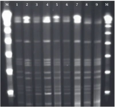
patterns were similar to those of the isolates recovered from a recent epidemic in Rio de Janeiro, Southeastern Brazil (strain CRM 0020, Figure 1).13 M. massiliense isolates
ob-tained from sputum samples (CRM 0270 and CRM 0273) showed no identical eletrophoretic patterns when compared to those isolates from Curitiba and Rio de Janeiro (Figure 2).
Figure 1: PFGE pattern of M. massiliense isolates from
Curitiba outbreak. M, molecular size markers (Lambda
DNA concatemers ranging from 48.5 to 1,018.5 kb); lane 1,
CRM-587; lane 2, CRM-591; lane 3, CRM-592; lane 4, CRM-593; lane 5, CRM-595, lane 6, CRM- 596; lane 7, CRM-598; lane 8,
CRM-602 and lane 9, CRM-604.
Figure 2: Map of rapidly growing mycobacteria confirmed cases by the Brazilian Public Health Surveillance System
from 2001 to 2008. The map indicates the cities of Goiânia,
Belém, Rio de Janeiro and Curitiba where outbreaks have been reported.
M 1 2 3 4 5 6 7 8 9 M
No confirmed cases
1 - 100 cases
101 - 250 cases
251 - 500 cases
Curitiba
Rio de Janeiro
Goiânia
DISCUSSION
The Brazilian Public Health Surveillance System has pre-sented cases of RGM infections from 2001 to date. Since 2001, a total of 1,937 confirmed cases were registered, with an evident increase of outbreaks in the period (Figure 3). However, there are only three reports of M. massiliense out-breaks in Brazil, all related to surgical infection following video-assisted surgeries.
This study reports the identification and molecular epi-demiological features of a single clone of M. massiliense iso-lated from a new outbreak of surgical site infections caused by RGM in Curitiba, Southern region of Brazil. Partial rpoB sequence analysis was considered discriminatory for identifi-cation of M. massiliense clinical isolates described here, since we obtained the highest similarity index (100%) when com-paring our sequences to that of the M. massiliense type strain.
The rpoB partial sequencing and PFGE analysis confirmed
the similarity of our isolates with those of previously report-ed outbreaks in Brazil.2,13,14 The molecular patterns obtained
suggest a common source of infection and spread of a single clone of M. massiliense in different regions of the country.
There were some limitations in this study. First a re-call bias might have occurred since only 39 cases were in-cluded causing the reduced availability of other strains for analysis. Additionally, the lack of standardized procedures or protocols for isolation of RGM from surgical supplies made it difficult to determine the infection sources and its relation with surgical devices and equipments. Since no environmental isolates were obtained, we were not able to definitely identify the source of the infection. In this outbreak surgical equipments were disinfected by immer-sion in 2% glutaraldehyde and were used in different hos-pitals by different surgical teams that brought along their own instruments and performed surgeries in other cities and states. Inspections made by public health authorities evidenced that the disinfection protocol was unsettled by some of the hospitals in different ways. Thus, strictly moni-toring concerning disinfection in 2% glutaraldehyde solu-tion may explain why some hospitals in Curitiba have not had cases of surgical infections caused by RGM.
The sources of the infections for the surgical cases have not been identified in the first outbreak, in Northern Brazil. Patient procedures were performed by different surgeons, who used their own laparoscopic equipment, referred to be disinfected by immersion in 2% glutaraldehyde between surgeries and which were used in different hospitals.2
Incon-sistencies in equipment cleaning, glutaraldehyde concentra-tions, or contact times were suggested as the cause of the M. massiliense strain selection. The disinfection procedure used in arthroscopic and laparoscopic surgeries in Central Brazil outbreak was also performed by immersion in 2% glutaraldehyde. According to inspections made by public health authorities, the disinfection protocol was incorrectly
implemented in some hospitals. So, inadequate aseptic tech-niques during surgeries could have been the possible cause.13
In the Southeastern outbreak, all hospitals that presented cases of M. massiliense infection used 2% alkaline glutaral-dehyde solution to sterilize surgical instruments. Interest-ingly, all M. massiliense isolates consistently presented toler-ance to 2% glutaraldehyde.14 The authors suggested that 2%
glutaraldehyde tolerance may partially explain the occur-rence of outbreaks in three different regions in Brazil.
Based on reported RGM outbreaks, potential source of infection was either the lack of adequate disinfection proce-dures (that could result in biofilm formation) or resistance to common disinfectants, or even both. Biofilms have been described in a high number of human infections, especially those related to biomaterials.23 Although biofilm formation
may contribute to tolerance to biocide solutions,24 biofilm
itself could not explain the single clone found on differ-ent time periods in distinct Brazilian regions.13 However,
since a significant relationship between biofilm develop-ment ability and clinical infection has been experidevelop-mentally demonstrated, biofilm development may be an important pathogenic risk factor for RGM, contributing to develop-ment of human infections.25 Moreover, biofilms are a
well-known form of bacterial resistance against antibiotics,26
and therefore the facility to develop these structures can explain treatment failures.27
Non-tuberculosis mycobacteria are often resistant to standard antituberculosis drugs and can be very difficult to treat. If RGM infection is identified and treated early, ade-quate recovery is possible, otherwise death can ensue. In this report, 33 patients (n = 39, 84.6%) were treated with a com-bination of amikacin, clarithromycin and terizidone. Treat-ment with multiple agents is preferable because of a high rate of relapse and emergence of drug resistance.28 Amikacin
and clarithromycin exhibited the greatest activity against all RGM isolates in this study and demonstrated to be
effec-tive when used in a multidrug regimen. All M. massiliense
isolates from Rio de Janeiro outbreak tested in vitro were susceptible to amikacin and clarithromycin and resistant to cefoxitin, ciprofloxacin and doxycycline;13 the same
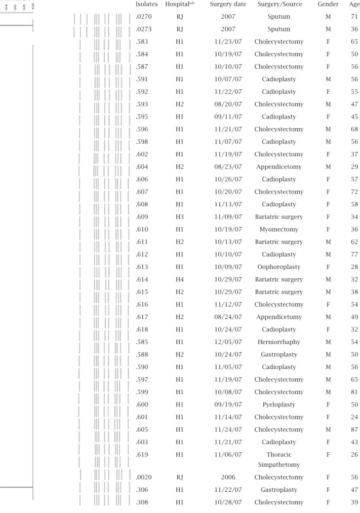
per-Figure 3: PFGE patterns of 36 M. massiliense isolates from Curitiba outbreak. Identical patterns clustered the isolates from
surgical patients. Comparison was performed with a commercial software (Applied Maths). The percentages of similarity among the profiles were calculated using Dice coefficient. The PFGE patterns of M. massiliense isolated from Rio de Janeiro outbreak (CRM 0020) and two epidemiologically unrelated strains from Rio de Janeiro patients (CRM 0270 and CRM 0273) were included
in the analysis. a Hn, mycobacterial isolate from patient who undergone surgery at hospital number n; b RJ, mycobacterial isolate
collected from Rio de Janeiro city.
Isolates Hospitala,b Surgery date Surgery/Source Gender Age
.0270 RJ 2007 Sputum M 71
.0273 RJ 2007 Sputum M 36
.583 H1 11/23/07 Cholecystectomy F 65
.584 H1 10/19/07 Cholecystectomy F 50
.587 H1 10/10/07 Cholecystectomy F 56
.591 H1 10/07/07 Cadioplasty M 56
.592 H1 11/22/07 Cadioplasty F 55
.593 H2 08/20/07 Cholecystectomy M 47
.595 H1 09/11/07 Cadioplasty F 45
.596 H1 11/21/07 Cholecystectomy M 68
.598 H1 11/07/07 Cadioplasty M 56
.602 H1 11/19/07 Cholecystectomy F 37
.604 H2 08/23/07 Appendicetomy M 29
.606 H1 10/26/07 Cadioplasty F 57
.607 H1 10/20/07 Cholecystectomy F 72
.608 H1 11/13/07 Cadioplasty F 58
.609 H3 11/09/07 Bariatric surgery F 34
.610 H1 10/19/07 Myomectomy F 36
.611 H2 10/13/07 Bariatric surgery M 62
.612 H1 10/10/07 Cadioplasty M 77
.613 H1 10/09/07 Oophoroplasty F 28
.614 H4 10/29/07 Bariatric surgery M 32
.615 H2 10/29/07 Bariatric surgery M 38
.616 H1 11/12/07 Cholecystectomy F 54
.617 H2 08/24/07 Appendicetomy M 49
.618 H1 10/24/07 Cadioplasty F 32
.585 H1 12/05/07 Herniorrhaphy M 54
.588 H2 10/24/07 Gastroplasty M 50
.590 H1 11/05/07 Cadioplasty M 56
.597 H1 11/19/07 Cholecystectomy M 65
.599 H1 10/08/07 Cholecystectomy M 81
.600 H1 09/19/07 Pyeloplasty F 50
.601 H1 11/14/07 Cholecystectomy F 24
.605 H1 11/24/07 Cholecystectomy M 87
.603 H1 11/21/07 Cadioplasty F 43
.619 H1 11/06/07 Thoracic F 26
Simpathetomy
.0020 RJ 2006 Cholecystectomy F 56
.306 H1 11/22/07 Gastroplasty F 47
.308 H1 10/28/07 Cholecystectomy F 39
oxide plasma or low-temperature steam with formaldehyde gas. Since these measures have been adopted by the hospitals no additional case of RGM infection was registered by the Brazilian Public Health Surveillance System.
The obtained results suggest that a single M. massiliense clone might be responsible for the infections that have oc-curred in Northern, Central and Southeastern regions of Brazil and reinforce the concept of M. massiliense BRA100 as an emergent pathogen in Brazilian hospital surgical en-vironment.
REFERENCES
1. Trupiano JK, Sebek BA, Goldfarb J et al. Mastitis due to
Myco-bacterium abscessus after body piercing. Clin Infect Dis 2001; 33:131-4.
2. Viana-Niero C, Lima KV, Lopes ML et al. Molecular
charac-terization of Mycobacterium massiliense and Mycobacterium
bolletii in isolates collected from outbreaks of infections after laparoscopic surgeries and cosmetic procedures. J Clin Micro-biol 2008; 46:850-55.
3. Brown-Elliott BA, Wallace RJ. Clinical and taxonomic status of pathogenic nonpigmented or late-pigmenting rapidly growing mycobacteria. Clin Microbiol Rev 2002; 15:716-46.
4. Zhibang Y, BiXia Z, Qishan L et al. Large-scale outbreak of
infection with Mycobacterium chelonae subsp. abscessus after
penicillin injection. J Clin Microbiol 2002; 40:2626-28. 5. Esteban J, Martın-de-Hijas NZ, Fernandez AI et al.
Epidemiol-ogy of infections due to Non-pigmented Rapidly Growing My-cobacteria diagnosed in an urban area. Eur J Clin Microbiol Infect Dis 2008; 27:951-57.
6. Kim HY, Kook Y, Yun YJ et al. Proportions of Mycobacterium
massiliense and Mycobacterium bolletii strains among Korean Mycobacterium chelonae-Mycobacterium abscessus group iso-lates. J Clinl Microbiol 2008; 46:3384-90.
7. Falkinham JO. Epidemiology of infection by nontuberculous mycobacteria. Clin Microbiol Rev 1996; 9:177-215.
8. Tiwari TS, Ray B, Jost Jr KC et al. Forty years of disinfectant failure: outbreak of postinjection Mycobacterium abscessus in-fection caused by contamination of benzalkonium chloride. Clin Infect Dis 2003; 36:954-62.
9. Wilson RW, Steingrube VA, Bottger EC et al. Mycobacterium
immunogenum sp. nov., a novel species related to
Mycobac-terium abscessus and associated with clinical disease, pseudo-outbreaks and contaminated metalworking fluids: an interna-tional cooperative study on Mycobacterial taxonomy. Int J Syst Evol Microbiol 2001; 51:1751-64.
10. Fraser VJ, Jones M, Murray PR et al. Contamination of flexible
fibreoptic bronchoscopes with Mycobacterium chelonae linked
to an automated bronchoscope disinfection machine. Am Rev Resp Dis 1992; 145:853-55.
11. Freitas D, Alvarenga L, Sampaio J et al. An outbreak of
Myco-bacterium chelonae infection after LASIK. Ophthalmol 2003; 110:276-85.
12. Sampaio JL, Chimara E, Ferrazoli L et al. Application of four
molecular typing methods for analysis of Mycobacterium
for-tuitum group strains causing post-mammaplasty infections. Clin Microbiol Infect 2006; 12:142-49.
13. Duarte RS, Lourenço MC, Fonseca L et al. An Epidemic of
Postsurgical Infections Caused by Mycobacterium massiliense.
J Clin Microbiol 2009; 47: 2149-55.
14. Cardoso AM, Martins de Sousa E,Viana-Niero C et al.
Emer-gence of nosocomial Mycobacterium massiliense infection in
Goias, Brazil. Microb Infect 2008; 10:1552-57.
15. McMurray DN. Mycobacteria and nocardia. In: Roberts GD. Laboratory Procedures in Clinical Microbiology. Springer-Verlag, Mayo Foundation, 1985.
16. Boom R, Sol CJ, Salimans MM et al. Rapid and simple method for purification of nucleic acids. J Clin Microbiol 1990; 28:495-503.
17. Adekambi T, Colson P, Drancourt M. rpoB-based
identifica-tion of nonpigmented and late-pigmenting rapidly growing mycobacteria. J Clin Microbiol 2003; 41:5699-708.
18. Hall TA. BioEdit: a user-friendly biological sequence align-ment editor and analysis program for Windows 95/98/NT. Nucleic Acids Symp 1999; 41:95-8.
19. CLSI. Clinical and Laboratory Standards Institute Quality Manual, 3rd edn. CLSI, 940 West Valley Road, Suite 1400, Wayne, Pennsylvania 19087-1898 USA, 2003.
20. Coleman NV, Spain JC. Distribution of the coenzyme M path-way of epoxide metabolism among ethene- and vinyl chloride-degrading Mycobacterium strains. Appl Environ Microbiol 2003; 69:6041-46.
21. Sampaio JL, Viana-Niero C, de Freitas D et al. Enterobacterial repetitive intergenic consensus PCR is a useful tool for typ-ing Mycobacterium chelonae and Mycobacterium abscessus iso-lates. Diagn Microbio Infect Dis 2006; 55:107-18.
22. Tenover FC, Arbeit RD, Goering RV et al. Interpreting chro-mosomal DNArestriction patterns produced by pulsed-field gel electrophoresis: criteria for bacterial strain typing. J Clin Microbiol 1995; 33:2233-39.
23. Donlan RM. Biofilm formation: a clinically relevant microbio-logical process. Clin Infect Dis 2001; 33;1387-92.
24. Simoes M, Pereira MO, Vieira MJ. Effect of mechanical stress on biofilms challenged by different chemicals. Water Res 2005; 39:5142-52.
25. Martín-de-Hijas NZ, García-Almeida D, Ayala G et al. Bio-film development by clinical strains of non-pigmented rapidly growing mycobacteria. Clin Microbiol Infect 2009; 15:931-36. 26. Mah TC, O’Toole GA. Mechanisms of biofilm resistance to
an-timicrobial agents. Trends Microbiol 2001; 9:34-9.
27. De Groote MA, Huitt G. Infections due to rapidly growing my-cobacteria. Clin Infect Dis 2006; 42:1756-63.