Respiratory bronchiolitis-associated interstitial lung
disease*
SÍLVIA CS. RODRIGUES, MAURI M. RODRIGUES, ESTER MC COLLETA, NAILÊ S ROCHA, CARLOS AC PEREIRA(TE SBPT)
Respiratory bronchiolitis-associated interstitial lung disease is one of many within the spectrum of smoking-related diffuse infiltrative lung diseases. The clinical and functional characteristics are typically subtle. Herein, we describe two cases of diagnosed through open-lung biopsy, and characterized by insidious evolution of dyspnea, digital clubbing, cystic lesions on computed tomography scans, and hipoxemia upon exertion. We emphasize that, when smokers are evaluated, it is imprtant to consider a diagnosis of respiratory bronchiolitis-associated interstitial lung disease in the context of interstitial cystitis, as well as in that of lymphangioleiomyomatosis, eosinophilic granuloma and idiophatic pulmonary fibrosis.
J Bras Pneumol 2004; 30(6) 585-7.
*Study carried out at the Hospital do Servidor Público Estadual - São Paulo
Correspondence to: Dra. Sílvia Carla Sousa Rodrigues. Av. Onze de Junho, Nº 625 - AP. 85 – Vila Clementino - CEP 04041-052 –São Paulo, SP, Brazil. Tel: 55 11 5539 1732. E-mail: [email protected]
Submitted: 13 January 2004. Accepted, after review: 13 April 2004.
Key words Smoking. Interstitial lung diseases. Bronchiolitis.
INTRODUCTION
Respiratory bronchiolitis is the most commonly found interstitial alteration in smokers(1) and may
be an incidental finding in the anatomopathological analysis of pulmonary tissue in asymptomatic smokers. It is characterized by the presence of macrophages in the lumen of membranous and respiratory bronchioles(2). In some cases, the patient
presents respiratory symptoms and tomographic alterations, which may regress completely upon smoking cessation. Respiratory bronchiolitis-associated interstitial lung disease (RB-ILD) is characterized by greater accumulation of macrophages in the bronchioles and accentuated peribronchiolar interstitial inflammatory process(3).
Although insidious, respiratory symptoms are more common. Pulmonary function is discretely affected and the clinical and functional characteristics are typically subtle(4). The most common tomographic
alterations are bronchial wall thickening, ground-glass patterns and centrilobular nodules. Reticular infiltrates and cystic lesions may also be observed(5).
Jornal Brasileiro de Pneumologia 30(6) - Nov/Dez de 2004
CASE REPORTS
Case 1
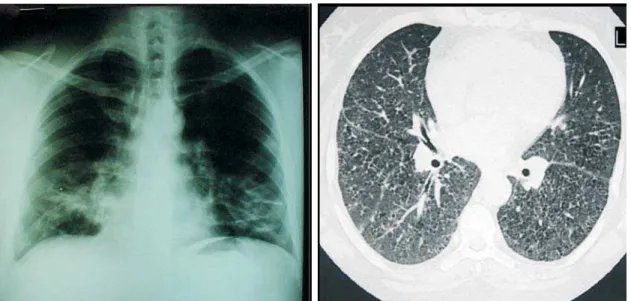
A white, female, 53-year-old kitchen assistant complained of slowly-progressive dyspnea, together with productive cough and nocturnal wheezing, for eight years. The patient also reported a current smoking habit and a 42-pack-year smoking history. She was suffering from diabetes and systemic arterial hypertension, controlled with metformin and captopril, respectively. Upon physical examination, digital clubbing was observed, although cardiac and pulmonary function was normal. The chest X-ray and HRCT scan are shown in Figure 1. The patient was submitted to a complete hemogram, blood biochemistry and immunological profile, the results of which were all within normal limits. Pulmonary function was evaluated through spirometry, in which no alterations were observed, and a six-minute walk test, which showed a significant reduction in arterial oxygen saturation by pulse oximetry (Table 1). Pulmonary diffusing capacity for carbon monoxide (DLCO) values were not available. The diagnosis of RB-ILD was established through
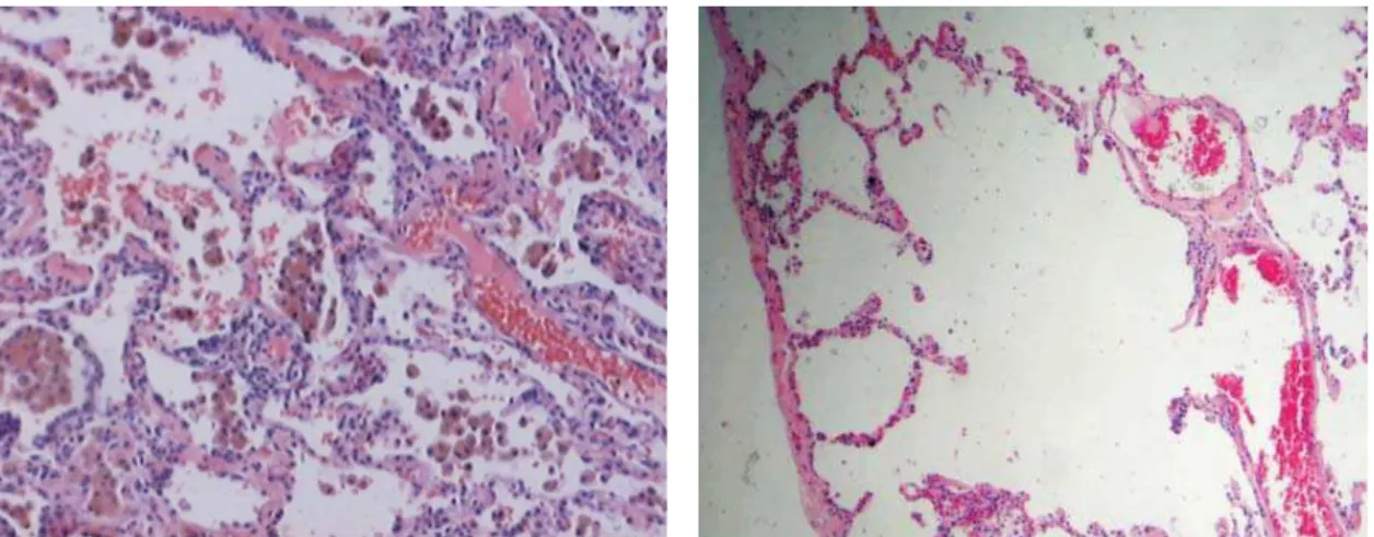
microscopic analysis of the pulmonary tissue obtained by surgical biopsy (Figure 2).
Case 2
A white, female, 45-year-old dressmaker presented progressive digital clubbing for ten years.
TABLE 1
Pulmonary function testing and SpO2(before and after the 6-minute walk test)
Case 1 Case 2
absolute % absolute %
FVC (L) 2,22 88 2,91 90
FEV1(L) 1,70 81 2,43 98
FEV1/FVC (%) 76 83
DLCO (ml/min/mmHg) 10,1 39
TLC (L) 3,45 71
RV (L) 1,00 62
RV/TLC (%) 29 initial final initial final
SpO2 (%) 96 87 98 94
SpO2: arterial oxygen saturation by pulse oximetry; FVC:
forced vital capacity; FEV1: forced expiratory volume in one second; DLCO: diffusing capacity for carbon monoxide; TLC: total lung capacity; RV: residual volume.
She complained of dyspnea upon moderate exertion (Mahler 9) and a dry cough. She reported a current smoking habit and a 30-pack-year smoking history. The patient presented no comorbidities.
Pronounced digital clubbing (Figure 3) was observed in the macroscopic examination and, upon auscultation, crackling rales were detected at the lung bases. Results of the radiological study of the lungs are presented in Figure 4. The patient was submitted to a complete evaluation of pulmonary function, including plethysmography. Mild restrictive ventilatory defect, with an accentuated reduction in pulmonary DLCO, was observed (Table 1). The diagnostic investigation included transbronchial biopsy, which was inconclusive, as well as subsequent surgical lung biopsy and anatomopathological study, through which the condition was defined as RB-ILD (Figure 5).
DISCUSSION
Although respiratory bronchiolitis is the most common type of smoking-related disease, it has, in general, few or no clinical or functional repercussions(1). In chest X-rays, the radiological
aspect varies from normal to a nodular or ground-glass pattern and, in HRCT scans, presents nodules with centrilobular distribution, as well as a ground-glass pattern(6). The main anatomopathological
characteristic is the accumulation of pigmented
macrophages in the lumen of membranous and respiratory bronchioles (a finding required for diagnosis), extending to the adjacent alveolar ducts and alveoli(2).The injury may be accompanied by a
minimal inflammatory process in the submucosa and peribronchiolar region. In some patients, almost exclusively heavy smokers, a more severe form, RB-ILD, appears. In such cases, microscopy of the lung reveals greater deposition of pigmented macrophages and an excess of connective tissue with or without greater infiltration by inflammatory cells into the peribronchiolar and alveolar spaces(3).
The patient usually complains of symptoms(5). The
desquamative interstitial pneumonia is also characterized by the presence of intra-alveolar
Figure 2. (a) Discrete fibrotic septal thickening and minimal mononuclear cell infiltration (thin arrow). Alveolar spaces containing numerous pigmented macrophages (thick arrow). H&E, x100. (b) Severed alveolar septa (arrow), characterized by areas of pulmonary emphysema. H&E, x100.
Jornal Brasileiro de Pneumologia 30(6) - Nov/Dez de 2004
macrophages. However, in this case, the lesions lose their bronchiolocentric preference and are more diffuse than those described in RB-ILD. The septae are thickened by fibrosis and interstitial inflammation is minimal. Desquamative interstitial pneumonia is related to smoking in 90% of cases(1).
Respiratory bronchiolitis is invariably associated with current or previous smoking. Fraig et al. examined 156 surgical samples of pulmonary tissue obtained under various conditions and observed findings typical of respiratory bronchiolitis in 109(3).
Of these 109 cases, 107 were smokers or ex-smokers. These findings support the idea that respiratory bronchiolitis is a highly sensitive and specific morphological marker of pulmonary injury caused by tobacco smoke. The authors also observed a correlation between smoking intensity and the degree of macrophage pigmentation and peribronchiolar fibrosis, finding that, in some patients, the damage may persist for many years
after smoking cessation. Another study(2) identified
signs of respiratory bronchiolitis in 70 out of 79 smokers (88.6%) submitted to some type of surgical treatment for spontaneous pneumothorax. The authors reported interstitial alterations in 53 (67%) of the 79 cases. In 9 patients, the anatomopathological lesions were considered severe and were consistent with desquamative interstitial pneumonia(2). Both studies showed that
respiratory bronchiolitis is usually an incidental finding in asymptomatic smokers and that, even in such cases, interstitial lesions consistent with RB-ILD or desquamative interstitial pneumonia may be evident.
The clinical presentation of RB-ILD is similar to that of other diffuse lung diseases. The patients are typically young, between 30 and 50 years of age, and the disease is more common in male patients(1), although epidemiological data show that
incidence of the disease is increasing in the female
population. When present, the symptoms are insidious and are characterized by cough and dyspnea. Crackling rales are detected in 76% of patients(5). The clinical and functional presentation
of the disease is rarely pronounced. In the literature, two cases of RB-ILD with incapacitating dyspnea and hypoxemia upon exertion have been reported(4). Digital clubbing, as illustrated in this
study, has been reported in at least four cases(4,7).
Digital clubbing in RB-ILD, albeit rare, calls attention to the fact that RB-ILD should be considered in the differential diagnosis of lung diseases presenting this condition upon macroscopic examination.
In a study of 21 individuals with RB-ILD(5), the
most common alterations seen in HRCT scans of the chest were thickening of the central bronchi (90%), thickening of the peripheral bronchi (86%), centrilobular nodules (71%) and ground-glass pattern (67%). Other findings were: centrilobular
emphysema, predominantly in the upper lobes (57%) and low-attenuation areas (38%), predominantly in the lower lobes. The authors observed septal and reticular lines in 7 patients (33%) and honeycombing in only 1 case(5).
Although uncommon, cystic lesions have also been described in RB-ILD(8,9). Koyama et al., in reviewing
92 tomographic images of chronic cystic lung disease patients, observed 8 cases of RB-ILD/ desquamative interstitial pneumonia(9). Scully et
al.(10) published a case of a young female smoker
presenting airway obstruction and cysts found on tomography of the chest. During the clinical discussion, lymphangioleiomyomatosis was considered the primary diagnostic hypothesis. However, the pathologist observed no cysts in the lung parenchyma and made a diagnosis of respiratory bronchiolitis. The authors interpreted the tomographic images of hypoattenuation as smoking-related centrilobular emphysema. Fibrosis
Jornal Brasileiro de Pneumologia 30(6) - Nov/Dez de 2004
is not typically seen in the emphysematous areas, although, when present, fibrotic wall formation may, on tomographic images, give the impression of cysts(11).
The case presented herein as case 1 is similar to that reported by Scully et al.(10). We observed
cystic lesions among areas of ground-glass pattern, which were classified as centrilobular emphysema under pulmonary microscopy (Figure 2). In case 2, there were concomitant large subpleural cysts (paraseptal emphysema), and a real cyst (characterized by a thin, well-defined wall) was seen in the HRCT scan of the chest (Figure 4) and confirmed in the histopathological analysis of the lung (Figure 5). These data emphasize the need to include RB-ILD in the differential diagnosis of chronic cystitis. Cysts are radiological markers for Langerhan’s cell histiocytosis, or eosinophilic granuloma, which is another disease strictly associated with smoking(9). Radiologists have
observed a superimposition of findings between respiratory bronchiolitis, RD-ILD and desquamative interstitial pneumonia(12). This was ratified by
pathologists, who have found, within the same tissue sample, areas with signs of RB-ILD, desquamative interstitial pneumonia and histiocytosis of Langerhan’s cell histiocytosis(1).
Therefore, the current tendency is to consider the four syndromes only as different expressions of the same smoking-related aggravation of the small airways and pulmonary parenchyma.
In RB-ILD patients, significant functional alterations have not been demonstrated, and spirometry may vary from normal to any type of respiratory disorder, depending on the interrelationships between elastic recoil pressure in the lungs and thoracic cavity and the total respiratory system compliance (a balance between bronchiolitis and alveolitis). In a study conducted by Park et al. and involving 21 cases of RB-ILD, 38% presented normal spirometry and total lung capacity, 35% presented a restrictive pattern (total lung capacity < 75%) and 24% had airway obstruction. The authors also found that arterial oxygen tension and pulmonary DLCO were reduced(5). Ideally, as has been suggested for other
interstitial lung diseases, the patient should be submitted to a complete evaluation of respiratory function, including whole-body plethysmography and estimation of pulmonary DLCO, in order to
accurately quantify the type and severity of the respiratory disorder. The respiratory physiology study of the patient described in case 2 is a good example of this. The spirometry results were considered normal, although plethysmography revealed a discrete reduction in total lung capacity, indicating mild restrictive ventilatory defect, in addition to the pronounced reduction in pulmonary DLCO. It is interesting to note the reduction in total lung capacity with no concomitant alteration in forced vital capacity, since the latter is responsible for 70% to 75% of the total lung volume. This is because the measurement of total lung capacity is determined by the balance between inspiratory force and distensibility of the respiratory system, rather than being an isolated analysis of any of its components(13). The reduction
in pulmonary DLCO documented in case 2 is as likely to be due to the emphysema as to the interstitial disease accompanying the bronchiolar injury or to the bronchiolitis itself. The mechanism through which bronchiolitis per se may affect DLCO is not clear(14). Both patients presented significantly
reduced arterial oxygen saturation by pulse oximetry after the 6-minute walk test, which is another indicator of the severity of these cases and a demonstration of the value of this test as a complement to the gas exchange analysis. Plethysmography and DLCO determination were not carried out in case 1, since not all tests were available at the institution of origin.
The diagnosis of RB-ILD follows the routine of diagnosing other diffuse pulmonary diseases, generally established through surgical pulmonary biopsy and requiring close interaction among the pulmonologist, radiologist and pathologist. The essential treatment is smoking cessation, which will determine the prognosis of the disease(1). The use
of corticosteroids is debatable. However, for the more severe presentations, it is prudent to use prednisone or an equivalent, at a dose of 0.5 mg/ kg/day, for a period of six to twelve months. Immunosuppressive drugs are rarely needed. When smoking is discontinued, the evolution of the disease is usually favorable; respiratory symptoms, tomographic findings and pulmonary function test results improve and stabilize(4,7). Complete recovery
is possible(1).
forms of interstitial cystitis, together with lymphangioleiomyomatosis, eosinophilic granuloma and idiopathic pulmonary fibrosis. The disease has a variable clinical presentation and may be an incidental finding in chest X-rays or in the anatomopathological study of pulmonary tissue obtained by lung biopsy or resection (procedures indicated due to a cause other than bronchiolitis). Another, less common spectrum of the disease is characterized by signs and symptoms of severe respiratory problems, including digital clubbing, as described in both cases reported in this study. The functional effect will depend on the balance between bronchiolitis and alveolitis, therefore, any type of respiratory disorder and gas exchange may be observed.
REFERENCES
1. Ryu JH, Colby TV, Hartman TE, Vassalo R. Smoking-related interstitial lung diseases: a concise review. Eur Respir J 2001; 17: 122-132.
2. Cottin V, Streichenberger N, Gamondès J-P, Thévenet F, Loire R, Cordier J-F. Respiratory bronchiolitis in smokers with spontaneous pneumothorax. Eur Respir J 1998; 12: 702-4. 3. Fraig M, Shreesha U, Savici D, Katzenstein AA. Respiratory bronchiolitis study in current smokers, ex-smokers and never-smokers.Am J Surg Pathol 2002; 26: 647-53. 4. Sadikot RT, Johnson D, Loyd JE, Christman JW. Respiratory
bronchiolitis associated with severe dyspnea, exertional hypoxemia, and clubbing. Chest 2000; 117: 282-5. 5. Park JS, Brown KK, Tuder RM, Hale VAE, King Jr. TE,
Lynch DA. Respiratory bronchiolitis-associated interstitial lung disease: radiologic features with clinical and pathologic correlation. J Comput Assist Tomogr 2002; 26: 13-20.
6. Müller NL, Fraser RS, Colman NC, Paré PD. Radiologic Diagnosis of Diseases of the Chest. W.B. Saunders Company. 2001; 452-520.
7. Moon J, Du Bois RM, Colby TV, Hansell DM, Nicholson AG. Clinical significance of respiratory bronchiolitis on open lung biopsy and its relationship to smoking related interstitial lung disease. Thorax 1999; 54: 1009-14.
8. Lee K, Lee JS, Lynch DA, Song K, Lim T. The radiologic differential diagnosis of diffuse lung diseases characterized by multiple cysts or cavities. J Comput Assist Tomogr 2002; 26: 5-12.
9. Koyama M, Johkoh T, Honda O, Tsubamoto M, Kozuka T, Tomiyama N, Hamada S, Nakamura H, Akira M, Ichikado K, Fujimoto K, Rikimaru T, Tateishi U, Müller NL. Chronic cystic lung disease: diagnostic accuracy of high-resolution CT in 92 patients. AJR 2003; 180: 827-35.
10. Scully RE, Mark EJ, Ebeling SH, Phillips LD. Case Records of the Massachusetts General Hospital. N Engl J Med 1998; 338: 1051-7.
11. Cardoso WV, Sekhon HS, Hyde DM, Thurlbeck WM. Collagen and elastin in human pulmonary emphysema. Am J Respir Crit Care Med 1994; 149: 1382-3. 12. Heyneman LE, Ward S, Lynch DA, Remy-Jardin M,
Johkoh T, Müller NL. Respiratory bronchiolitis, respiratory bronchiolitis-associated interstitial lung disease and desquamative interstitial pneumonia: different entities or part of the spectrum of the same disease process? AJR 1999; 173: 1617-22.
13. Barreto SM. Diretrizes para Testes de Função Pulmonar. Volumes Pulmonares. Jornal Brasileiro de Pneumologia 2002; 28: 83-94.