Lung volume reduction surgery in an experimental rat
model of emphysema
LAERTE BRASILIENSE FUSCO(TE SBCT), MARCELO HELENO FONSECA, PAULO MANUEL PÊGO-FERNANDES(TE SBCT), ROGÉRIO PAZETTI, VERA CAPELOZZI, FABIO BISCEGLI JATENE(TE SBCT), SERGIO ALMEIDA OLIVEIRA
* Study carried out in the Pulmonology Sector of the InCor Thoracic Surgery Department - HC FMUSP
Correspondence to: Paulo M. Pêgo-Fernandes. Rua Dr. Enéas de Carvalho Aguiar, 44 2o andar, CEP: 05403-000 São Paulo SP.Phone: 55-11 3069 5248. E mail: [email protected]
Submitted: 18 May 2004. Accepted, after review: 20 September 2004. J Bras Pneumol 2005; 31(1): 34-40.
Key words: Pulmonary emphysema. Papain/drugs efects. Case-control studies. Disease models, animal. Respiratory mechanics/ drugs efects. Lung/sugery. Lung/anatomy & histology.
Background: Lung volume reduction surgery may be a viable treatment alternative for emphysema patients suffering from severe respiratory insufficiency.
Objectives: To evaluate functional and morphological aspects of emphysematous rat lungs, prior to and following lung volume reduction surgery.
Method: Wistar rats were divided into two experimental groups (papain without surgery and papain with surgery) and three control groups (saline without surgery, saline with surgery and papain without mechanical ventilation). After approximately 40 days of endotracheal instillation of papain or saline solution, animals in the papain with surgery and saline with surgery groups were submitted to bilobectomy of the middle lobes by right thoracotomy along the posterior border of the superior vena cava. After 1 week, the same animals were submitted to a mechanical ventilation study, which involved measurement of lung elasticity and airway resistance. For all of the animals studied, lung tissue was analyzed in order to determine alveolar diameter and the elastic fiber quantity.
Results: Morphometric analysis revealed higher mean alveolar diameter in the lungs of all animals exposed to papain as compared to those exposed to saline. Elastic fiber counts in the alveolar septa of animals treated with papain were lower than those of animals receiving saline. In the animals submitted to bilobectomy and papain, lung elasticity was greater than in those receiving papain without surgery and was statistically equal to that seen in animals receiving saline (with or without surgery).
INTRODUCTION
In the mid 1990s, a viable treatment alternative to lung transplant for emphysema patients suffering from severe respiratory insufficiency re-entered the panorama of procedures available to thoracic surgeons(1). Lung volume reduction surgery
b e g a n t o c a l l o u r a t t e n t i o n d u e t o t h e e t i o p a t h o g e n i c m e c h a n i s m o f p u l m o n a r y emphysema and the alterations to the ventilatory mechanics brought about by this type of surgery. Hyperinflation, which causes a reduction in elastic recoil and expiratory flow limitation, increases the respiratory work and the demand for oxygen, negatively affecting gas exchanges and increasing oxygen consumption by cells, which leads to progressive incapacity to perform physical activities.
Various types of emphysema can be defined according to the pattern of involvement of the pulmonary acini, indicating the etiology and the physiopathological behavior of the disease presentation.
Panacinar emphysema presents pulmonary acini affected by a diffuse enlargement that usually extends from the hilar region to the lung periphery. This type of emphysema is found in patients with alpha-1 antitrypsin deficiency and is also the standard form seen in experimental models of emphysema induced through the use of proteolytic enzymes such as papain and elastase, being directly related to the dosage of the enzyme used(2).
Experimental models of pulmonary emphysema induced by proteolytic enzymes such as papain and elastase, which are instilled or nebulized in the airways of animals, are based on the theory that there is an imbalance in the production of aggressive and protective substances in the pulmonary acini(3).
In 1964, Gross et al.(4) first reproduced experimental
emphysema in rats through intratracheal instillation of papain, a proteolytic enzyme extracted from the fruit and sap of the papaya tree (Carica papaya). These experimental models resulted in histomorphological and physiological alterations to the lungs that were equivalent to the alterations found in emphysema in humans(5-8).
Lung volume reduction surgery is a generic term that encompasses various techniques intended to offer relief of symptoms, especially dyspnea, for patients with chronic obstructive pulmonary disease caused by emphysema(9-12).
Lung volume reduction surgery can be performed using thoracotomy, sternotomy or, more re c e n t l y, w i t h t h e h e l p o f v i d e o - a s s i s t e d t h o r a c o s c o py. T h is t e c h n i q u e c o ns is t s o f resectioning 20% to 30% of the pulmonary parenchyma that has diffuse involvement in each lung, using surgical staples buttressed with strips of bovine pericardium. It is possible to perform this surgery in one or both lungs. The surgery results in considerable improvement in ventilatory parameters and in lung elastic recoil as well as providing significant relief of dyspnea(13).
I n o r d e r t o e v a l u a t e f u n c t i o n a l a n d morphological aspects of emphysematous rat lungs prior to and following lung volume reduction surgery, we created an experimental rat model of pulmonary emphysema using tracheal instillation of a papain-based solution. We compared the study group lungs to normal rat lungs, submitted or not to mechanical ventilation in order to potentiate the effect of the substance.
METHODS
Male Wistar rats (n = 47), weighing 292.24 g ± 35.76 g, were obtained from the animal laboratory of the University of São Paulo School of Medicine and were used in this study after approval by the university Animal Research Ethics Committee.
The animals were divided into five experimental groups (two study groups and three control groups): group 1 (study animals submitted to endotracheal instillation of papain solution and mechanical ventilation; n = 10); group 2 (study animals submitted to endotracheal instillation of papain solution and mechanical ventilation, followed by bilobectomy; n = 12); group A (control animals submitted to endotracheal instillation of 0.9% saline solution and mechanical ventilation; n = 10); group B (control animals submitted to endotracheal instillation of 0.9% saline solution and right bilobectomy, followed by mechanical ventilation; n = 10); group C (control animals submitted to endotracheal instillation of papain solution but not to mechanical ventilation; n = 5).
Our objective in forming control group C was to monitor the possible effects of mechanical ventilation on the lung parenchyma, in which it is known to cause marked distention and alterations. The animals in groups 1, 2 and C were submitted to instillation of 3.5 mL/kg of papain solution in 0.9% isotonic saline solution (crystallized, lyophilized powder/10-20 units per mg of Sigma® protein), corresponding to 20 mg/kg of rat weight (approximately 6 mg of papain per animal). Instillation was performed over a period of approximately 2 minutes, using an orotracheal tube at a slow infusion rate.
The animals in groups A and B were submitted to instillation of 3.5 mL/kg of isotonic saline solution. Instillation was also performed over a period of approximately 2 minutes, using an orotracheal tube at a slow infusion rate.
Surgery was carried out after approximately 40 (40 ± 3) days of instillation of saline or papain solution(14).
We decided to standardize the resection of the middle and accessory lobes of the right lung, r e d u c i n g t h e r i g h t l u n g p a r e n c h y m a b y approximately 20% to 30% in accordance with certain anatomical criteria. The lungs remained in their respective thoracic cavities and were covered by a thin layer of visceral pleura. The right lung comprises four lobes: the cranial lobe, the middle lobe, the caudal lobe, and a fourth lobe, lying on the diaphragm and pierced by the inferior vena cava, commonly referred to as the accessory lobe. The left lung consists of only one lobe(2). We
weighed 10 right lungs and their respective lobes separately in order to quantify the parenchymal resection obtained through the bilobectomy. This quantification revealed that the middle and accessory lobes together represented 36.79% ± 2.16% of the total right lung weight of the Wistar rats studied.
Access to the right pleural cavity was gained through right lateral thoracotomy, and the resection of lobes was performed using a single ligation of the corresponding vessels and bronchi w i t h 5 - 0 n y l o n ( m o n o f i l a m e n t ) s u t u r e . Thoracotomy closure was carried out in three planes with 2-0 and 3-0 Polycot. We used a 2-mm-diameter water-sealed polyethylene tube to drain fluid and remove air from the pleural cavity, which
r e q u i r e d a p p r o x i m a t e l y 5 m i n u t e s u n d e r mechanical ventilation.
After 50 days of the intratracheal instillation of papain or saline solution, the animals in groups 1, 2, A and B were submitted to anesthesia with 0.5 g of sodium thiopental (Thionembutal®), at a dose
of 30 mg/kg, and submitted to orotracheal intubation and mechanical ventilation (respiratory frequency of 80 cycles per minute and tidal volume of 10 mL/kg) for determination of ventilatory parameters such as tracheal flow, volume and pressure in order to determine respiratory system elastance and resistance.
T h e t u b e w a s c o n n e c t e d t o a F l e i s c h pneumotachograph (model 0000; OEM Medical, Richmond, VA, USA) coupled to a Validyne differential pressure transducer (model DP 45-16-2114; Validyne Corp. Northridge, CA, USA) in order to measure the respiratory flow. Tracheal pressure was measured with a Validyne pressure transducer (model DP 45-16-2114) attached to a T-shaped metal piece connecting the tracheal tube and the pneumotachograph. Tracheal flow and pressure data were registered on a polygraph (Gould model RS 3400, Gould Inc., Cleveland, OH, USA) through a 12-byte analog-digital converter (model DT2801A, Data Translation, Marlboro, MA, USA) at a f r e q u e n c y o f 2 0 0 H z a n d s t o r e d i n a microcomputer. Each measurement was obtained with a mean of 13 respiratory cycles. Flow data were integrated so that variations in lung volume were determined.
Respiratory system resistance and elastance were obtained from the movement equation adapted for the respiratory system:
TP = E .V(t) + R .V(t)
where TP is tracheal pressure, E is respiratory system elastance, V is lung volume, (t) is time, and R is respiratory system resistance.
After measurement of ventilatory mechanics, the animals in groups 1, 2, A, B and C were sacrificed and the samples were processed.
After the removal of the heart and lungs, the right lungs were immediately fixed in liquid nitrogen for 2 minutes and subsequently immersed in Carnoy’s solution (ethanol-chloroform-acetic acid in a 60:30:10 proportion) and stored at -20oC for 24 hours. At the end of this period,
up to absolute ethanol. After 24 hours, samples were placed in 10% formaldehyde solution at -4oC, and 5-µm-thick sections were cut from the
lung parenchyma and stained with hematoxylin and eosin for qualitative and quantitative analysis of the effect of papain.
Pulmonary emphysema was quantified by the presence of alveolar destruction determined by the measure of the mean alveolar diameter in m i c r o m e t e r s . T h i s t e c h n i q u e c o n s i s t s o f determining how many times gas exchange structures in the parenchyma intersect a series of grid lines(15).
The lung tissue was submitted to analysis and quantification of the elastic fibers present in the interalveolar septa, with the random selection of 10 fields, 400 times increased, of 5 animals submitted to the instillation of the papain solution and of 4 animals submitted to the instillation of saline solution. Fibers of the elastic system that contain elastin were identified using Weigert’s resorcin-fuchsin stain. The quantification of elastic fibers was carried out with the aid of a digital system using a specific software program (Bioscan-Optimas; Bioscan Incorporated, Edmont, WA, USA).
The area occupied by elastic fibers was determined by digital densitometry(16). We were
thereby able to determine the percent of elastic fibers in the alveolar septa of lungs submitted to the effect of papain or 0.9% saline solution.
All variables were initially analyzed descriptively. The analysis of continuous variables was carried out through observation of minimum and maximum values, means, standard deviations and medians. We calculated absolute and relative frequencies f o r c a t e g o r i c a l v a r i a b l e s . W e u s e d t h e nonparametric Kruskall-Wallis test with Dunn correction for multiple comparisons in order to test the hypothesis of equality of the means among groups. The significance level for the tests was set at 5%. We used the non-parametric Mann-Whitney test specific for small samples for the statistical analysis of the quantification of elastic fibers (p = 0.006).
RESULTS
The mortality rate in study groups 1 and 2, which were submitted to instillation of papain solution, was 20% (6 animals), compared with 7% (2 animals) in the control groups A and B, which
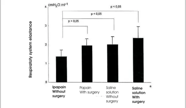
Figure 1 – Demonstration of variation and elastance by group
Ipapain Without surgery
Papain With surgery
Saline solution Without surgery
Saline solution With surgery
R
espiratoty
system
were submitted to instillation of 0.9% saline solution. We identified the enzymatic aggression caused by papain as the direct cause of mortality in some animals since these animals presented respiratory distress followed by hemoptysis.
Using the nonparametric Kruskall-Wallis test, we found the following results for respiratory system resistance: there differences between group A (saline solution without surgery), group B (saline solution with surgery), group 1 (papain solution with surgery) and group 2 (papain solution without surgery) were not statistically significant (p < 0.05). As for respiratory system elastance, we determined that there were differences between the groups (p = 0.001) (Figure 1).
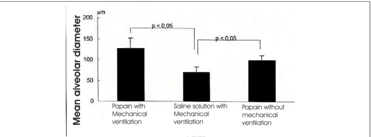
Using multiple paired comparison (Dunn’s method), we found the following: respiratory system elastance in group 1 (papain without surgery) was statistically lower than that of group A (saline solution without surgery) (p < 0.05); respiratory system elastance in group 1 (papain without surgery) was statistically lower than that of group 2 (papain with surgery) (p < 0.05); there were no statistically significant differences among the other groups. Using morphometric studies to determine alveolar diameter (Lm) we obtained the following alveolar diameter means (Figure 2): group 1 (papain solution without surgery): 146.61 mm; group 2 (papain solution with surgery): 109.67 mm; group A (saline solution without surgery):
Figure 2 – Demonstration of variation in the measurement of mean alveolar diameter by group
Figure 3 – Demonstration of the effect of papain in the quantification of elastic fibers in lung tissue. Papain with
Mechanical ventilation
Saline solution with Mechanical ventilation
Papain without mechanical ventilation
Mean
alveolar
diameter
papain Saline solution
Amount
of
area
occupied
b
y
elastic
fibers
in
75.90 mm; group B (saline solution with surgery): 62.68 mm; group C (papain solution without surgery and without mechanical ventilation): 100.56 mm.
We found statistically significant differences among the 3 groups (p < 0.0001). Group C (papain without mechanical ventilation) presented no significant difference when compared to group 1 (papain with mechanical ventilation). Together, group 1 (papain with mechanical ventilation) and group C (papain without mechanical ventilation) presented a significant difference when compared to group B (saline solution with mechanical ventilation) (p < 0.05).
Histological sections from the lungs of rats receiving papain (groups 1, 2 and C) revealed panacinar emphysema with histologically preserved lobular architecture, with recognizable terminal bronchioles, respiratory bronchioles and adjacent alveolar tissue. These changes were seen at the same intensity in all groups submitted to papain (with or without mechanical ventilation).
The mean ratio of the area occupied by elastic fibers in the lungs of the animals receiving papain was 5.36%, whereas that of the animals receiving saline solution was 17.09% (Figure 3).
Comparison of the means found in the two groups revealed that the mean ratio of the area occupied by elastic fibers was 70% lower in the lungs of animals receiving papain. Using the Mann-Whitney test, the difference in the mean ratio of the area occupied by elastic fibers was found to be statistically significant (p = 0.006).
DISCUSSION
Studies in the literature have shown that models of pulmonary emphysema that make use of the airway instillation or nebulization of proteolytic enzymes are efficacious in reproducing this disease(2,5-7,14,17). Therefore, we chose to use
intratracheal instillation of papain to produce pulmonary emphysema in rats.
Histopathological(2,18) and morphometric(19)
changes found in the lungs submitted to the effect of papain revealed enlarged alveolar spaces and rupture of the alveolar septa, meeting the morphological criteria for the reproduction of panacinar pulmonary emphysema. The mean alveolar diameter in rats free of lung diseases is on the order of 70 mm(20).
In relation to the histopathological changes found, questions arose regarding a possible effect of mechanical ventilation. We decided to create a control group to study this variable, group C, composed of animals submitted to papain instillation but not to mechanical ventilation. This group presented changes compatible with panacinar emphysema, although presenting lower mean alveolar diameters when compared to those of the animals submitted to papain instillation and mechanical ventilation (groups 1 and 2) and higher mean alveolar diameters when compared to those of the animals submitted to saline solution and mechanical ventilation (control groups A and B). These results revealed that, although papain produced emphysematous changes, there might have been an adjuvant effect of the mechanical ventilation on the parenchyma submitted to papain, producing marked alveolar distention and resulting in higher mean alveolar diameters.
The etiopathogenesis of pulmonary emphysema may be related to enzymatic digestion, especially that of elastic and collagen fibers, caused by polymorphonuclear cells and alveolar macrophages in the alveolar spaces(5). The hypothesis of
homeostatic imbalance between enzymes and inhibiting substances responsible for alveolar destruction is known as the hypothesis of protease-antiprotease imbalance(3). Osman et al.(18), using
nebulized papain solution in dogs, reported a decrease in total elastin in the lungs. In emphysema, the aggression process takes place in the alveolar spaces and involves destruction of the interalveolar septa, which leads to a change of the cytoarchitecture caused by the destruction of elastic and collagen fibers. Cigarette smoke has been reported to increase elastolytic activity, inhibit the proliferation of pulmonary fibroblasts, increase elastase susceptibility and inactivate antielastases(3).
Consequently, the main functional alteration caused by emphysema is a decrease in the elastic recoil pressure, or rather the tissue elastance, of the lungs(21).
to surgery. Respiratory system elastance was higher in group 2 (papain with surgery) than in group 1 (papain without surgery). Likewise, the elastance in group 2 was similar to that found in the groups receiving saline solution. This improved elastic recoil capacity in the respiratory system of the rats, translating to greater elastance, experimentally reinforced the concept of improved ventilatory mechanics after lung volume reduction of emphysematous lungs(22). In this model using
papain, respiratory system resistance suffered no significant alterations, revealing that airway changes did not produce functional repercussions. These data reinforced the histological and morphological findings confirming the fact that we had reproduced a model for the study of pulmonary emphysema, rather than a model of bronchitic-pattern chronic pulmonary obstructive disease with functional alteration of airway resistance caused by inflammation and bronchial hyperresponsiveness.
The object of lung volume reduction surgery is the resection of defunctionalized target areas, reducing the quantity of gas trapped in the lungs and thus improving ventilatory mechanics(13).
This reduction in pulmonary hyperinflation optimizes ventilatory mechanics through improving respiratory muscle performance, achieving this by restoring a geometric pattern in the chest wall and muscular fibers that is closer to normal(23).
This experimental investigation confirmed that experimental emphysema can be reproduced in rats through airway instillation of papain. It also revealed that this type of emphysema presents morphological and physiopathological characteristics similar to those seen in humans. Finally, we demonstrated that even the bilobectomy technique of lung volume reduction might reverse the functional alterations found in experimental emphysema.
REFERENCES
1. Cooper JD, Trulock EP, Triantafillou NA, Patterson GA, Pohl MS, Deloney PA, et al. Bilateral pneumonectomy (volume reduction) for chronic obstructive lung disease. J Thorac Cardiovasc Surg. 1995;109:106-19. 2. Snider GL, Lucey EC, Stone PJ. Animal models of
emphysema. Am Rev Respir Dis. 1986;133:149-69. 3. Janoff A. Elastases and emphysema: current assessment
of the protease-antiprotease hypothesis. Am Rev Respir Dis. 1985;132:417-33.
4. Gross P, Bajak MA, Tolker E, Kaschak M. Enzymatically produced pulmonary emphysema; a preliminary report. J Occup Med. 1964;6:481-4.
5. Hayes JA, Korthy A, Snider GL. The pathophysiology of elastase-induced panacinar emphysema in hamster. J Pathol. 1975;117:1-14.
6. Martorana PA, Wusten B, Van Even P, Gobel H, Schaper J. A six-month study of the evolution of papain-induced emphysema in the dog. Am Rev Respir Dis. 1982;126:898-903.
7. Pushpakon R, Hogg JC, Woolcock AJ, Angus AE, MaCklem PT, Thurlbeck WM. Experimental papain-induced emphysema in dogs. Am Rev Respir Dis. 1970;102:778-89.
8. Fusco LB, Pêgo-Fernandes PM, Xavier AM. Modelo experimental de enfisema pulmonar em ratos induzido por papaína. J Pneumol. 2002;28(1):1-7.
9. B r a n t i g a n O C . S u r g i c a l t r e a t m e n t o f p u l m o n a r y emphysema. W Virginia Med J. 1954;50:283-5. 10. Takaro T, White SM. Emphysema. Am Rev Respir Dis.
1973;108:334-42.
11. Crenshaw GL, Rowles DF. Surgical management of pulmonary emphysema. J Thorac Cardiovasc Surg. 1952;24:398-410.
12. Naef A.P. History of emphysema surgery. Ann Thorac Surg. 1997;64:1506-8.
13. National Emphysema Treatment Trial Research Group. A randomized trial comparing lung-volume-reduction surgery with medical therapy for severe emphysema. N Engl J Med. 2003;348:2059-73.
14. Johanson Jr., WG, Pierce AK, Reynolds RC. The evolution of papain emphysema in the rat. J Lab Clin Med. 1971;78:599-607.
1 5 . W e i b e l E R . P r i n c i p l e s a n d m e t h o d s f o r t h e morphometric study of the lung and others organs. Lab Invest. 1963;12:131-55.
16. Uitto J., Paul JL, Brockley K, Pearce RH, Clark JG. Elastic fibers in human skin: quantitation of elastic fibers by computerized digital image analyses and determination of elastin by radioimmunoassay of desmosine. Lab Invest. 1983;49:499-505.
17. Weinbaum G, Marco V, Ikeda T, Mass B, Meranze DR, Kimbel P. Enzymatic production of experimental emphysema in the dog. Route of exposure. Am Rev Respir Dis. 1974;109: 351-7.
18. Osman M, Keller S, Cerreta JM, Levenberger P, Mandl I, Turino GM. Effect of papain induced emphysema on canine pulmonary elastin. Proc Soc Exp Biol Med. 1980;164:471-7.
19. Johanson Jr. WG, Pierce A. Lung structure and function w i t h a g e i n n o r m a l r a t s a n d r a t s w i t h p a p a i n emphysema. J Clin Invest. 1973;52:2921-7.
20. Baker HJ, Lindsey JR, Weisbroth SH. The laboratory rat. New York: Academic Press; 1979. v.1: Biology and diseases, p. 83-6.
21. Stead WW, Fry DL, Ebert RV. The elastic properties of the lung in normal men and in patients with chronic pulmonary emphysema. Am Rev Respir Dis. 1952;674-81 .
22. Gelb AF, McKeena Jr. RJ, Brenner M, Fischel R, Baydur A, Zamel N. Contribution of lung and chest wall mechanics following emphysema resection. Chest. 1996;110:11-7.