SURGICAL EXPLORATION OF THE INJURED KIDNEY: CURRENT
INDICATIONS AND TECHNIQUES
MICHAEL J. METRO, JACK W. McANINCH
Department of Urology, University of California School of Medicine, San Francisco, California, and Urology Service, San Francisco General Hospital,San Francisco,
California, USA
ABSTRACT
When treating renal injuries, the goals of the urologic surgeon are preservation of maximal renal function with a minimal risk of complications. To meet these, accurate staging is essential. The combined use of clinical and radiologic findings, with intra-operative information where avail-able, will enhance the practitioner’s ability to detect, classify, and treat renal injuries appropriately. We discuss our current approach to renal trauma and current indications and techniques for surgical exploration of the injured kidney.
Key words: kidney; wounds and injuries; practice management; reconstructive surgical procedures
Int Braz J Urol. 2003; 29: 98-105
CLASSIFICATION OF RENAL INJURIES
In patients sustaining abdominal trauma, ap-proximately 10% will have an injury of the genitouri-nary tract. Of these injuries, one half will be to the kidney (1,2). Renal injuries traditionally have been classified by mechanism: blunt trauma (constituting 80 - 90%), occurring most commonly in falls, motor vehicle accidents, and assaults (3); and penetrating trauma, occurring most commonly from gunshot and stab wounds. The majority of blunt renal injuries are minor and can be managed conservatively (at our in-stitution, only 2.5% have required exploration and surgical repair [2,4]), while penetrating injuries more often require operative intervention owing to the fre-quency of severe damage and associated intra-abdomi-nal injuries (5).
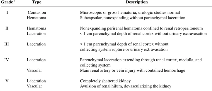
Accurately determining the grade of renal injury is a key factor in deciding the mode of man-agement. The Organ Injury Scaling Committee of the American Association for the Surgery of Trauma has classified five grades of traumatic renal injuries (6,7) (Table-1).
INDICATIONS FOR SURGICAL
EXPLORATION
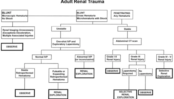
Before a renal injury can be selected for nonoperative management, it must be radiographically imaged and accurately staged (Figure-1). Incomplete staging mandates surgical exploration. Our indications for renal imaging have been well described (3,8). In adults, the presence of gross hematuria, microhematuria with shock, or microhematuria in patients with major deceleration injury warrants imaging with computed tomography (CT). In the pediatric population, any degree of hematuria, with or without shock, or a mechanism of injury to suggest a possible renal in-jury (e.g. deceleration inin-jury or flank contusion) man-dates imaging. The widespread use of CT and accu-mulated experience with the non-operative manage-ment of high-grade renal injuries have led to decreased rates of renal exploration.
Absolute Indications
Table 1 - American Association for the Surgery of Trauma. Organ injury severity scale for the kidney *
Grade † Type Description
I Contusion Microscopic or gross hematuria, urologic studies normal Hematoma Subcapsular, nonexpanding without parenchymal laceration
II Hematoma Nonexpanding perirenal hematoma confined to renal retroperitoneum Laceration < 1 cm parenchymal depth of renal cortex without urinary extravasation
III Laceration > 1 cm parenchymal depth of renal cortex without collecting system rupture or urinary extravasation
IV Laceration Parenchymal laceration extending through renal cortex, medulla, and collecting system
Vascular Main renal artery or vein injury with contained hemorrhage
V Laceration Completely shattered kidney
Vascular Avulsion of renal hilum, devascularizing the kidney
* Data drawn from reference 6; reprinted with permission from reference 7 † Advance one grade for bilateral injuries up to Grade III.
indicates persistent bleeding, usually from major pa-renchymal or vascular injury, and exploration is man-dated (9). In grade 5 injuries, for instance, the sever-ity - either pedicle avulsion or extensive parenchy-mal destruction - will require intervention (see Vas-cular Injury, below).
If adequately staged, many major renal inju-ries can be managed expectantly. Expectant manage-ment is not necessarily nonoperative: it is a period of close observation (with repeat radiographic studies in some cases), which determines when the injury might require surgical intervention.
Incomplete Staging
Often the instability of associated injuries will hinder complete staging, and in these cases a more aggressive approach is warranted. When a suspected renal injury has not been adequately staged preopera-tively, an intraoperative single-shot high-dose intrave-nous urogram should be obtained. Injection of 2 mL/ kg of intravenous contrast is given as a bolus and a single film is obtained at 10 minutes (10). Any abnor-mal or incomplete finding warrants renal exploration.
Thus, exploration is indicated in patients with unstaged blunt renal trauma, a retroperitoneal he-matoma, or equivocal findings on single-shot intra-venous urography. In addition, all patients with pen-etrating renal trauma with a retroperitoneal hematoma in whom adequate preoperative staging is not pos-sible should undergo exploration. This approach has resulted in a high rate of renal salvage and has not increased the rate of unnecessary nephrectomy (11).
Relative Indications
peri-nephric abscess, infected urinoma, and delayed hem-orrhage), requiring open surgical management. When immediate exploration with renal repair was per-formed in similar patients with associated pancreatic or bowel injuries, morbidity was reduced to 23% (13). On this basis, grades 3 and 4 injuries with significant devitalized fragments and concomitant intraperitoneal organ injuries should undergo immediate surgical re-pair.
In our experience, patients with a large, non-viable fragment and urinary extravasation or retro-peritoneal hemorrhage, even without significant in-traperitoneal injury, may also benefit from early re-nal exploration. The intervention is usually partial nephrectomy, which minimizes potential post-trau-matic complications.
Urinary extravasation alone does not neces-sitate surgical intervention, but it commonly reflects a major renal injury (grade 4) from either a lacera-tion of the renal pelvis or parenchyma or an avulsion of the ureteropelvic junction (UPJ). If the latter, im-mediate exploration is indicated. Suspicion of UPJ avulsion is raised by nonvisualization of the ipsilat-eral ureter on CT or intravenous urography (IVU) and
by the presence of significant contrast extravasation both medially and perirenally on the imaging study. These injuries are fairly rare and are more common in children with rapid deceleration injuries (14). They rarely heal spontaneously.
Blunt trauma can lead to forniceal rupture and significant urinary extravasation without associated parenchymal injury (15). When the degree of extrava-sation is small, most cases will resolve spontaneously. Larger degrees of extravasation may still subside with-out intervention, but monitoring with serial CT scans is indicated because of the risk of complication with-out spontaneous resolution. Intervention is indicated in persistent leakage, significant urinoma formation, or sepsis development.
Recent literature has shown more than 75% spontaneous resolution rate of urinary extravasation associated with grade 4 renal injuries. Percutaneous or endoscopic treatment was successful in most cases (16,17). Of 47 patients with major renal lacerations and urinary extravastion reported by Glenski & Husmann (16), 15% required endoscopic stenting for persistent leakage and only 9% of these required fur-ther intervention, i.e. exploration.
Figure 1 - Algorithm for treating patients with renal trauma. (Reprinted with permission from: Meng MV, Brandes SB, McAninch JW.
Gunshot wounds to the kidney often result in significant tissue damage and an increased risk of delayed complications, owing to the “blast effect” of the projectile’s temporary and permanent cavities. High-velocity missiles or close-range shotgun blasts are particularly devastating. Thus, the threshold for exploration for urinary extravasation from gunshot wounds should be lower than that for stab wounds or blunt trauma (5).
Vascular Injury
In cases of renovascular injury, prompt diag-nosis and immediate operative repair are mandatory for renal preservation. However, the detection of re-nal pedicle injuries is frequently delayed because as-sociated life-threatening injuries take precedence. Over 50% of trauma victims with renal vascular in-juries present in shock and the mortality rate ranges from 10 - 50% (18).
Renal pedicle injuries are seen more com-monly in children because of their relatively larger kidneys and lower amount of perinephric fat and de-gree of musculoskeletal development. During decel-eration injuries the inelastic intima of the artery can be disrupted, leading to thrombosis of a segmental or main renal artery with consequent parenchymal is-chemia or infarction. Main renal artery injuries have the lowest rate of repair and salvage (19). If surgical repair is undertaken within 12 hours, the chance of salvage is greatest; nevertheless, revascularization has demonstrated only a modest 10 - 30% success rate in multiple reports (19-22). Even with intervention within 5 hours, Cass et al. (19) have reported signifi-cantly reduced function in the few kidneys appropri-ate for vascular repair. Such patients are always criti-cally ill, and attempted repair subjects them to in-creased operative time and risks the complications of hypertension and delayed nephrectomy. Thus, renal preservation is best attempted within 12 hours of in-jury and in patients with bilateral inin-jury or solitary renal units. Patients in whom the injury appears to be incomplete or perfusion seems intact intraopera-tively can also be considered for reconstruction.
When the diagnosis of renal artery thrombo-sis is delayed or repair is not otherwise indicated, nephrectomy should be performed at exploration for
associated injuries. Patients with isolated renal ar-tery thrombosis who otherwise do not require explo-ration can be safely observed. The kidney can be allowed to atrophy slowly over time; complications of bleeding, infection and hypertension requiring ne-phrectomy are rare (23).
RENAL EXPLORATION
Although an in-depth description ofspecific renal reconstructive techniques is not within the pur-view of this article, principles regarding renal expo-sure must be borne in mind to enexpo-sure good salvage rates. When exploring an injured kidney, nephron preservation is the primary goal. Because uncontrolled hemorrhage is often the cause of total nephrectomy, we advocate preliminary proximal vascular control in all cases of renal trauma (24,25).
Early Vascular Control
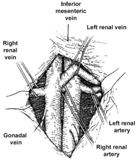
Proximal vascular control was initially de-scribed by Scott & Selzman (26). A transabdominal midline incision from the xyphoid to the pubic sym-physis provides the best access to the abdominal vis-cera and vasculature. The transverse colon is lifted from the abdomen and placed on the chest under moist laparotomy sponges. The root of the small bowel mesentery and the underlying retroperitoneum are exposed by lifting the bowel superiorly and to the right. A vertical incision is made over the aorta supe-rior to the supesupe-rior mesenteric artery and into the retroperitoneum, and this is extended upwards to the ligament of Treitz. Often, the aorta is difficult to pal-pate owing to the presence of retroperitoneal he-matoma. In these cases, the inferior mesenteric vein is used as a guide: the incision is made just medial to it, and the dissection is carried down to the anterior surface of the aorta (Figure-2).
first occluded and, if bleeding persists, the vein is clamped to reduce back bleeding. Warm ischemia time should be held to less than 30 minutes if possible (27). In our experience, occlusion of the renal vessels was required in only 17% of cases, but there is no reliable method for identifying such patients before exploration. On average it takes only 12 minutes to isolate the renal vessels.
Once vascular control has been achieved, the colon is reflected medially and the retroperitoneal hematoma is evacuated after Gerota’s fascia is incised laterally (Figure-4). The kidney is then exposed and assessed for injuries. The entire kidney must be well exposed to examine the renal pelvis, parenchyma and vessels fully.
RECONSTRUCTIVE PRINCIPLES
The first step in reconstruction involves ad-equate debridement: all nonviable tissue should be sharply excised and removed. Preservation of one-third of one kidney provides sufficient renal function to avoid dialysis. The renal capsule should be pre-served if at all possible, as it makes eventual closure more successful. Parenchymal vessels should be su-ture-ligated with 4-0 chromic sutures. Persistent, smaller venous bleeding will usually stop after the parenchymal defect is closed.
Lacerations in the collecting system should be closed in a watertight fashion with running 4-0 chromic suture. Careful injection of dilute methyl-ene blue into the renal pelvis after gentle occlusion of the proximal ureter can aid identification of inju-ries and confirm adequate closure of the collecting system. Additional drainage by internal stent or nephrostomy tube is not routinely required.
Figure 2 - Surgical approach to the renal vessels and kidney.
The retroperitoneal incision is made over the aorta medial to the inferior mesenteric vein. (Reprinted with permission from: McAninch JW: Surgery for Renal Trauma. In: Novick AC, Streem SB, Pontes JE (eds.), Stewart’s Operative Urology. Baltimore, Williams & Wilkins. 1989; 234-9).
Figure 3 - Anatomic relationship of the renal vessels. (Reprinted
After reconstruction, the defect should ide-ally be covered with renal capsule by reapproximation of the parenchymal edges. This is done with inter-rupted 3-0 vicryl sutures tied over gelfoam bolsters. This improves hemostasis and reduces the risk of uri-nary extravasation. We place titanium surgical clips on the sutures to aid identification of the suture line on postoperative CT scans. If the renal defect is sig-nificant, it can be packed with a hemostatic agent such as Avitene (microfibrillar collagen hemostat; Bard; Murray Hill, NJ) or with perinephric fat (Figure-5).
In rare cases, a devitalized polar segment will require partial nephrectomy with amputation and clo-sure of the collecting system. Omentum is a good choice to cover the polar defect if renal capsule is not available. In all renorrhaphies, a one-inch Penrose drain is left dependently to drain the retroperitoneum. A suction drain should not be used as it can promote urinary leakage from the repaired collecting system. Vicryl mesh can be placed around the kidney to sta-bilize the renorrhaphy repair or when large or mul-tiple parenchymal defects are difficult to cover.
Figure 4 - The retroperitoneal incision lateral to the colon,
exposing the kidney. (Reprinted with permission from: McAninch JW: Surgery for Renal Trauma. In: Novick AC, Streem SB, Pontes JE (eds.), Stewart’s Operative Urology. Baltimore, Williams & Wilkins. 1989; 234-9).
Figure 5 - Technique of renorrhaphy after midpole grade IV injury. (Reprinted with permission from: McAninch JW: Surgery for Renal
CONCLUSIONS
Our treatment guidelines and algorithms for the management of renal trauma are based on our 25-year experience with more than 3150 renal injuries at San Francisco General Hospital as well as on the accumulated knowledge of other trauma centers. This experience has validated our approach and reconstruc-tive techniques. Renal exploration is necessary in only 2% of blunt injuries and in 57% of penetrating inju-ries (42% of stab wounds and 76% of gunshot wounds [28]). Early vascular control yields a high rate of nal salvage, with only 11% of renal explorations re-quiring nephrectomy in our hands.
REFERENCES
1. Meng MV, Brandes SB, McAninch JW: Renal trauma: indications and techniques for surgical exploration. World J Urol. 1999; 17: 71-7.
2. McAninch JW, Carroll PR, Klosterman PW, Dixon CM, Greenblatt MN: Renal reconstruction after injury. J Urol. 1991; 145: 932-8.
3. Miller KS, McAninch JW: Radiographic assessment of renal trauma: our 15 year experience. J Urol. 1995, 154: 352-5.
4. PA, Bruce JE, McAninch JW: Nephrectomy for traumatic renal injuries. J Urol. 1995; 153: 609-11. 5. McAninch JW, Carroll PR, Armenakas N, Lee P:
Renal gunshot wounds: methods of salvage and reconstruction. J Trauma 1993; 35: 279-83.
6. Moore EE, Shackford SR, Pachter HL, McAninch JW, Browner BD, Champion HR, et al.: Organ injury scaling: spleen, liver, and kidney. J Trauma 1989; 29: 1664-6.
7. Brandes SB, McAninch JW: Reconstructive surgery for trauma of the upper urinary tract. Urol Clin North Am. 1999; 26: 183-99.
8. Nicolaisen GS, McAninch JW, Marshall GA, Bluth RF, Carroll PR: Renal trauma: re-evaluation of indications for radiographic assessment. J Urol. 1985; 133: 183-7.
9. Holcroft JW, Trunkey DD, Minagi H, Korobkin MT, Lim RC: Renal trauma and retroperitoneal hematomas: indications for exploration. J Trauma 1975; 15: 1045-52.
10. Morey AF, McAninch JW, Tiller BK, Duckett CP, Carroll PR: Single shot intraoperative excretory
urography for the immediate management or renal trauma. J Urol. 1999; 161: 1088-92.
11. McAninch JW, Carroll PR, Klosterman PW, Dixon CM, Greenblatt MN: Renal reconstruction after injury. J Urol. 1991; 145: 932-7.
12. Husmann DA, Morris JS: Attempted non-operative management of blunt renal lacerations extending through the corticomedullary junction: the short-term and long-term sequelae. J Urol. 1990; 143: 682-4. 13. Husmann DA, Gilling PJ, Perry MO, Morris JS, Boone
TB: Major renal lacerations with a devitalized fragment following blunt abdominal trauma: a comparison between nonoperative (expectant) versus surgical management. J Urol. 1993; 150: 1774-7. 14. Townsend M, DeFalco AJ: Absence of ureteral
opacification below ureteral disruption: a sentinel CT finding. AJR Am J Roent. 1995, 164: 253-4. 15. Borirakchanyavat S, Nash PA, McAninch JW: Renal
forniceal rupture following blunt abdominal trauma. J Urol. 1995; 153: 315A.
16. Glenski WJ, Husmann DA: Non-surgical management of major renal lacerations associated with urinary extravasation. J Urol. 1995; 153: 315A.
17. Matthews L, Smith EM, Spirnak JP: Nonoperative management of major blunt renal lacerations with urinary extravasation. J Urol. 1997; 157: 2056-8. 18. Carroll PR, Klosterman PW, McAninch JW: Surgical
management of renal trauma: Analysis of risk factors, technique and outcome J Trauma 1988; 28: 1071-7. 19. Cass AS, Burbick M, Luxenberg M, Gleich P, Smith
C: Renal pedicle injury in patients with multiple injuries. J Trauma 1985; 25: 892-6.
20. Cass AS: Renovascular injuries from external trauma. Urol Clin North Am. 1989; 16: 213-20.
21. Clark DE, Georgitis JW, Ray FS: Renal artery injuries caused by blunt trauma. Surgery 1981; 90: 87-96. 22. Maggio AJ, Brosman S: Renal artery trauma. Urology
1978; 11: 125-30.
23. Peterson NE: Complications of renal trauma. Urol Clin North Am. 1989; 16: 221-36.
24. McAninch JW, Carroll PR: Renal trauma: kidney preservation through improved vascular control - a refined approach. J Trauma 1982; 22: 285-90. 25. Klosterman PW, McAninch JW: Early vascular control
for renal trauma: a critical review. J Urol. 1989; 141: 826-9.
27. Carroll PR, McAninch JW, Wong A, Wolf JS Jr, Newton C: Outcome after temporary vascular occlusion for the management of renal trauma. J Urol. 1994; 151: 1171-3.
28. McAninch JW, Carroll PR: Renal Exploration after Trauma: Indications and Reconstructive Techniques. In: McAninch JE (ed.), Traumatic and Reconstructive Urology. Philadelphia, Saunders. 1996; p. 105.
Received: July 27, 2002 Accepted: August 10, 2002
Correspondence address:
Dr. Jack W. McAninch Urology Service, 3A20
San Francisco General Hospital 1001 Potrero Avenue