VASCO DA GAMA UNIVERSITARY SCHOOL
VETERINARY MEDICINE MASTER´S DEGREE
A GUIDE FOR FERRET LYMPHOMA
Daniela Sofia Neves Soares
VASCO DA GAMA UNIVERSITARY SCHOOL
VETERINARY MEDICINE MASTER´S DEGREE
A GUIDE FOR FERRET LYMPHOMA
Coimbra, April 2015
Author
Daniela Sofia Neves Soares
Supervisors
i
Abstract
Ferrets popularity as pets is increasing in North America and Europe and appear more frequently in veterinary practice. Therefore, it is important to study this specie in order to provide advanced medical care.
After insulinoma and adrenocortical neoplasia, lymphoma is the third more common neoplasia in domestic ferrets, and this condition appears to be increasing in frequency. Lymphoma can be described as a proliferation of neoplastic lymphoid cells, and can occur in ferrets of all ages and in practically all organ systems. Despite various theories, the exact etiology of lymphoma remains unknown.
Although the increased incidence of lymphoma, there are still difficulties in obtaining an accurate diagnose of this neoplasia, lack of uniformity in its classification, and protocols to ensure an appropriate treatment.
This article aims to present guide that gathers the information published about lymphoma in ferrets, and to provide an orientation on the diagnosis and treatment of ferrets with this neoplasia.
ii
Resumo
Os furões são cada vez mais populares como animais de companhia na América do Norte e Europa, surgindo mais frequentemente na prática veterinária. Portanto, é cada vez mais importante conhecer esta espécie e estar apto a prestar cuidados médicos avançados.
Depois do insulinoma e da neoplasia adrenocortical, o linfoma é a terceira neoplasia mais comum nos furões domésticos, estando a sua frequência a aumentar. O linfoma pode ser descrito como uma proliferação de células linfóides neoplásicas, e pode ocorrer em todas as faixas etárias e em praticamente todos os sistemas de órgãos. Apesar das várias teorias, a verdadeira etiologia do linfoma permanece desconhecida.
Apesar da elevada incidência do linfoma, há ainda dificuldades em obter um diagnóstico preciso desta neoplasia, falta de uniformidade na sua classificação, e de protocolos que assegurem um tratamento apropriado.
Este artigo tem como objectivo apresentar um guia que reúna a informação publicada acerca de linfoma em furões, e fornecer uma orientação no diagnóstico e tratamento de furões com esta neoplasia.
iii
Index
1. Introduction 1
2. Lymphoma in ferrets 1
a. Etiology 1
b. Classification 2
3. Diagnosis 5
a. Clinical signs 5
b. Differential diagnosis 9
c. Complementary exams 10
i. Hematology and serum biochemistry 10
ii. Imagiology 11
iii. Cytology/biopsy/immunohistochemistry 12
4. Treatment 13
a. Surgical 13
b. Chemotherapy 13
c. Radiation therapy 18
d. Ancillary treatment 18
5. Prognosis 20
6. Future directions 20
7. Conclusions 21
8. Acknowledges 22
iv
Figuresindex
Figure 1. Clinical signs of mediastinal lymphoma
Figure 2. Clinical signs of multicentric lymphoma
Figure 3. Clinical signs of gastrointestinal lymphoma
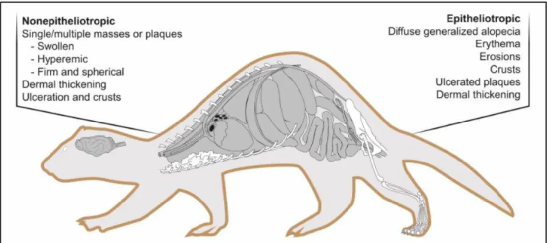
Figure 4. Clinical signs of nonepitheliotropic and epitheliotropic cutaneous lymphoma
Figure 5. Clinical signs of leukemia
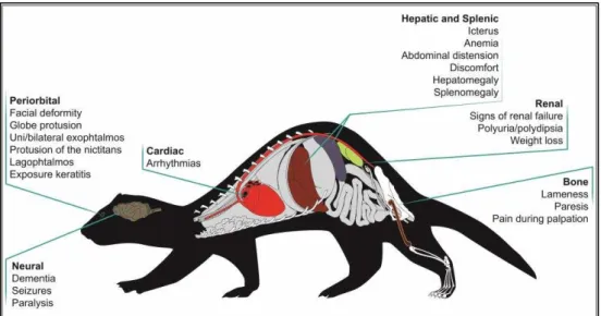
Figure 6. Clinical signs of extranodal lymphomas
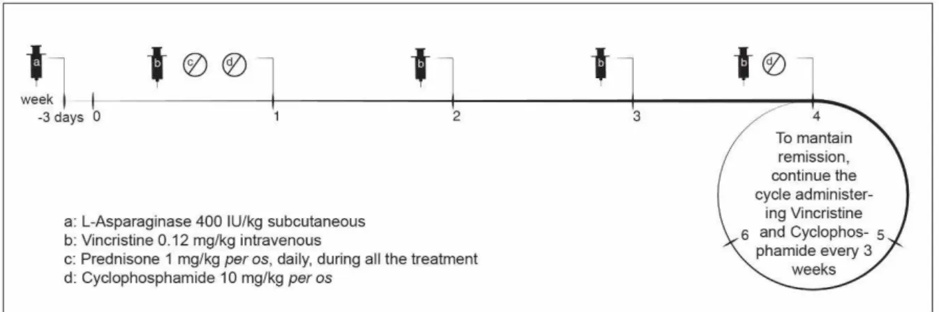
Figure 7. L-COP protocol for ferret lymphoma
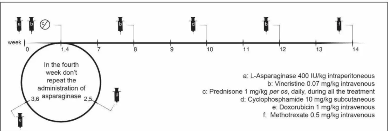
Figure 8. L-VCAMP protocol for ferret lymphoma
Figure 9. Noninvasive protocol for ferret lymphoma
Tables index
v
List of abbreviations
ADV Aleutian disease parvovirus
FeLV Feline leukemia virus
MALT Mucosa-associated lymphoid tissue
RBC Red blood cell
CD Cluster of differentiation
PCR Polymerase chain reaction
COP Cyclophosphamide, Vincristine, Prednisone
CHOP Cyclophosphamide, Doxorubicin, Vincristine, Prednisone
L-COP L-Asparaginase, Cyclophosphamide, Vincristine, Prednisone
1
1. Introduction
The domestic ferret (Mustela putorius furo) belongs to the family Mustelidae of the order Carnivora1 and is considered an obligate carnivore2 with a life expectancy of 5 to 8 years.37 The most probable ancestor of ferrets is the European polecat (Mustela putorius). However, ferrets may also be related to the steppe polecat (Mustela eversmannii). They are thought to have been domesticated 2000 years ago but there are references to the domestic use of ferrets since 63 BC.3 Nevertheless, most of the changes resulting from the domestication have been behavioral, with few or no biological changes.4 The ferrets were used to hunt rodents and rabbits (ferreting), to transport cables through long lengths of conduit, to entertain (ferret-legging), as animal models in biomedical research and, more recently, ferrets were also considered pets.3 The popularity of ferrets as pets is increasing, especially in North America and Europe,5 which turns their presence in veterinary clinics more frequent,6 and requires a knowledge of diseases associated with these animals to provide advanced medical care.
2. Lymphoma in ferrets
Ferrets are susceptible to a variety of diseases throughout their lives, being the development of neoplasia one of the main common conditions. After insulinoma and adrenocortical neoplasia, the lymphoma is the most common neoplasia in domestic ferrets,7-10 and it is their most frequent hematopoietic neoplasia.7,10 This condition appears to be increasing in frequency, probably due to the growing popularity of ferrets as pets, to their increased longevity, to the improved diagnostic testing, and to the increased disease reporting.11,12 The fact is that lymphoma represents 10% to 15% of all neoplastic presentations in ferrets from the United States and Europe,12 and it can occur in ferrets of all ages and in practically all organ systems.13 Lymphoma can infiltrate several organs, such as lymph nodes, thymus, lungs, spleen, liver, intestines, kidneys, and bone marrow. Other less common locations may include stomach, pancreas, nervous system, orbit and skin.7,14,15
a) Etiology
2
between virus infection (ADV, FeLV) and lymphoproliferative disease in ferrets, there are also recent studies reporting that ferret’s lymphoma may occur without these two virus infection.7,14,16 Regarding the retroviral cause, there is a study demonstrating an horizontal transmission of lymphoma by inoculating recipient ferrets with cells from a ferret with lymphoma. The presence of reverse transcriptase activity and retrovirus-like particles in cultivated cells from the inoculated ferrets suggested a retroviral cause,17 but no specific virus has been identified.13Other studies identified an association between gastric lymphoma and gastrointestinal infection by Helicobacter mustelae,7 and cases of H. mustelae-associated gastric adenocarcinoma.18 In fact, Hess et al7 showed that chronic infection due to H. mustelae bacterium resulted in extensive lymphoproliferative responses in the gastric mucosa. Additionally, Fox et al.,19 reported that primary gastric lymphomas may develop in ferrets naturally infected by H. mustelae. The study of gastric mucosa-associated lymphoid tissue (MALT) suggested that this Gram-negative bacterium may play an important role in the development of lymphoma.18 The genome of H. mustelae was sequenced in order to detect genetic alterations that might explain the association between the ulcerogenic and the carcinogenic activity induced by this bacterium,20 but specific genes associated to gastric lymphoma have not yet been identified.14
b) Classification
Previously, the ferret lymphoma was simply classified according to age at onset, cell type, and organ system affected.7 Therefore, lymphoma was commonly divided into lymphoblastic type or juvenile, lymphocytic type or adult/chronic7,21 and into an immunoblastic polymorphous type. The lymphoblastic type seems to affect predominantly young ferrets (under 2 years of age), with acute onset of clinical signs, and is characterized by the presence of large, immature and atypical lymphoblasts. The lymphocytic type is seen in ferrets older than 3 years of age, and has a chronic development, being characterized by the presence of mature and well differentiated neoplastic lymphocytes. The immunoblastic polymorphous type is more unusual, can occur in all ages, and is characterized by the presence of large atypical lymphocytes, lymphoblasts, immunoblasts, and small lymphocytes.7,14
As in others pathologies, it is essential to establish a standardized classification of ferret lymphoma that allows the systematization of all the information about lymphoma cases. This classification should take into account the macroscopic and microscopic properties, the cellular and the molecular alterations that characterize these tumours,12,14 and should have prognostic value.16 A recent proposal for ferret lymphoma classification enables an effective categorization of ferret lymphoma, and allows the creation of a database of tumour classification and respective response to treatment.12,14 The main points of this classification include staging, grading and immunophenotyping, which were considered the main points to achieve the description of the lymphoma.12,14,16
3
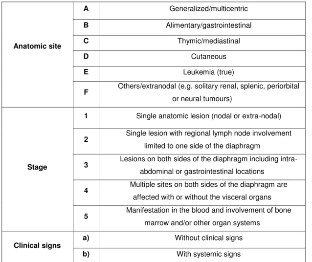
nodal versus extranodal lesions, and involvement of the blood or bone marrow.14,16 Thereby, this recent lymphoma classification proposed the following staging system (table 1),12 wherein the stage is obtained by conjugating a letter (anatomic site) with a stage number (location of lesions relative to the diaphragm, nodal versus extranodal lesions, and involvement of the blood or bone marrow). In addition to these, it is also possible to considerer the existence or absence of clinical signs.Table 1. Staging system of ferret lymphoma
Anatomic site
A Generalized/multicentric
B Alimentary/gastrointestinal
C Thymic/mediastinal
D Cutaneous
E Leukemia (true)
F Others/extranodal (e.g. solitary renal, splenic, periorbital
or neural tumours)
Stage
1 Single anatomic lesion (nodal or extra-nodal)
2 Single lesion with regional lymph node involvement
limited to one side of the diaphragm
3 Lesions on both sides of the diaphragm including
intra-abdominal or gastrointestinal locations
4 Multiple sites on both sides of the diaphragm are
affected with or without the visceral organs
5 Manifestation in the blood and involvement of bone
marrow and/or other organ systems
Clinical signs
a) Without clinical signs
b) With systemic signs
Adapted from Mayer J, Burgess K: An uptdate on ferret lymphoma: a proposal for a standardized classification on ferret lymphoma. Journal of Exotic Pet Medicine 21:343-346, 2012
4
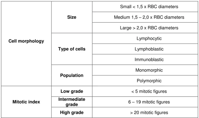
considered “low grade”, between 6 and 19 mitotic figures lymphoma is considered “intermediate grade”, and above 20 mitotic figures is considered “high grade”.11 According to this system, it seems that young ferrets are more likely to have high-grade lymphoma, while older ferrets can present either high- or low-grade lymphoma.14Table 2. Grading system of ferret lymphoma
Cell morphology
Size
Small < 1,5 x RBC diameters Medium 1,5 – 2,0 x RBC diameters
Large > 2,0 x RBC diameters
Type of cells
Lymphocytic Lymphoblastic Immunoblastic
Population
Monomorphic Polymorphic
Mitotic index
Low grade < 5 mitotic figures
Intermediate
grade 6 – 19 mitotic figures
High grade > 20 mitotic figures
Lastly, the imunophenotyping, which defines the tumour as B-cell or T-cell lymphoma, can be achievedthrough the analysis of the expression of CD3 (Cluster of Differentiation) and of CD79α by immunohistochemistry or flow cytometry.8,11,12,14,16,22,23 CD3 is considered a T-cell marker, and CD79α is a B-cell marker.11,13,14,23 There is a study that reported a larger mean survival time in ferrets with B-cell lymphoma than in ferrets with T-B-cell lymphoma. Despite being a small study, this can be the beginning of a database that correlates the phenotype of the lymphoma with prognosis.15
5
those observed in Hodgkin’s-like lymphoma.14 Furthermore, it is also described a clinical case of Hodgkin’s-like lymphoma in a ferret associated to a hyperesosinophilic syndrome.26 Nevertheless, the classification of the lymphoma in ferrets does not take in consideration the existence of the Reed-Sternberg cells.3. Diagnosis
a. Clinical signs
There are no pathognomonic clinical signs for ferret lymphoma and the clinical presentation can be very unspecific.14,16 Ferrets with lymphoma may present with anorexia, lethargy, and weight loss. The occurrence of these clinical signs can be intermittent and the diagnosis can be obtained unintentionally during routine visit, or evaluation for another disease process in ferrets clinically asymptomatic. In some cases, multiple palpable masses can be found during the physical examination.10,14,16,27 The presence of enlarged lymph nodes without pyrexia may suggest neoplasia.28
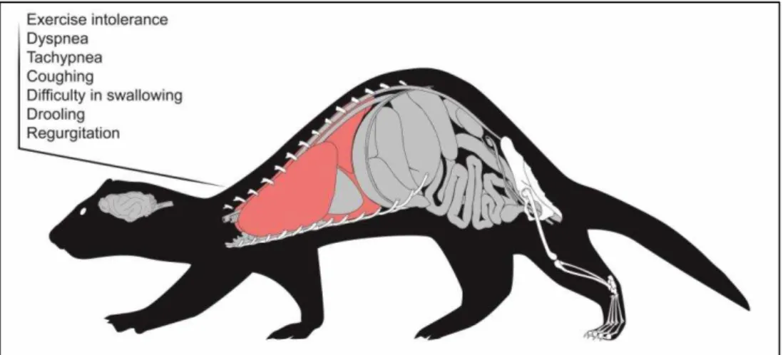
Considering that lymphoma may affect multiple organs, clinical signs will depend on which organ(s) is(are) affected. If lymphoma is mediastinal (figure 1), the most probable clinical presentation will include exercise intolerance and respiratory distress, with coughing, dyspnea, tachypnea, and possibly cyanosis. Difficulty in swallowing and regurgitation may also be observed.14,27 These clinical signs are normally the result of thoracic masses and/or pleural effusion, which may muffle heart sounds.27 This type of lymphoma has typically an acute presentation.14
Figure 1. Clinical signs of mediastinal lymphoma
6
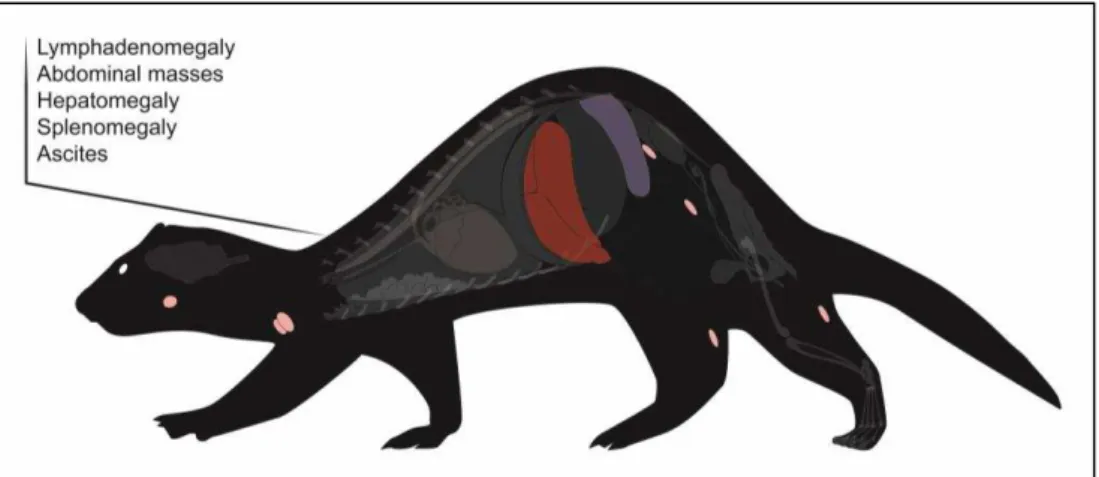
Figure 2. Clinical signs of multicentric lymphoma
If the infiltrated system organ is the gastrointestinal tract (figure 3), the clinical history can include anorexia, weight loss, lethargy, and abdominal discomfort. When it is a gastric lymphoma, the clinical signs can be very nonspecific, but if there is ulceration, the ferret can present melena and, in severe cases, pale mucous membranes. If the tumours location is the pyloric antrum, it can occur an obstruction of gastric emptying, and consequently vomiting. On the other hand, if lymphoma affects small and large intestines, diarrhea could occur. If there is intestinal infiltration of lymphoma or sublumbar lymphadenopathy, the ferret can also present tenesmus due to intestinal straining, or even obstruction.7,13,14,27
Figure 3. Clinical signs of gastrointestinal lymphoma
7
The epitheliotropic malignant lymphoma (figure 4) is a neoplasm of T lymphocytes, and normally arises with diffuse generalized alopecia, erythema, erosions, crusts, and ulcerated plaques. It seems to have predilection for the epithelium.29 This type of lymphoma is not very frequent, but a previous study reported a case of mycosis fungoides, a cutaneous epitheliotropic T-cell lymphoma. This cutaneous epitheliotropic T-cell lymphoma was characterized by crusting, scaling, and erythema, and presented an infiltration of the epidermis by neoplastic lymphocytes that could extend into the dermis and hair follicles.7 This condition may be pruritic, with dermal thickening, and ulceration.14,27 A preputial lymphoma has also been reported as an ulcerated mass around the prepuce.30Ferrets with lymphocytic leukemia (figure 5) are usually very lethargic, anorexic or inappetant, debilitated, and may be febrile.13 When the bone marrow is affected, it can occur anemia, and the ferret may also present pale mucous membranes.27
Figure 4. Clinical signs of nonepitheliotropic and epitheliotropic cutaneous lymphoma
Figure 5. Clinical signs of leukemia
8
lymphoma, and cardiac lymphoma. In case of periorbital lymphoma, the ferret can have facial deformity, and globe protusion.27 The orbital and retro-orbital lymphoma are rarely seen in ferrets, and can course with unilateral or bilateral exophthalmos, protrusion of the nictitans, lagophthalmos, and exposure keratitis.7,14 There is also a case report of infiltrative lymphoplasmacytic keratitis in a 2-years-old ferret with multicentric lymphoma.31In neural lymphoma, the clinical signs can include dementia, seizures, and paralysis.27 There is a report of a ferret with a diffuse central nervous system lymphoma characterized by the involvement of the meninges, the choroid plexus, the brain, and the spinal cord.10 If the location of the lymphoma is the spinal cord, ferrets can present a fast progressing paraparesis.7,14,27 In fact, Hanley et al.32 reported a T cell lymphoma in the lumbar spine of a ferret that was paraparetic and with neurologic alterations consistent with lesion in L4 to S3 spinal cord segments. However, primary spinal lymphoma is rare. Normally, it is a consequence of the release of neoplastic lymphocytes from the bone marrow to the peripheral circulation, and occurs predominantly in juvenile ferrets with mediastinal lymphoma.7
Additionally, lymphoma can affect the bone structure, which may be associated with lameness and paresis.14 It has been described a clinical case of myelo-osteolytic plasmablastic lymphoma of the femur in a ferret that presented lameness of the right hind limb and pain during palpation.33 There are also described clinical cases of lymphoma in the tibia. When there is skeletal involvement, the lymphoma can cause aggressive osseous lesions that have a predominantly lytic-destructive pattern, and cortical disruption.10,14
Other lymphoma locations include the hepatic and splenic forms that can cause icterus, anemia, abdominal distention, and discomfort due to the development of splenomegaly, and renal lymphoma with renomegaly and signs of renal failure. Lastly, it can also occur cardiac lymphomas that are associated to the development of arrhythmias.13,27 The most common extranodal lymphomas are the splenic, the renal, and the hepatic.14
9
Although the association between disease presentation and age of the ferret is not completely demonstrated,16 there are some types of lymphomas that occur tendentiously in certain ages. Mediastinal lymphoma is more common under 1 year of age, and may occur until 5 years of age, leukemia tends to occur between 1 and 3 years of age, multicentric lymphoma is more frequent between 4 and 7 years of age, and solitary lymphomas occur mainly between 6 and 7 years of age.14 Moreover, Antinoff et al13 reports that approximately 71% of ferret lymphomas diagnosed in his practice were affecting the abdomen, 33% were affecting the peripheral lymph nodes, and only 17% were located in the thorax.Finally, ferrets with lymphoma are immunocompromised, and therefore they become vulnerable to opportunistic infections and may develop secondary infections by Actinomyces sp., Cryptococcus sp., severe periodontal disease, and septicemia.14 There are also reports of mycobacterial infections in ferrets with lymphoma which has been associated to the immunosuppression.34,35 There is a case report of demodecic mange also associated with lymphoma in a ferret. Demodex sp. are commensals in the hair follicles and sebaceous glands, and can multiply with a immunological deficiency condition.36
b. Differential diagnosis
Due to the lack of pathognomonic signs, several differential diagnoses must be investigated. Considering that one of the most frequent clinical signs is lymphadenomegaly, it is important to differentiate it from other conditions, namely subcutaneous fat that surrounds the peripheral lymph nodes especially in obese ferrets, and reactive lymph nodes.27 The gastric lymph nodes are frequently reactive to any gastrointestinal process, whereby it is important to differentiate between lymphadenopathy and reactive nodules due to chronic gastrointestinal inflammatory or infiltrative disease, such as Helicobacter mustelae gastritis, lymphoplasmacytic gastroenteritis, eosinophilic gastroenteritis, ferret coronavirus, or epizootic catarrhal enteritis.13,27 In addition, it is also important to consider that, when the mesenteric lymph nodes are the only affected area, hyperplasia is more likely than neoplasia.27
Splenomegaly, that can sometimes lead to suspicions of lymphoma, should be differentiate from the splenomegaly that occurs gradually with age and is apparently normal in ferrets, being extramedullary hematopoiesis the most common cause of an enlarged spleen in ferrets.7,27,28 Therefore, cytological evaluation of an aspirate of the spleen should be cautiously interpreted, since bizarre-looking lymphocytes found on spleen aspirates with polymorphic lymphomas may sometimes be confused with megakaryocytes seen in hematopoiesis, or multinucleated giant cells seen in granulomas.14
10
intussusception, or other gastrointestinal tumours,27 such as pyloric adenocarcinoma, leiomyoma, and leiomyosarcoma.13The mediastinal form of lymphoma with pleural effusion may also be confused clinically with congestive heart failure, pneumonia, chylothorax, hemothorax, or thymoma.14,27
The cutaneous lesions associated to lymphoma may be related to other processes, such as mast cell tumours or other cutaneous neoplasms, such as sebaceous epitheliomas, and cutaneous hemangiomas, or chronic inflammatory disease.27,29
If the ferret present an aggressive bone lesion, it is important to differentiate it from other neoplastic diseases, such as osteosarcoma, fibrosarcoma, chondrosarcoma, primary soft tissue tumours with bone invasion, and infectious diseases.33
c. Complementary exams
i. Hematology and serum biochemistry
Considering the nonspecific clinical signs and the extensive list of differential diagnoses, it is important to evaluate hematologic and biochemical alterations. The first step should be blood collection. The cranial vena cava may be used to collect blood, but usually needs sedation to avoid vessel laceration.37 However, it must be considered that some anesthetics, such as isofluorane, may induce splenic sequestration of red blood cells, inducing an erroneous interpretation of the hematologic parameters.14 For this reason, a previous study pointed that the most accessible and convenient site to obtain a blood sample is the jugular vein,37 which can be easily used in awake ferrets.14
To avoid hemodynamic imbalance, a safe blood sample volume shall not exceed 7.5% to 10% of whole blood volume (whole blood volume is 5% to 6% of body weight), and a smaller volume in geriatric, anemic, hypoproteinemic, or ill ferrets (0.5% of body weight).37
Although the diagnosis of lymphoma should not be based just on hematologic parameters, it is common present some alterations,14 such as a mild nonregenerative anemia (under 45%)7,11,14,16,27 that may be severe in ferrets with leukemia.27
Bone marrow involvement can lead to leukocytosis or leukopenia,13 with lymphocytosis or lymphopenia. Young ferrets with mediastinal lymphoma tend to have atypical lymphocytosis that may vary between 10 000 and 70 000 cells/µL.7,14 On the other hand, older ferrets may present a normal lymphocyte count, or even lymphopenia.7 In fact, lymphocytosis is considered prodromal, and as the disease progresses it may give rise to lymphopenia.14 Neutropenia and thrombocytopenia are unusual findings, at least in an initial phase.16,27 Considering that lymphopenia and anemia can also be the consequence of chronic stress or viral infection,7 and lymphocytosis could be chronic and associated to a smoldering infection, the diagnostic of lymphoma needs more than a complete blood count.16
11
impaired kidney function can lead to azotemia.7,13,14 Occasionally, ferrets presenting T-cell lymphoma may develop hyperproteinemia, hyperglobulinemia, and hypercalcemia, although these are rare findings.7,13,14,16,27 Hypoalbuminemia can also occur in association to small intestinal lymphoma.16 Hypercalcemia and hypoglycemia are paraneoplastic syndromes occasionally seen, but rare.7,13,14,16 Urinalysis is usually normal, unless there is a renal failure.27ii. Imagiology
Radiographs can be useful, but should never be considered diagnostic.16 In ferrets with mediastinal lymphoma, radiographs may exhibit signs of pleural effusion, sternal or thracheobronchial lymphadenomegaly, widened mediastinum and thoracic masses.7,13,14,16,27 Mediastinal masses are normally radiodense masses in the cranial thorax, and cause dorsocaudally deviation of the heart and lungs.14 In a study of 14 ferrets with lymphoma, the most common thoracic finding was mild pleural effusion.10
Sublumbar or mesenteric lymphadenomegaly may be identified by abdominal radiography,27 as well as hepatomegaly, renomegaly, and splenomegaly.13,16,27 Radiographs can equally be useful to detect intestinal masses or abdominal effusion,27 and to evaluate bone lesions that in lymphoma tend to be lytic with bone dissolution.14
However, it is important to be aware that the absence of radiographic changes does not discard the possibility of lymphoma, as well as their presence does not ensure the existence of lymphoma.16
When spinal lymphoma is suspected, it can be still relevant to perform a myelography,13 or a computed tomography that can show bone lysis and the presence of masses.32
Ultrasonography seems to be the most valuable imaging method to evaluate ferrets with lymphoma.167 Considering that lymphadenopathies and masses are normally hypoechoic,10 ultrasonography allows the evaluation of abdominal and mesenteric lymph nodes,16 the detection of mesenteric or sublumbar lymphadenomegaly, nodules,27 as well as the existence of peritoneal effusion.10
In ultrasonography of the mesenteric lymph nodes, it is important to moderate the pressure in their search because excessive pressure can displace the lymph node dorsally, and cause confusion with the adrenal glands. According to an ultrasonography study performed in 28 healthy ferrets, the mean dimensions of the mesenteric lymph nodes (+/- standard deviation) were 12.6 (+/- 2.6) mm in length by 7.6 (+/- 2.0) mm in width.38
By ultrasonography it is also possible to assess abdominal organs as the liver, spleen, kidney, and mediastinum, in order to evaluate alterations on their size and structure, which could be associated to an infiltrative disease.7,13,16,27 The gastrointestinal tract can also be evaluated in order to detect enlarged lymph nodes and masses.27
12
infiltrated, ultrasonography should be considered only a complementary method in the diagnosis of lymphoma.13iii. Cytology/biopsy/immunohistochemistry
In order to achieve a more accurate diagnostic, it is possible to obtain a fine needle aspirate from lymphadenopathies and to perform a cytological evaluation. If a lymphoma is present, the cytological analysis may evidence the presence of monomorphic and atypical lymphoid cells, without components of peripheral blood. Otherwise, if the population of cells is variable in cell size and type, or if there are other types of white blood cells, it is probably not a lymphoma.16 It is also important to take in consideration that cytology using the fine needle aspirate, even though being a useful noninvasive and precocious method of diagnostic for lymphoma in ferrets,11 is associated to the occurrence of false-negative results.12,13,21,27
Therefore, to confirm the diagnostic, it is necessary to perform an histopathological analysis using tissue from incisional or excisional biopsies.7,10,12-14,21,27,28,39 The samples can be harvested from lymph nodes, bone marrow, liver, or spleen,7 as well as from masses or from effusions.40
In the case of popliteal lymph node, due to its accessibility, it is possible to perform a nodectomy.21,27,28 The nodectomy can be easily done by finding the lymph node that is close to the surface, and probably enclosed by fat, and dissecting it out.28
The histopathological analysis of the biopsy may evidence the existence of a monomorphic population of neoplastic lymphocytes,7,14,16,27 with pronounced nuclei, protuberant nucleoli, and small amount of cytoplasm,16 that destroys the architecture of the affected tissue, and replaces the normal parenchyma,7,14,27 as well as an increased number of cells in mitosis.16
Since hyperplastic lymph nodes can also present alterations of their architecture, sometimes it can be difficult to distinguish malignant lymphoma from a simple benign proliferation of lymphocytes based on the histopathological analysis.14,41,42 In these situations, it is important to use molecular methods, such as polymerase chain reaction (PCR), in order to evaluate the existence of clonality among cells. Assuming that clonality defines malignancy, since all malignant cells provide from one single malignant clone, the presence of a polyclonal population may indicate a reactive lymphocytosis.41,42 In addition, considering that cells from lymphoma often demonstrate altered expression of antigens, it is also possible to detect by flow cytometric immunophenotyping, the existence of aberrant antigen expression.12,18,43
13
In a spleen aspirate of a ferret with lymphoma, it is also expected to find an uniform population of abnormal lymphocytes and mitotic figures, but the presence of red blood cells precursors, megakaryocytes, or a large amount of peripheral blood is more suspicious of extramedullary hematopoiesis.164. Treatment
After reaching a definitive diagnosis, the next step will be the choice of the most adequate treatment. There are many possibilities for the treatment of lymphoma, such as surgery, chemotherapy, radiation therapy, or a combination of two or more types of therapy14,15,32,33 The treatment should be chosen according to remission rates, effectiveness, and adverse effects, as well as ferret’s age, tumour location, and concurrent disease.7,16
a. Surgical indications
If there is a solitary abdominal or cutaneous tumour easily accessible without the compromise of main vessels or nerves, or focal masses that are possible to remove without impair the involved organs function, surgery is considered the first line treatment since it may allow the removal of the tumour.7,14,15,27,33,44 Furthermore, surgery is also the first option when tumour induces intestinal obstruction.27 Even when lymphoma affects the bone, it can be useful to perform surgery. In fact, in myelo-osteolytic plasmablastic lymphoma in the femur, a caudal hemipelvectomy could be the adequate treatment.33 Splenectomy may also be a possibility when the spleen is infiltrated in such a way that occupies half of the abdominal cavity.7,14,15 However, before performing a splenectomy, it is advisable to analyze a bone marrow aspirate to ensure that the extramedullary hematopoietic function achieved by spleen is not essential.7 Nevertheless, it may also be benefic to associate other modalities of treatment, such as chemotherapy, to prevent potential postoperative development of micrometastasis.45
b. Chemotherapy options and protocols
The treatment of lymphoma in ferrets is often based in chemotherapeutic drugs,33 being this neoplasia considered one of the most responsive to chemotherapy.13 Ferrets have a good tolerance to continued treatments,28 with good response and minor adverse effects.13 Chemotherapy can be used as adjuvant therapy to treat lymphoma systemically after surgical removal of a solitary mass, as neoadjuvant therapy in an attempt to reduce tumour size before surgery, or sometimes as the only therapy.45 In any case, chemotherapy aims to reduce the proliferation ability of the tumour cells and to increase the tumour cell death through apoptosis, in order to prevent tumour growth and metastasis, and to extend the lifetime with quality.15,45
14
should also take into account the age of the ferret,7,15 type and location of lymphoma, and concurrent pathologies.7,13,15 Special attention must be given when more than 50% of bone marrow is compromised,7,40 or when the patient has concomitant pathologies, such as insulinoma or hyperadrenocorticism, which is common in ferrets.14,27 In spite of the chemotherapy advantages, it is crucial to balance the benefits and the risks. This is why the veterinarian should perform a complete evaluation of the ferret before initiate the chemotherapy protocol, including a complete blood count, serum biochemistry, radiographs, ultrasonography, cytology of fine needle aspirates, and analysis of bone marrow aspirate.7,28,40 Ferrets medicated for a long time with corticosteroids, which is common in patients with insulinoma or inflammatory bowel disease, are more likely to develop multidrug resistance and, therefore, are poor candidates for chemotherapy.7,13-16,27,40Whenever the chemotherapy protocol involves intravenous administrations, it is essential to place an intravenous catheter, and sedate the patient.7,13,27,28,33,40 This may avoid the risk of extravasation, especially when using irritants substances that can cause necrosis.7,13,33 It is also important to ensure patency before administering the chemotherapeutic agents by flushing heparinized saline,28,40 except when the drug is doxorubicin, that requires the use of non-heparinized saline,28 since it precipitates when contacts with heparin.40 Recently, vascular access ports were used as an alternative to the conventional intravenous catheter.7,13-15,27,32,46 Vascular access ports are subcutaneous implants that enable recurrent administration of intravenous drugs in the jugular vein, in a more safe process, with much less stress for the ferret, and without the necessity of sedation for each administration.13,14,46
Chemotherapy can be based on the use of a single drug, or in a combination of drugs.13,16 However, better results are usually obtained when several drugs with different mechanisms of action are combined, in order to achieve the maximum tumour destruction.15,16 There are numerous published protocols of chemotherapy to the treatment of ferret lymphoma, many of them extrapolated from dog and cat. However, in ferrets there is little feedback of results, which turn difficult the comparison of results.7,15,16 With chemotherapy, the highest reported survival in ferrets is about 11 to 23 months.7,33 Ammersbach et al11 has determined in his study that the mean survival time in ferrets treated with chemotherapy was 4.3 months in T-cell lymphoma cases, and 8.8 months in B-cell lymphoma cases, but more studies are still needed, especially with more uniformity in diagnosis and treatment.
The first chemotherapy protocol reported in ferret lymphoma included L-asparaginase, prednisone and cyclophosphamide.15 asparaginase is an enzyme that causes depletion of L-asparagine, thereby preventing the protein synthesis and causing death of the tumour cells which lack asparagine synthetase.47 Prednisone is particularly useful in hematopoietic tumours due to its effect of cytoxicity. The mechanism of action of this corticosteroid involves binding to glucocorticoids receptors of hematological cells to induce apoptosis.45,48 Cyclophosphamide is an alkylating agent that promotes DNA cross-link, preventing its replication and cell division.45,49 This protocol resulted in complete remission of lymphoma after 21 weeks of treatment in the case report.15
15
cytotoxicity of vincristine, a vinca alkaloid, is linked to its ability to prevent the formation of the mitotic spindle.45,49 Afterward, it was suggested a modified COP protocol, the L-COP protocol (figure 7), that added the administration of L-asparaginase three days before initiating the COP protocol.13,15,27 This alteration can be useful in cases of peripheral lymphadenomegaly.27 Antinoff et al13 described improvement in clinical signs and quality of life in 85% of the ferrets treated with this protocol.The CHOP (cyclophosphamide, doxorubicin, vincristine and prednisone) protocol, also adapted from the treatment of dogs and cats, offers a more complete approach by adding doxorubicin.16 Doxorubicin is an antitumour antibiotic that has multiples mechanisms of cytotoxicity, including DNA damages by alkylation, and inhibition of topoisomerase II which impedes DNA replication.45,49
Another chemotherapy protocol (figure 8) was recommended by Rosenthal,51 adding methotrexate which is an antimetabolite that replaces natural metabolites, and inhibits cell division.45,49 This protocol includes vincristine, L-asparaginase, prednisone, cyclophosphamide, doxorubicin, and methotrexate (L-VCAMP protocol).51
However, even being recommended intravenous chemotherapy for ferrets,33 sometimes it is difficult to implement these protocols. Some owners do not accept the use of the invasive and aggressive techniques which may turn impractical to submit weekly the ferret to the risks of sedation or accidental extravasation of chemotherapeutical agents.21,33 In response to the need for a noninvasive protocol that would simplify chemotherapy, a non-intravenous protocol (figure 9) was developed particularly for ferrets.14-16,21,28,33 This protocol uses prednisone, L-asparaginase, cyclophosphamide, cytarabine, methotrexate, chlorambucil, and procarbazine.14-16,28 Cytarabine, as methotrexate, is an antimetabolite that inhibits the synthesis of DNA during the S phase of the cell cycle. Chlorambucil is an alkylating agent and its cytotoxicity is related to the bifunctional alkylating capacity, which origins DNA cross-link and impedes replication.45,49 Procarbazine is also an alkylating agent that generates toxic metabolites to inhibit DNA and RNA synthesis, and cause DNA methylation.45
Figure 7. L-COP protocol for ferret lymphoma
16
Figure 8. L-VCAMP protocol for ferret lymphoma
Adapted from Rosenthal K: Ferrets. Vet Clin North Am Small Anim Pract 24:1-23, 1994.
Figure 9. Noninvasive protocol for ferret lymphoma
Adapted from Mayer J, Erdman S, Fox J: Diseases of the Hematopoietic System, in Fox J, Marini R (eds): Biology and Diseases of the Ferret (ed 3), Ames, IA, Wiley-Blackwell, pp 311-327, 2014
17
When the ferret relapses after achieving remission, it is possible to attempt a rescue protocol, which can be obtained through the combination of doxorubicin with prednisone, or through the use of other method of treatment, such as radiotherapy.13,16,27In ferrets in which chemotherapy is contraindicated by the associated risks, it may be preferable to opt by a palliative treatment with prednisone alone, avoiding more aggressive drugs. This option can also be considered when the owners are not willing to allow chemotherapy, either because they can’t afford or because they are against the use of these drugs. Lymphoma responds well to the treatment with corticosteroids, which decrease tumour size, help to control clinical signs and improve quality of life at short term.7,13,14,16,27,32,40 Besides, ferrets tolerate well the use of high-dose corticosteroids, without gastrointestinal side effects.7 The main problem of using this type of treatment is, as previously mentioned, multidrug resistance development, which complicates posterior treatments with chemotherapeutic agents.7,16,27,40,45 It may also be useful to combine prednisone with cyclophosphamide at lower doses, or with longer pauses, in order to minimize adverse effects.13,16
Chemotherapy is associated to numerous side effects, which can be immediate (24 to 48 hours after the treatment), acute (a few days after) or chronic (weeks to years after). In any case, the owners should be previously informed. Immediate side effects include hypersensitivity reactions.45 Therefore, before the treatment with L-asparaginase and doxorubicin, it is advisable to administrate diphenhydramine (0.5 to 2 mg/kg intramuscular) to avoid anaphylactic effects.13,27
Other secondary effects of chemotherapy may include lethargy, fever, anorexia, vomiting and diarrhea, mild alopecia (frequently beginning with whiskers loss), and weakness.7,13,15,16,27,28,40 The ferret can also present dyspnea and, less frequently, collapse.7,28,40 These signals of toxicity may happen a few days after the treatment,28,40 and for this reason it is important to separate administrations, in order to conclude which is the causative chemotherapy agent, and reduce the dose in 20%.16
Beyond these side effects, corticosteroids can induce polyuria, polydipsia, and weight gain,13 doxorubicin has cumulative and dose-related cardiotoxic effects,27,45 and cyclophosphamide was associated with the development of cystitis in one ferret.13
18
Lastly, it is important to remember that ferrets treated with chemotherapy should suspend all the vaccinations for the rest of their life, to avoid relapses by stimulating the immune system.16c. Radiation therapy
Hematopoietic tumours are the most sensitive to radiation therapy.52 Being lymphoma highly radiosensitive,16,52 adjuvant radiotherapy assumes a significant role in the treatment of this tumour.53 Radiation therapy may be useful firstly to reduce large masses in thorax causing dyspnea, or abdominal and intestinal masses causing obstruction,13,16 wherein only one dose can significantly reduce the mass.16 Nonetheless, it is preferable to apply multiple treatments and, depending on case, it may be used up to 21 treatments in 6 weeks.13,16
It may also be recommended as adjunct therapy,27 or as rescue therapy.13,16 The combination of radiation therapy with doxorubicin has once resulted in 23 months of survival.15 Nevertheless, this treatment is responsible by different periods of remissions in ferret lymphoma.14 It has also been suggested that radiation therapy could successfully treat multidrug resistant lymphoma.28
Half-body radiotherapy has been used,13,15 and in one case it was achieved a total of 12 months of remission with two treatments,15 but this method can present severe adverse effects. Local radiation therapy is safer and effective.13
Adverse effects of radiation therapy can be grouped into two types: acute effects that tend to occur with low doses per treatment, and appear soon after the radiotherapy, and delayed effects that are more associated with higher doses, and usually start 12 months after the treatment.52,53 Acute toxicity is usually self-limiting, and involves mainly dermatitis, mucositis, alopecia, humid desquamation, and conjunctivitis.52-54 The more important caution is to avoid self-trauma, to provide pain management,53 and in some cases it may be necessary to use antibiotics and anti-inflammatory drugs.52,54 Late toxicity effects can be difficult to treat,53,54 and may include vascular damages, fibrosis, bone necrosis, and myelopathy.52-54
d. Ancillary therapy
19
Another very important ancillary therapy is pain management. Pain control is particularly important in oncologic patients, because pain can affect the survival, and also interferes with the life quality of the animal. Moreover, pain may be derived from the tumour itself, but may also be caused by the treatment (surgery, chemotherapy, or radiotherapy). Pain may vary depending on the tumour location and the animal, thus one the best ways to find out if the tumour is causing pain is through palpation, and evaluating the activity and behavior of the patient.55 Thereby, it becomes essential to know how to recognize signs of discomfort and pain in ferrets.A ferret with pain may vocalize with squeals and screams, cry and whimper. The absence of play patterns and decreased activity can also be used as indicators of discomfort, stress, and pain.56,57 Other signs of pain in ferrets may include anorexia, weight loss, apathy, hiding behaviour, abnormal aggression, absence of grooming behavior, bruxism, tail piloerection, and trembling.1,56,57
The key to an effective pain management in oncologic patients is multimodal analgesia, providing different drugs with an additive or synergistic effect. This will allow decreasing doses of each analgesic, and consequently minimize side effects.55,57 Guidelines to cancer pain treatment were created by the World Health Organization, and include the use of nonsteroidal anti-inflammatory drugs, opioid drugs, and adjuvant drugs, such as N-methyl D-aspartate antagonists.55 Nonsteroidal anti-inflammatory drugs, such as meloxicam and carprofen, are widely used in ferrets, and therefore considered safe analgesics in this specie. The use of opioids, such as buprenorphine, hydromorphone, oxymorphone, and tramadol, has also been reported in ferrets. In order to assess the necessity of adding more drugs or dose adjustment, it is very important to perform repeated evaluations of the level of pain that the patient presents.57
Following nutrition and pain management, other supportive cares may be required. After chemotherapy, the ferret possibly will need gastrointestinal protectants, histamine H2-receptor antagonists, and antiemetics to control the gastrointestinal side effects of this treatment. It is advisable to use antiemetics that act on the central nervous system, where is the chemoreceptor trigger zone, such as metoclopramide or maropitant.13,15,16 Ferrets that become dehydrated should receive fluid therapy, either intravenously or subcutaneously.7,27 In case of anemia, erythropoietin could be used, as well as blood transfusions in severe cases.13
20
retinoids such as isotretinoin (2 mg/kg orally every 24 hours), despite not being curative, seem to have a satisfactory effect as a palliative treatment of cutaneous epitheliotropic lymphoma in ferrets13,27,29,59Acupuncture, with or without electrical stimulation, has also been widely mentioned. Electroacupuncture inhibits pain by activating nerve fibers, and causing liberation of endogenous opioid in the local of the inflammation.60 There are numerous studies in humans demonstrating analgesia induced by electroacupuncture, nevertheless there is lack of studies proving the analgesic efficacy of acupuncture in veterinary medicine.55 A recent study has utilized photoacoustic tomography on mice to assess and monitor the effects of acupuncture, and this could be a start to reveal the exact mechanism of actuation and to measure its effects.61
5. Prognosis
Not being possible to achieve a cure, the prognosis of a ferret with lymphoma tends to be poor.7,39 The worst prognostics are related to lymphomas with multicentric distribution,40 young ferrets with lymphoblastic lymphoma,7 and acute onset, in which the response to the treatment is poor.15,50 When the primary tumour is located in gastrointestinal tract, liver, lymph nodes, bone marrow, central nervous system, or in multiple organs, the ferrets are less treatment-responsive as well.7,14,15,27 As previously mentioned, there is also a relation between the phenotype and prognosis, wherein T-cell lymphomas tend to have a worst prognosis.12
Oppositely, older ferrets with chronic onset of lymphoma and with lymphocytic form, maybe for the slower growth of the tumour, seem to present a better prognosis,7,40,50 as well as ferrets with mediastinal, splenic, and cutaneous lymphoma.27
Above all, it is very important to explain to the owners that, although treatment is not curative, during therapy and remission time the quality of life is usually good, and the survival rate is increased when compared to that in non-treated ferrets.13,27 Moreover, it is important to consider that, for an animal with a life expectancy of 5 to 8 years,37 2 years of survival may be nearly or more than a quarter of lifetime.
6. Future directions
21
lymphoma diagnosis and contribute to the establishment of a more adequate therapeutic strategy, and of an accurate prognosis.417. Conclusions
Lymphoma is one of the most common neoplasias in domestic ferrets,7-10 and it is their most frequent malignant neoplasia.7,10 However, the data regarding the cellular and molecular characterization of lymphoma is scarce, which may limit the establishment of an adequate treatment protocol, and of the prognosis. Until the moment, the selection of the treatment is made according to each case necessities and expectations, which may limit the survival rate.16 Therefore, more studies are needed to implement appropriate treatment protocols that take in consideration the stage, the grade, and the immunophenotype of the tumour, as well as the characteristics of each patient.
22
8. Acknowledges
I would like to express my sincere acknowledges to all of those who somehow made contributions to this dissertation. My sincerely gratitude to my supervisors, Prof. Dr.ª Anália do Carmo and Dr.ª Ana Luísa Vieira, for all the support during this long process. My deepest acknowledges to Prof. Dr.ª Anália do Carmo for all the patience, constant dedication and availability, and to Dr.ª Ana Luísa Vieira for all the advices and recommendations.
I am also grateful to all the team of Centro Veterinário Conimbricence, especially to Prof. Dr. Nuno Cardoso, Dr.ª Andreia Freire and Dr. Pedro Silva, for receiving me so well, and always share their knowledge.
I would also like to extend my sincerest acknowledgments to all the team of Hospital Veterinario El Bosque, especially to Dr. Antonio Rodríguez, for all the hospitality and availability.
My sincere thanks to all my friends and colleagues for helping and supporting me during this process, and especially to Inês, who have accompanied me throughout almost all my life, for all her precious friendship.
My deepest gratitude to all my family, especially to my mother Rosa for her unconditional love, friendship, patience, for being my examples of strength and perseverance, and for always believe in me.
Last but not least, I am very grateful to my boyfriend Francisco Semião, who made important contributions by creating the artwork of this article, and for all the comprehension, support, and love.
23
9. References
1. Bixler H, Ellis C: Ferret care and husbandry. Vet Clin North Am Exot Anim Pract 7:227-255, 2004 2. Johnson-Delaney CA: Ferret nutrition. Vet Clin North Am Exot Anim Pract 17:449-470, 2014
3. Fox J: Taxonomy, History, and Use, in Fox J, Marini R (eds): Biology and Diseases of the Ferret, Ames, IA, Wiley-Blackwell, pp 5-16, 2014
4. Church B: Ferret Biology, Natural History, and Behavior, in Proceedings of the North American Veterinary Conference, Vol 21. Orlando, Florida, pp 1636 – 1639, 2007
5. Castanheira de Matos R, Morrisey J: Common procedures in the pet ferret. Vet Clin North Am Exot Anim Pract 9:347-365, 2006
6. Wenker C, Christen C: Ferrets in veterinary practice. Schweiz Arch Tierheilkd 144:575-584, 2002 7. Hess L: Ferret Lymphoma: The Old and the New. Seminars in Avian and Exotic Pet Medicine
14:199-204, 2005
8. Onuma M, Kondo H, Ono S, et al: Cytomorphological and immunohistochemical features of lymphoma in ferrets. J Vet Med Sci 70:893-898, 2008
9. Gupta A, Gumber S, Schnellbacher R, et al: Malignant B-cell lymphoma with Mott cell differentiation in a ferret (Mustela putorius furo). J Vet Diagn Invest 22:469-473, 2010
10. Suran JN, Wyre NR: Imaging findings in 14 domestic ferrets (Mustela putorius furo) with lymphoma. Vet Radiol Ultrasound 54:522-531, 2013
11. Ammersbach M, Delay J, Caswell JL, et al: Laboratory findings, histopathology, and immunophenotype of lymphoma in domestic ferrets. Vet Pathol 45:663-673, 2008
12. Mayer J, Burgess K: An uptdate on ferret lymphoma: a proposal for a standardized classification on ferret lymphoma. Journal of Exotic Pet Medicine 21:343-346, 2012
13. Antinoff N, Hahn K: Ferret oncology: diseases, diagnostics, and therapeutics. Vet Clin North Am Exot Anim Pract 7:579-625, 2004
14. Mayer J, Erdman S, Fox J: Diseases of the Hematopoietic System, in Fox J, Marini R (eds): Biology and Diseases of the Ferret (ed 3), Ames, IA, Wiley-Blackwell, pp 311-327, 2014
15. Fisher PG, Lennox A: Therapeutic Options for Ferret Lymphoma: A Review. Journal of Exotic Mammal Medicine and Surgery 1:1-5, 2003
16. Antinoff N, Williams B: Neoplasia, in Quesenberry K, Carpenter J (eds): Ferrets, Rabbits, and Rodents: Clinical Medicine and Surgery (ed 3), St. Louis, MO, Elsevier/Saunders, pp 106-115, 2012
17. Erdman SE, Reimann KA, Moore FM, et al: Transmission of a chronic lymphoproliferative syndrome in ferrets. Lab Invest 72:539-546, 1995
18. Erdman SE, Correa P, Coleman LA, et al: Helicobacter mustelae-associated gastric MALT lymphoma in ferrets. Am J Pathol 151:273-280, 1997
24
20. O'Toole PW, Snelling WJ, Canchaya C, et al: Comparative genomics and proteomics of Helicobacter mustelae, an ulcerogenic and carcinogenic gastric pathogen. BMC Genomics 11:164, 201021. Mayer J: Update on Ferret Lymphoma, in Proceedings of the The North American Veterinary Conference, Vol 20. Orlando, Florida, pp 1748 – 1749, 2006
22. Coleman LA, Erdman SE, Schrenzel MD, et al: Immunophenotypic characterization of lymphomas from the mediastinum of young ferrets. Am J Vet Res 59:1281-1286, 1998
23. Hammer AS, Williams B, Dietz HH, et al: High-throughput immunophenotyping of 43 ferret lymphomas using tissue microarray technology. Vet Pathol 44:196-203, 2007
24. Valli VE, San Myint M, Barthel A, et al: Classification of canine malignant lymphomas according to the World Health Organization criteria. Vet Pathol 48:198-211, 2011
25. Kuppers R, Hansmann ML: The Hodgkin and Reed/Sternberg cell. Int J Biochem Cell Biol 37:511-517, 2005
26. Blomme EA, Foy SH, Chappell KH, et al: Hypereosinophilic syndrome with Hodgkin's-like lymphoma in a ferret. J Comp Pathol 120:211-217, 1999
27. Oglesbee B: Lymphosarcoma, in Oglesbee B (ed): Blackwell's Five-Minute Veterinary Consult Small Mammal (ed 2), Ames, IA, Wiley-Blackwell, pp 141-143, 2011
28. Lewington J: General neoplasia, in Lewington J (ed): Ferret Husbandry, Medicine and Surgery (ed 2), Philadelphia, PA, Elsevier/Saunders, pp 319-327, 2007
29. Kanfer S, Reavill DR: Cutaneous neoplasia in ferrets, rabbits, and guinea pigs. Vet Clin North Am Exot Anim Pract 16:579-598, 2013
30. Parker GA, Picut CA: Histopathologic features and post-surgical sequelae of 57 cutaneous neoplasms in ferrets (Mustela putorius furo). Vet Pathol 30:499-504, 1993
31. Miller PE: Ferret Ophthalmology. Seminars in Avian and Exotic Pet Medicine 6:146-151, 1997 32. Hanley CS, Wilson GH, Frank P, et al: T cell lymphoma in the lumbar spine of a domestic ferret
(Mustela putorius furo). Vet Rec 155:329-332, 2004
33. Eshar D, Wyre NR, Griessmayr P, et al: Diagnosis and treatment of myelo-osteolytic plasmablastic lymphoma of the femur in a domestic ferret. J Am Vet Med Assoc 237:407-414, 2010
34. Nakata M, Miwa Y, Tsuboi M, et al: Mycobacteriosis in a domestic ferret (Mustela putorius furo). J Vet Med Sci 76:705-709, 2014
35. Saunders GK, Thomsen BV: Lymphoma and Mycobacterium avium infection in a ferret (Mustela putorius furo). J Vet Diagn Invest 18:513-515, 2006
36. Beaufrere H, Neta M, Smith DA, et al: Demodecic Mange Associated With Lymphoma in a Ferret. Journal of Exotic Pet Medicine 18:57-61, 2009
37. Smith SA, Zimmerman K, Moore DM: Hematology of the Domestic Ferret (Mustela putorius furo). Vet Clin North Am Exot Anim Pract 18:1-8, 2015
25
39. Zaffarano B: Ferrets: Examination and Standards of Care. Journal of Exotic Pet Medicine19:73-81, 2010
40. Wolf T: Ferrets, in Mitchell M, Tully T (eds): Manual of Exotic Pet Practice, Vol. St. Louis, MO, Elsevier/Saunders, pp 364-367, 2009
41. Avery A, Olver C, Khanna C, et al: Molecular diagnostics, in Withrow S, Vail D, Page R (eds): Withrow & MacEwen's Small Animal Clinical Oncology (ed 5), St. Louis, MO, Elsevier/Saunders, pp 133-135, 2013
42. Avery PR, Avery AC: Molecular methods to distinguish reactive and neoplastic lymphocyte expansions and their importance in transitional neoplastic states. Vet Clin Pathol 33:196-207, 2004
43. Craig FE, Foon KA: Flow cytometric immunophenotyping for hematologic neoplasms. Blood 111:3941-3967, 2008
44. Li X, Fox G, Erdman SE, et al: Cutaneous lymphoma in a ferret (Mustela putorius furo). Vet Pathol 32:55-56, 1995
45. Gustafson DL, Page RL: Cancer chemotherapy, in Withrow S, Vail D, Page R (eds): Withrow & MacEwen's Small Animal Clinical Oncology (ed 5), St Louis, MO, Elsevier/Saunders, pp 157-179, 2013
46. Powers LV: Techniques for Drug Delivery in Small Mammals. Journal of Exotic Pet Medicine 15:201-209, 2006
47. Keating MJ, Holmes R, Lerner S, et al: L-asparaginase and PEG asparaginase-past, present, and future. Leuk Lymphoma 10 Suppl:153-157, 1993
48. Greenstein S, Ghias K, Krett NL, et al: Mechanisms of glucocorticoid-mediated apoptosis in hematological malignancies. Clin Cancer Res 8:1681-1694, 2002
49. Couto C: Practical chemotherapy, in Nelson R, Couto C (eds): Small Animal Internal Medicine (ed 4), St. Louis, MO, Mosby/Elsevier, pp 1156-1157, 2009
50. Erdman SE, Moore FM, Rose R, et al: Malignant lymphoma in ferrets: clinical and pathological findings in 19 cases. J Comp Pathol 106:37-47, 1992
51. Rosenthal K: Ferrets. Vet Clin North Am Small Anim Pract 24:1-23, 1994
52. Mauldin GN, Shiomitsu K: Principles and Practice of Radiation Therapy in Exotic and Avian Species. Seminars in Avian and Exotic Pet Medicine 14:168-174, 2005
53. LaRue S, Gordon I: Radiation therapy, in Withrow S, Vail D, Page R (eds): Withrow and MacEwen’s Small Animal Clinical Oncology (ed 5), St Louis, MO, Elsevier/Saunders, pp 180-197, 2013
54. McEntee MC: Principles of adjunct radiotherapy and chemotherapy. Vet Clin North Am Small Anim Pract 25:133-148, 1995
55. Duncan B, Lascelles X: Supportive Care for the Cancer Patient, in Withrow S, Vail D, Page R (eds): Withrow & MacEwen’s Small Animal Clinical Oncology (ed 5), St Louis, MO, Elsevier/Saunders, pp 245-259, 2013
26
57. Van Oostrom H, Schoemaker NJ, Uilenreef JJ: Pain management in ferrets. Vet Clin North AmExot Anim Pract 14:105-116, 2011
58. Block KI, Koch AC, Mead MN, et al: Impact of antioxidant supplementation on chemotherapeutic toxicity: a systematic review of the evidence from randomized controlled trials. Int J Cancer 123:1227-1239, 2008
59. Rosenbaum MR, Affolter VK, Usborne AL, et al: Cutaneous epitheliotropic lymphoma in a ferret. J Am Vet Med Assoc 209:1441-1444, 1996
60. Zhang R, Lao L, Ren K, et al: Mechanisms of acupuncture-electroacupuncture on persistent pain. Anesthesiology 120:482-503, 2014