J
ournal of Epilepsy and ClinicalNeurophysiology
J Epilepsy Clin Neurophysiol 2012;18(1):7-11
Original Article
a Department of Neuropediatrics, Hospital Pequeno Príncipe, Brazil. b University Tuiuti of Paraná, Otoneurology Research Center, Brazil.
Received Jan. 10, 2012; accepted Feb. 01, 2012.
Epilepsy and Electroencephalographic Features.
Comparative Study of Down Syndrome and
Non-Syndromic Mental Retardation
Karyn Regina Jordão Koladicza, Paulo Breno Noronha Liberalessoa,b, Bianca Simone Zeigelboimb, Jair Mendes Marquesb, Ari Leon Jurkiewiczb
Department of Neuropediatrics, Hospital Pequeno Príncipe, Brazil
ABSTRACT
Introduction: Down syndrome (DS) is the most common chromosomal abnormality causing mental retardation and its association with epilepsy is highly variable in childhood. Although the irst descriptions of the syndrome did not report seizures, their association with epilepsy is relatively common. Methods: were evaluated 68 individuals with DS and 83 with non-syndromic mental retardation (N-SMR). All patients underwent digital EEG, lasting at least 30 minutes and electrodes positioned according to the International 10-20 System of Electrode Placement. Data were analyzed using descriptive statistics and proportions were compared with Student’s t-test and test of Differences between Proportions with p<0.05 considered statistically signiicant. Results: DS:27.9% had epilepsy (irst seizure with 2.2±3.7 years). Fifteen (22.1%) patients had epileptiform discharges, 5 (7.4%) hypsarrhythmia, 5 (7.4%) focal pattern, 3 (4.4%) generalized pattern and 2 (2.9%) multifocal pattern. N-SMR: 33.7% patients had epilepsy (irst seizure with 1.2±4.5 years). Twenty-three (27.7%) patients had epileptiform discharges, 10 (12.0%) focal pattern, 5 (6.0%) generalized pattern and 8 (9.6%) multifocal pattern. Conclusion: The difference between the occurrence of epilepsy in DS and N-SMR
was not statistically signiicant, as well as between normal EEG, EEGs with focal pattern, generalized pattern and multifocal pattern. In SD group 7.4% have shown hypsarrhythmia.The comparison with N-SMR was not possible because none of these has shown this EEG abnormality.
Keywords: Epilepsy; Down syndrome; mental retardation.
RESUMO
Epilepsia e aspectos eletrencefalográficos. Estudo comparativo entre síndrome de Down e deficiência mental não sindrômica
Introdução: síndrome de Down (SD) é a anormalidade cromossômica que mais comumente causa deiciência mental e sua associação com epilepsia é muito variável na infância. Embora as descrições iniciais da síndrome não relatassem crises, sua associação com epilepsia é relativamente comum. Métodos: foram avaliados 68
indivíduos com SD e 83 com retardo mental não sindrômico (RMNS). Todos os pacientes foram submetidos à EEG digital, com duração mínima de 30 minutos e com eletrodos posicionados segundo o sistema internacional 10-20 de posicionamento de eletrodos. Dados foram analisados usando estatística descritiva e proporções foram comparadas com o teste t de Student e teste de Diferença entre Proporções com p<0,05 sendo considerado estatisticamente signiicativo. Resultados: SD: 27,9% tinham epilepsia (primeira crise 2,2±3,7 anos). Quinze
(22,1%) pacientes tinham descargas, 5 (7,4%) hipsarritmia, 5 (7,4%) padrão focal, 3 (4,4%) padrão generalizado, 2 (2,9%) padrão multifocal. N-SMR:33,7% pacientes tinham epilepsia (primeira crise com 1,2±4,5 anos). Vinte e três (27,7%) pacientes tinham descargas, 10 (12,0%) padrão focal, 5 (6,0%) padrão generalizado e 8 (9,6%) padrão multifocal. Conclusões: a diferença entre a ocorrência de epilepsia no grupo SD e RMNS não
foi estatisticamente signiicativa, assim como o EEG normal, com padrão focal, generalizado e multifocal. No grupo SD, 7,4% apresentaram hipsarritmia. A comparação com o grupo RMNS não foi possível por que ninguém neste grupo apresentou esta anormalidade no EEG.
INTRODUCTION
Down syndrome (DS) is the most common chro- mosomal abnormality cause of mental retardation, with an incidence varies according to maternal age between 1/40-1000 live births. Risk factors include advancing maternal age (because older eggs have a greater risk of improper chromosome division), parents carry the genetic translocation for DS, and having had a child with the syndrome.1
In 1959, with the advent of karyotype tests, the etiology of DS was determined as the presence of an abnormal extra chromosome 21. If an egg or sperm carrying 24 chromosomes combines with an egg or sperm carrying 23 chromosomes, the result would be a children with cells in which there are 47 chromosomes. This is known a genetic trisomy and almost all cases are caused by non disjunction mechanism causing trisomy 21, although unbalanced translocation involving chromosome 21 and another chromosome may occur. Rarely, less than 1% of the cases, the change in the distribution of chromosomes in the cell division occur shortly after fertilization. In these cases, the individual display cells with a combination of different patterns of chromosomes. This condition is called genetic mosaicism.1-3
DS is clinically characterized by mental retardation, dysmorphic facial features like slanting palpebral fissures, epicanthal folds, broaden the base of the nose, small ears, absence of the central fissure of the tongue and gray spots around the iris, particularly in neonates. Often occurs growth retardation, developmental delay, hypotonia (floppy baby), brachycephaly, single palmar crease, clinodactyly, dysplastic or hypoplastic hips, neck lymphedema, hypo- gonadism, gastrointestinal and cardiac malformations, diabetes, tumors, leukemia, premature degeneration of the musculoskeletal system, atlantoaxial subluxation, thyroid abnormalities, cataracts, refractive optical defects and immune deficiency. With increased life expectancy in DS, become more frequent the psychiatric disorders such as depression and dementia. Other neuropsychiatric manifestations can also occur are disruptive behavior disorders and autism.1,2
In 1866, seizures are not included in original report by British physician John Langdon Haydon Down. When the syndrome was described was thought to be due to a process of degeneration of the human race. At that time, physicians and researchers were more concerned with the physical and mental aspects, and therefore, the seizures were undervalued4. Other older researchers as Ireland (1877), Wilmarth (1890), Comby (1903) and Herrman (1905) do not reported seizures in DS5.
Although the seizures were considered infrequent for many years, currently we know that are significantly
more prevalent in DS than the general population, and less prevalent than in non-syndromic mental retardation (N-SMR).4
The aim of this study is to compare the incidence of seizures and electroencephalographic abnormalities in individuals with DS and N-SMR.
METHODS
The sample consisted of 68 individuals with DS (30 females and 38 males, aged 7.4±5.7 years) and 83 with N-SMR (37 female and 46 males, aged 9,4±4,8 years), all from special education programs in Brazil. Data were collected between January 2006 and December 2011. All subjects were evaluated clinically by the same physician and, where possible, underwent standardized psychometric tests. In specialized educational institutions, all participants were also evaluated by a psychologist, phonoaudiologist and occupational therapist.
In the DS group, 56 (82.4%) patients had genetic diagnosis (trisomy of chromosome 21) and all had physical characteristics that allowed the clinical diagnosis.
All patients underwent EEG in equipment Neuropmap EEG-40i, Neurofax Nihon Kohden EEG-1200 or EEG Brain Wave II, lasting at least 30 minutes and electrodes positioned according to the International 10-20 System of Electrode Placement.
To standardize the research, the terms “epilepsy” and “mental retardation” have been previously defined. Epilepsy was defined as the presence of at least one seizure, caused by a brain disorder caused by an enduring predisposition of the brain to generate recurrent seizures and their neurobiological, cognitive and psychosocial consequences.6 Mental retardation was defined as a neurobiological disorder, characterized by significantly subaverage intellectual functioning and impairments in at least two of the following areas: communication, self-care, activities of daily living, social/interpersonal skills, and use of community resources, self direction, functional academic skills, work, leisure, health and safety.7
generalized, but it never appears as a rhythmically repetitive and highly organized pattern”8.
Data were analyzed using descriptive statistics and proportions were compared with Student’s t-test and test of Differences between Proportions with p<0.05 considered statistically significant. All data are reported as means ± SDs (standard deviations).
All aspects of this research project were approved by the Ethics Committee on Research Involving Human Subjects at our institution (number registration – 1065-12).
RESULTS
Individuals withDS
Sample of 68 individuals, 19 (27.9%) had epilepsy, and the mean age at first seizure of 2.2±3.7 years. In 15 (22.1%) patients were identified epileptiform discharges (Table 1). Five patients (7.4%) had hypsarrhythmia, 5 (7.4%) focal pattern, 3 (4.4%) generalized pattern and 2 (2.9%) multifocal pattern.
Table 1. Electroencephalographic abnormalities in patients with Down syndrome
Patients Age Gender Firstseizure Electroencephalogram
1 0,2 male 0,1 Sharp wave frontal – right
4 0,5 female 0,4 Hypsarrhythmia
5 0,6 male 0,3 Hypsarrhythmia 6 0,6 female 0,5 Hypsarrhythmia 9 0,9 female 0,6 Hypsarrhythmia 10 1,0 female 0,4 Hypsarrhythmia
15 1,4 female 1,2 Sharp wave parietal/
occipital left
28 4,7 female 4,0 Generalized spike/ polyspike
40 8,2 male 5,0 Sharp wave parietal/ occipital right
41 8,6 female 4,0 Generalized spike/ polyspike wave
50 12,4 female 2,0 Sharp wave temporal/
frontal right 57 15,0 male 0,5 Multifocal sharpwave
59 15,2 female 0,8 Multifocal sharpwave 61 15,7 female 3,3 Sharp wave frontal left/
central right 67 16,7 male 9,2 Generalized spike/
polyspike wave
Note: age and first seizure in years.
Individuals with N-SMR
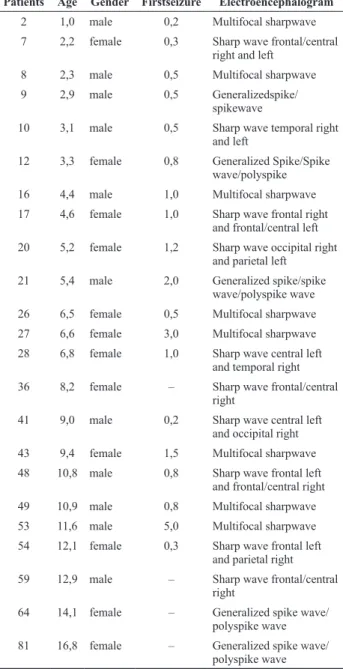
Sample of 83 patients, 28 (33.7%) patients had epilepsy, and the mean age at first seizure of 1.2±4.5 years. In 23 (27.7%) patients were identified epileptiform discharges (Table 2). Ten patients (12.0%) had focal pattern, 5 (6.0%) generalized pattern and 8 (9.6%) multifocal pattern.
Table 2. Electroencephalographic abnormalities in patients with N-SMR
Patients Age Gender Firstseizure Electroencephalogram
2 1,0 male 0,2 Multifocal sharpwave 7 2,2 female 0,3 Sharp wave frontal/central
right and left
8 2,3 male 0,5 Multifocal sharpwave 9 2,9 male 0,5 Generalizedspike/
spikewave
10 3,1 male 0,5 Sharp wave temporal right
and left
12 3,3 female 0,8 Generalized Spike/Spike wave/polyspike 16 4,4 male 1,0 Multifocal sharpwave 17 4,6 female 1,0 Sharp wave frontal right
and frontal/central left
20 5,2 female 1,2 Sharp wave occipital right
and parietal left
21 5,4 male 2,0 Generalized spike/spike wave/polyspike wave 26 6,5 female 0,5 Multifocal sharpwave 27 6,6 female 3,0 Multifocal sharpwave 28 6,8 female 1,0 Sharp wave central left
and temporal right 36 8,2 female – Sharp wave frontal/central
right
41 9,0 male 0,2 Sharp wave central left and occipital right
43 9,4 female 1,5 Multifocal sharpwave 48 10,8 male 0,8 Sharp wave frontal left
and frontal/central right 49 10,9 male 0,8 Multifocal sharpwave
53 11,6 male 5,0 Multifocal sharpwave
54 12,1 female 0,3 Sharp wave frontal left and parietal right
59 12,9 male – Sharp wave frontal/central right
64 14,1 female – Generalized spike wave/ polyspike wave 81 16,8 female – Generalized spike wave/
polyspike wave
Note: age and first seizure in years. N-SMR – non-syndromic mental retardation.
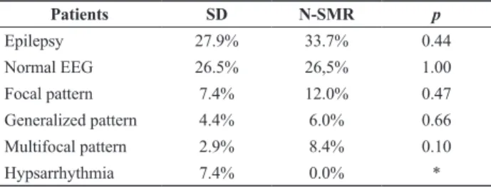
Statistical comparison between groups
Table 3. Statistical comparison between Down syndrome and N-SMR
Patients SD N-SMR p
Epilepsy 27.9% 33.7% 0.44 Normal EEG 26.5% 26,5% 1.00
Focal pattern 7.4% 12.0% 0.47 Generalized pattern 4.4% 6.0% 0.66
Multifocal pattern 2.9% 8.4% 0.10 Hypsarrhythmia 7.4% 0.0% *
N-SMR – non-syndromic mental retardation. * Statistical test is not applicable.
DISCUSSION
The incidence of seizures in DS is contradictory, varying between 1-17%.8,9 There are distinct peaks to the incidence of seizures. In the perinatal period seizures often occur because systemic complications, cardiovascular malformations and infectionsrelated to immunodeficiency. After the first six months of life, predominate infantile spasms (West syndrome – WS), and after the fourth decade of life crises are mainly related to Alzheimer’s dementia.11 The prevalence of epilepsy increased with age, reaching 46% after the fifth decade of life4.
Although there are many researches about the electroencephalographic abnormalities in this syndrome, no specific pattern was determined. There is a large variation in EEG of these patients, as the slowing of background activity, nonspecific burst of slow waves and epileptiform discharges with various morphologies.Even in individuals with DS without epilepsy, epileptiform discharges are common.1-4,10
The highest incidence of seizures has been classically attributed to changes in brain structure, although other clinical conditions such as heart defects, thyroid disease and dementia are also involved. Potential mechanisms to explain this increased frequency of seizure would be decrease neuronal density, abnormal neuronal lamination, dendritic spine dysgenesis, primitive synaptic profiles, fewer GABAergic interneurons in cortical layers II and IV, decrease voltage threshold for spike generation, abnormal potassium, calcium or sodium current, altered neurotransmitter systems GABAergic, serotoninergic, adrenergic, cholinergic or glutaminergic.11 Ross et al.12 compared the brain histological aspects of two patients (6-year-old DS girl and 25-year-old DS man) with normal controls, and showedcytoarchitetonic abnormalities characterized by significant poverty of granular cells in the DS brains, particularly in granular fields such as areas 3, 17 and 41.
In 1972, Morre13 conducted a very important multi- center study involving 22 medical institutions responsible for treating patients with mental disabilities. This study
evaluated 24,257 patients, 2748 with DS. The incidence of epilepsy in individuals with mental retardation without DS was 31%, and those with SD were 5.2%. In a classic paper, Seppäläinen and Kivalo.14 analyzed the electrographic patterns in a large group of patients with SD (8.7% with epilepsy), found electrographic abnormalities in only 12% of cases and interictal paroxysmal activity in 21%. In our patients with DS, the incidence of epilepsy (27.9%) was significantly higher thanto describe by Morre (5.2%)13 and Seppäläinen and Kivalo (8.7%).14 However, the incidence of epilepsy in individuals N-SMR (33.7%) was very similar (31%) as described by Morre.13 In our research, the occurrence of interictal paroxysmal activity in DS (22.1%) was resembles to report by Seppäläinen and Kivalo.14
The prevalence of reflex epilepsy appears to be higher in patients with DS than individuals with other forms of mental disability and the mentally healthy general population. Guerrini et al.15 in a retrospective study of 30 SD patients with epilepsy, reported6 (20%) with reflex epilepsy (three had Lennox-Gastaut syndrome, one had benign myoclonic epilepsy of infancy, one had reflex epilepsy and partial symptomatic epilepsy and one had generalized symptomatic epilepsy). The high incidence of reflex epilepsy in SD may suggest impairment of inhibitory phenomena in the cerebral cortex of these patients. Neither of our 19 epileptic patients with SD had reflex epilepsy.
All types of seizures andepileptogenic interictal paroxysmscan occur in DS children, although the tonic-clonic, myoclonic and infantile spasms (West syndrome – WS) are considered the most frequent.10,12 In our patients with DS, 7.4% had hypsarrhythmia, 7.4% focal pattern, 4.4% generalized pattern and 2.9% multifocal pattern. In N-SMR group, 12.0% had focal pattern, 6.0% generalized pattern and 9.6% multifocal pattern.
When we compared the incidence of epilepsy, age at first seizure, occurrence of focal, generalized or multifocal patterns, did not find statistically significant differences between DS and N-SMR groups. The clinical and electroencephalographic aspects have no consistent differences between individuals with DS and with N-SMR, except for the elevated incidence of WS in patients with DS. When we compared WS/hypsarrhythmia in EEG, observed incidence of 7.4% on DS group and none in SMR-N group.
Silva et al.18 reported 14 children with DS and WS, and no cardiac abnormalities or history of perinatal asphyxia. In this sample, the spasms started between 4 and 18 months (mean age of 8 months) and all patients had symmetrical hypsarrhythmia in EEG. Seven children exhibited other types of seizures after WS, including myoclonic seizures, tonic-clonic, atonic and atypical absence. In the seven cases who were recorded epileptic spasms, the ictal recordings was characterized with high voltage slow waves in both hemispheres followed by fast rhythmic activity of low-voltage. Lujic et al.19 reported 11 children at the age of 3 years to 10 years with DS and WS. In this group, the spasms began at the age of 5 to 10.5 months in 10 children, in one patient at the age of 16 months. All children had their spasms controlled with ACTH and hypsarrhythmia also disappeared in all cases after drug therapy.
In children without DS, the incidence of WS is estimated between 0.25 to 0.60 cases per 1000 live births, with prevalence between 0.15 and 0.20 cases per 1000 children.20 Therefore, although the WS is a frequentepileptic encephalopathy of childhood, it is considerably more common in children with DS than without DS.
Atypical or modified hypsarrhythmia have been reported in children with WS. However, the classic hypsarrhythmia pattern (“… random high-voltage slow waves and spikes that vary from moment to moment, both in location and duration; at times they appear to be focal, and a few seconds later they seem to originate from multiple foci; occasionally, the spikes becomes generalized, but in never appears as a rhythmically repetitive and highly organized patter; the abnormality is almost continuous…”)21 was observed in all patients with DS and WS in our research.
It is very important to know the comorbidities and complications related to this syndrome. Children with DS presenting seizures may have worse neurological outcome. To know the clinical aspects of seizures and electroencephalographic features provides a more accurate and earlier diagnosis.
REFERENCES
1. Pueschel SM. Clinical aspects of Down syndrome from infancy to adulthood. AJMG 1990;37(S7):52-6.
2. RoizenNJ, Patterson D. Down’s syndrome. The Lancet 2003;361 (9365):1281-9.
3. Lejeune J. 1959. Le Mongolism. Premier exempled’aberration autosomique humaine. Ann Genet 1:41-49.
4. McVicker RW, Shanks OEP, McClelland RJ. Prevalence and associated features of epilepsy in adults with Down’s syndrome. Br J Psychiatry 1994;164:528-32.
5. MacGillivray RC. Epilepsy in Down anomaly. J ment Def Res 1967;11(1):43-8.
6. Fisher RS, van Emde Boas W, Blume W, Elger C, Genton P, Lee P, Engel J. Epileptic seizures and epilepsy: definitions proposed by the International League Against Epilepsy and the International Bureau for Epilepsy. Epilepsia 2005;46(4):470-2.
7. American Psychiatric Association: Diagnostic and Statistical Manual of Mental Disorders. 4ª ed. Washington, DC: American Psychiatric Association, 1994.
8. Gibbs FA, Gibbs EL. Atlas of electroencephalography. Vol. 2: Epilepsy. Reading: Addison-Wesley; 1925.
9. Kirman BH. Epilepsy in mongolism. Archives of Disease in Childhood 1951; 26: 501-3.
10. Johannsen P. Christensen JE, Goldstein H, Nielsen VK, Mai J. Epilepsy in Down syndrome prevalence in three age groups. Seizure 1996;5:121-5.
11. Stafstrom CE. Epilepsy in Down syndrome: clinical aspects and possible mechanisms. American Journalon Mental Retardation 1993; 98: 12-26.
12. Ross MJ, Galaburda AM, Kemper TL. Down syndrome: Is there a decreased population of neurons? Neurology 1984; 34: 909-16. 13. Moore BC. Some characteristics of institutionalisedmongols. J ment
Defic Res 1973;17:46-51.
14. Seppäläinen AM, Kivalo E. EEG findings and epilepsy in Down’s syndrome. J MentDefic Res 1973;11(2):116-25.
15. Guerrini R, Genton P, Michelle B, Dravet C, Roger J. Reflex seizures are frequent in patients with Down syndrome and epilepsy. Epilepsia 1990;31(4):406-17.
16. Arya R, Kabra M, Gulati S. Epilepsy in children with Down syndrome. Epileptic Disord 2011;13(1):1-7.
17. Kajimoto M, Ichiyama T, Akashi A, Suenaga N, Matsufuji H, Furukawa S. West syndrome associated with mosaic Down syndrome. Brain Dev 2007;29(7):447-9.
18. Silva ML, Cieuta C, Guerrini R, Plouin P, Livet MO, Dulac O. Early clinical and EEG features on infantile spasms in Down syndrome. Epilepsia 1996;37(10):977-82.
19. Lujic L, Bosnjak VM, Delin S, Duranovic V, Krakar G. Infantile spasms in children with Down syndrome. CollAntropol 2011;35(1):213-8. 20. Mackay MT, Weiss SK, Adams-Webber T, Ashwal S, Stephens D,
Ballaban-Gill K, Baram TZ, Duchowny M, Hirtz D, Pellock JM, Shields WD, Shinnar S, Wyllie E, Snead OC. Practice parameter: medical treatment of infantile spasms. Report of the American Academy of Neurology and the Child Neurology Society. Neurology 2004; 62:1668-81.
21. Gibbs FA, Gibbs EL. Atlas of electroencephalography. Vol. 2: Epilepsy. Reading: Addison-Wesley; 1952.
Corresponding Author Paulo Breno Noronha Liberalesso
Department of Neuropediatrics, Hospital Pequeno Príncipe, Brazil Rua Desembargador Motta, 1070
CEP 80250-060, Curitiba, PR, Brazil