Printed in Brazil - ©2004 Sociedade Brasileira de Química 0103 - 5053 $6.00+0.00
Article
* e-mail: [email protected]
Polyprenylated Benzophenones with a Tricyclo [4.3.1.1
3,8]Undecane Skeleton
from
Clusia obdeltifolia
Frederico G. Cruz* and Josanaide S. R. Teixeira
Instituto de Química, Universidade Federal da Bahia,Campus Universitário de Ondina, 40170-290 Salvador- BA, Brazil
Do extrato hexânico do tronco de Clusia obdeltifolia foram isoladas três novas benzofenonas polipreniladas 7E-H-11-benzoil-5D-hidroxi-6,6,10,10-tetrametil-1-(3-metil-2-butenil)tetraciclo [7.3.1.13,1103,7]tetradecano-2,12,14-triona, 8-benzoil-4D
-(1-hidroxi-1-metiletil)-7,7-dimetil-1,3-di(3-metil-2-butenil)triciclo[4.3.1.13,8]undecano-2,9,11-triona e 7D
-H-1-benzoil-4-hidroxi-3-(3-hidroxi-3-metilbutil)-6,6,13,13-tetrametil-11-(3-metil-2-butenil)-5-oxatetraciclo[7.3.1.03,704,11
]tridecano-2,12-diona e duas benzofenonas polipreniladas conhecidas como sampsoniona B e sampsoniona G. Estes compostos apresentam um raro esqueleto do tipo triciclo[4.3.1.13,8]undecano e suas estruturas
foram determinadas a partir dos dados espectrais e por comparação destes com os dados relatados na literatura.
The hexane extract of Clusia obdeltifolia trunk yielded three new polyprenylated benzophenones 7E-H-11-benzoyl-5D-hydroxy-6,6,10,10-tetramethyl-1-(3-methyl-2-butenyl)tetracyclo [7.3.1.13,1103,7]tetradecane-2,12,14-trione, 8-benzoyl-4D
-(1-hydroxy-1-methylethyl)-7,7-dimethyl-1,3-di(3-methyl-2-butenyl)tricyclo[4.3.1.13,8]undecane-2,9,11-trione and 7D
-H-1-benzoyl-4- hydroxy-3-(3-hydroxy-3methylbutyl)-6,6,13,13-tetramethyl-11-(3-methyl-2-butenyl)-5-oxatetracyclo[7.3.1.03,704,11]tridecane-2,12-dione along with two known polyprenylated
benzophenones, sampsonione B and sampsonione G. These benzophenones exhibited a complex tricyclo [4.3.1.13,8]undecane skeleton and their structures were determined from spectral data and
comparison with those of previously reported compounds.
Keywords:Clusia obdeltifolia, Guttiferae, Clusiaceae, benzophenones
Introduction
As part of an ongoing investigation of the chemistry of Brazilian Clusiaceae,1,2 we have examined the hexane extract of Clusia obdeltifolia, a plant which occurs in Chapada Diamantina, Bahia, Brazil.
The genus Clusia comprises about 250 species that occur in tropical and subtropical regions of South and Central America. The species of this genus produce a large amount of latex rich in polyprenylated benzophenones.3 These substances exhibit a wide range of significant biological and pharmacological activities, e.g. anti-inflammatory, antimicrobial,4 antifungal and anti-HIV activity.5
This paper reports the isolation of five polyprenylated benzophenones with a tricyclo [4.3.1.13,8]undecane skeleton. The benzophenones with this carbon structure form a rare family and were previously reported only for
Hypericum sampsonii,6-9 Clusia plukenetii10 and C. havetiodes.11
Results and Discussion
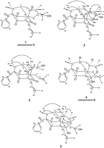
From the hexane extract of the trunk of C. obdeltifolia were isolated by silica gel column chromatography, gel permeation/adsorption on Sephadex LH-20 and TLC, three new polyprenylated benzophenones and two known compounds named sampsonione B,6 4 and sampsonione
G,71. The molecular formula were determined by EI mass spectrometry and by 1H and 13C NMR.
unsubstituted aromatic ring. IR spectra exhibited bands of hydroxyl groups and conjugated and unconjugated carbonyl groups. 1H NMR spectra showed signals between G 7.00 and G 8.00 that corresponded to a monosubstituted aromatic ring (Table 1). All molecular structures were deduced by analysis of 1H and 13C NMR, 1H-1H COSY, HMQC, HMBC and NOESY spectral data.
Compound1 was a yellow solid paste with molecular formula C33H42O5. All spectral data and DD value were in accordance with those related to sampsonione G, previously isolated from H. sampsonii.7
Compound2 has the molecular formula C30H36O5. Its NMR spectral data (Table1) were similar to those of 1, showing significant differences only in 1H and 13C signals
of five- membered ring due the replacement of the 2-E -hydroxyisopropyl group at C-13 in 1 by the D-hydroxyl group in 2.
The carbon skeleton of 2 was traced from HMBC correlations. The hydrogens of the gem-dimethyl groups atG 1.37 (H-26) and G 1.41 (H-25) correlated with each other and with the carbons at G 47.9 (C-9), G 81.1 (C-1), andG 42.4 (C-8). The signal at G 2.50 (H-10a) showed cross peaks with carbons at G 206.0 (C-4), G 68.2 (C-3), G 42.4 (C-8), G 29.1 (C-20), and G 22.9 (C-7) and that at G 2.24 (H-10b) with carbons at G 204.3 (C-2), G 68.2 (C-3), G 47.9 (C-9) and G 42.4 (C-8), establishing the six-membered ring C-1, C-9, C-8, C-10, C-3, C-2. Additionally, the correlations of the signal at G 2.30 (H-6) with the signals
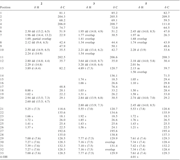
Table 1.1H and 13C NMR data of compounds 2,3 and 5
2 3 5
Position G H G C G H G C G H G C
1 81.1 85.2 82.7
2 204.3 205.5 209.5
3 68.2 69.1 59.5
4 206.0 208.7 111.0
5 74.7 72.0 64.5
6 2.30 dd (13.2; 6.5) 51.9 1.95 dd (14.8; 4.9) 51.2 2.45 dd (14.0; 8.5) 47.0
7 1.96 dd (14.4; 13.2) 22.9 1.77 overlap 30.5 1.97 m 26.3
1.69, partial overlap 1.41 overlap 1.68 overlap
8 2.12 dd (9.4; 6.5) 42.4 1.34 overlap 44.4 1.74 overlap 43.9
9 47.9 50.1 48.6
1 0 2.50 dd (14.9; 6.5) 35.5 2.21 dd (13.4; 6.2) 42.7 2.28 d (3.9) 33.6
2.24 d (14.9) 1.54 overlap
1 1 202.5 206.6 208.3
1 2 2.80 dd (14.8; 4.4) 35.7 3.64 dd (14.0; 8.7) 35.0 2.18 dd (14.0; 5.8) 30.4
2.29 d (14.8) 3.28 dd (14.0; 6.4) 2.01 bs
1 3 3.89 d (4.4) 82.2 4.89 t (7.3) 120.7 2.13 m 39.7
1.56 overlap
1 4 136.1 71.5
1 5 1.74 s 18.5 1.05 s 29.4
1 6 1.66 s 26.6 1.10 s 29.8
1 7 48.8 76.4 84.4
1 8 0.88 s 20.1 1.03 s 33.2 1.50 s 28.4
1 9 1.02 s 22.2 1.31 s 26.2 1.58 s 33.0
2 0 2.94 dd (13.5; 7.3) 29.1 2.84 dd (13.9; 6.8) 30.5 2.74 dd (14.0; 7.0) 29.2 2.60 dd (13.5; 4.7)
2.80 dd (13.9; 7.3) 2.45 dd (14.0; 8.5)
2 1 5.25 t (7.3) 118.6 5.55 t (7.0) 120.7 5.53 t (7.8) 120.8
2 2 135.6 134.6 135.9
2 3 1.66 s 18.1 1.92 s 18.5 1.72 s 18.3
2 4 1.72 s 26.0 1.85 s 26.6 1.76 s 26.5
2 5 1.41 s 25.4 1.43 s 25.6 1.43 s 25.5
2 6 1.37 s 22.5 1.56 s 24.1 1.21 s 22.5
2 7 192.6 195.6 195.4
2 8 135.0 138.8 137.3
2 9 7.00 d (7.6) 128.5 7.77 d (7.5) 129.9 7.61 d (7.4) 129.3
3 0 7.27 t (7.6) 128.3 7.16 t (7.5) overlap 7.34 t (7.4) 128.4
3 1 7.39 t (7.6) 132.3 7.10 t (7.5) 131.4 7.42 t (7.4) 132.2
3 2 7.27 t (7.6) 128.3 7.16 t (7.5) overlap 7.34 t (7.4) 128.4
3 3 7.00 d (7.6) 128.5 7.77 d (7.5) 129.9 7.61 d (7.4) 129.3
4-OH 4.01 s
atG 206.0 (C-4), G 74.7 (C-5), G 202.5 (C-11), G 22.9 (C-7), permitted to define the seven membered ring C-1, C-9, C-8, C-7, C-6, C-5, C-11. The dimethylpentacyclic moiety was defined by the correlations of the signal at G 2.30 (H-6) with the signals at G 48.8 (C-17), G 82.2 (C-13), G 22.2 (C-19) and G 20.1 (C-18), and by the signal at G 3.89 (H-13) with G 74.7 (C-5), G 48.8 (C-17) and G 20.1 (C-18).
The NOESY experiments were not conclusive about the C-6 stereochemistry. However, the NOE interactions permitted the localization of H-6, H-12a, H-13, and H-19 in the same face of molecule (Figure 1).
A careful comparison of 1H and 13C NMR data of 2 with those of sampsoniones C-H isolated from H. sampsonii,7 permitted the establishment of C-6 stereochemistry. Sampsoniones with D H-6 (C,D, and E) show C-7 chemical shifts between G 28.6 and G 29.0 ppm and C-10 chemical shifts between G 42.3 and G 43.9 ppm, while those with E H-6 (F,G, and H) show these signals between G 23.5 and G 24.7 ppm and between G 35.0 and G 35.3 ppm, respec-tively. Another observation was in relation to the chemical shifts of H-7a that were on average 0.36 ppm more shielded inF,G, and H (minimum value at G 1.92 and maximum value at G 2.04) than in C,D, and E (minimum value at
G 2.28 and maximum value at G 2.48), and of H-10b that were on average 0.28 ppm more shielded in C,D, and E
(minimum value at G 1.88 and maximum value at G 1.96) than in F,G, and H(minimum value at G 2.17 and maximum value at G 2.21). In conclusion, the H-6 stereochemistry of
2 was the same as that of sampsoniones F,G, and H. Compound 3, C33H42O5, presented some spectral features that resembled those of 1 and 2. However, the lack of the dimethylpentacyclic moiety and the presence of a second prenyl group at C-5 and one 2-hydroxyisopropyl group at C-6 produced, in the vicinity of these groups, significant differences in chemical shifts of 1H and 13C NMR signals in relation to 1 and 2.
The1H NMR spectrum of 3 obtained in CDCl
3 showed a complex overlap of signals between G 1.90 and G 2.20 making the assignment of important signals difficult. However, the spectrum obtained in C6D6 showed a better spreading of signals and facilitated their correct assignment (Table 1). The 13C NMR spectrum confirmed the presence of three nonconjugated carbonyls in addition to the 2-hydroxyisopropyl group and two prenyl groups. The carbon skeleton was traced from HMBC cross peaks in a way similar to that in 2. The NOESY data permitted the establishment of the relative stereochemistry (Figure 1). The cross peak observed between H-10a and H-25 defined the relative position of Me-25 and Me-26. The latter presented cross peak with H-7a, which defined the relative position of the H-7 methylene hydrogens. Finally, as H-6 showed cross peak with H-7b and not with H-7a, it was positioned on the same face of H-7b.
Compound4 has the molecular formula C33H42O5. The spectral data were in accordance with those related to sampsonione B, previously isolated from H. sampsonii.6
The molecular formula of compound 5 was deduced as C33H44O6. Its spectral features (Table 1) were closely related to those of sampsonione B,4, and revealed the presence of only one prenyl group and one 3-hydroxy-3-methylbutenyl group. The carbon skeleton was established from HMBC correlations with a method similar to that used to 1, 2,3 and 4. The relative stereochemistry was proposed from NOESY data (Figure 1). The relative positions of the two gem-dimethyl groups at C-9 and C-17 and that of the methylene hydrogens at C-10 were defined by the cross peaks observed between H-10a and H-25 and between H-10b and H-19. The correlations observed between 26 and 7a, 7a and 6 and of 6 and H-18, defined the relative stereochemistry at C-6. On the other hand, the molecular models revealed that the closing of the furan ring could just happen when H-6 occupies the D position in an intermediary like 3, what was in agreement with the proposed stereochemistry for 5.
Experimental
General procedures
Optical rotations were measured on a Perkin-Elmer 241 polarimeter. IR spectra were obtained with a JASCO FT-IR spectrophotometer. A Bruker Advance DRX-500 spectro-meter, operating at 500.13 MHz for 1H and 125.75 MHz for13C in CDCl
3 or C6D6 with TMS as int. standard were used to obtain NMR data. EIMS with direct probe insertion at 70 eV was obtained in an HP MSD 5973 apparatus.
Plant material
Clusia obdeltifolia Bittrich was collected in a “campo rupestre” (rocky field) area near Palmeiras in Parque Nacional da Chapada Diamantina, Bahia, Brazil, in April 1996, and was identified by Prof. Maria Lenise Silva Guedes. A voucher specimen, number ALCB-035997, was deposited in the “Alexandre Leal Costa” Herbarium, Instituto de Biologia, Universidade Federal da Bahia, Salvador, Brazil.
Extraction and isolation
Dried powdered trunk (4400 g) was extracted with hexane. Evaporation of solvent under reduced pressure yielded 39.2 g of extract. This extract was submitted to a chromatography on silica gel column using hexane-EtOAc (0-100%) to give 19 fractions.
Fraction 06 (6.8g) was rechromatographed on silica gel column using hexane-EtOAc (0-100%). Fractions 17 to 20; 27 to 30 and 31 to 40 were chosen for work. Fractions 17 to 20 were submitted again to a chromatography on silica gel column using CHCl3-EtOAc (0-100%) and then to preparative TLC (silica gel; hexane- EtOAc 9:1) to provide
4(10mg). Fractions 27 to 30 were rechromatographed on silica gel column using hexane-EtOAc (0-100%) to give 1
(18mg). Fractions 31-40 were submitted to chromatography on silica gel column using hexane-CH2Cl2 (0-100%) and then to TLC (silica gel; hexane-EtOAc 9:1) to yield 2(8mg). Fraction 07 (1.7g) was rechromatographed on silica gel CC using hexane- EtOAc (0-100%). Fractions 14 and 15 were submitted to preparative TLC (silica gel; hexane-EtOAc 7:3) to yield 3(11mg).
Fraction 08 (1.0g) was rechromatographed on silica gel column using hexane- EtOAc (0-100%). Fractions 44 to 47 were chosen for work and were submitted to Sephadex LH-20 CC using hexane-CH2Cl21:4 to yield 5(12mg).
7E-H-11-benzoyl-5E (1hydroxy1methylethyl)6,6,10,10t e (1hydroxy1methylethyl)6,6,10,10t r a m e (1hydroxy1methylethyl)6,6,10,10t h y l 1 ( 3 m e (1hydroxy1methylethyl)6,6,10,10t h y l 2
-butenyl)tetracyclo[7.3.1.13,1103,7
]tetradecane-2,12,14-trione, Sampsonione G(1)
C33H42O5. Yellow amorphous solid; [D]D25= +10.0o (c 0.04, CHCl3); IR (film CHCl3)Qmax/cm
-1: 3448; 2960; 2851;
1733, 1705, 1464; 1H and 13C NMR see reference 5; EIMS m/z 518(6), 500 (19), 105 (11), 472 (100).
7E-H-11-benzoyl-5D -hydroxy-6,6,10,10-tetramethyl-1-(3-methyl-2-butenyl)tetracyclo[7.3.1.13,1103,7
]tetradecane-2,12,14-trione (2)
C30H36O5. Yellow amorphous solid; IR (film CHCl3) Qmax/cm
-1: 3423, 2924, 2851; 1736, 1702, 1447; 1H and
13C NMR Table 1; EIMS m/z 476 (13), 448 (16), 105 (100),
77 (38).
8-benzoyl-4D -(1-hydroxy-1-methylethyl)-7,7-dimethyl-1,3-di(3-methyl-2-butenyl)tricyclo[4.3.1.13,8
]undecane-2,9,11-trione (3)
C33H42O5. Yellow amorphous solid; [D]D25= +10.8o (c 0.011, CHCl3); IR (film CHCl3)Qmax/cm
-1: 3422, 2924,
2853, 1734, 1697, 1463, 1449, 1243; 1H and 13C NMR Table 1. EIMS m/z 500 (5), 363 (30), 317 (35), 309 (46), 105 (100), 77 (6).
7D H1benzoyl4hydroxy6,6,13,13tetramethyl3,11d i ( 3 m e t h y l 2 b u t e n y l ) 5 -oxatetracyclo[7.3.1.03,704,11]tridecane-2,12-dione,
SampsonioneB(4)
C33H42O5. Yellow amorphous solid: [D]D25= +10.0o (c 0.018, CHCl3). IR (film CHCl3)Qmax/cm
-1: 3393, 2924,
2853,1719, 1685, 1457; 1H and 13C NMR (see reference 3). EIMSm/z 518 (4), 449 (29), 105 (100), 77 (24).
7D -H-1-benzoyl-4-hydroxy-3-(3-hydroxy-3-methylbutyl)- 6,6,13,13-tetramethyl-11-(3-methyl-2-butenyl)-5-oxatetracyclo[7.3.1.03,704,11]tridecane-2,12-dione (5)
C33H44O6. Yellow amorphous solid: [D]D25= -5.1o (c 0.012, CHCl3); IR (film CHCl3)Qmax/cm
-1: 3391, 2967,
2926, 1719, 1686, 1448, 1232, 768, 738, 694; 1H and 13C NMR Table 1; EIMS m/z 536 (12), 518 (32), 431 (82), 345 (55), 105 (100).
Acknowledgments
Andrade Uchoa for recording NMR spectra, to Dr. João H. G. Lago from IQ-USP for recording the DD values and to Roy Funch from Fundação Chapada Diamantina (Lençóis) for providing local support during plant collection, as well as to CNPq for the fellowships for JSRT. This work was supported by grants from CNPq and FINEP.
References
1. Cruz, F. G.; Moreira, L. de M.; Santos, N. A. S; Guedes, M. L. S.; J. Braz. Chem. Soc.2002,13, 704.
2. Cruz, F.G.; da Silva-Neto, J. T.; Guedes, M. L. S.; J. Braz.
Chem. Soc.2001, 12, 117.
3. Cerrini, S.; Lamba, D.; Monache, F. D.; Pinherio, R. M.;
Phytochemistry1993,32, 1023.
4. Iinuma, M.; Tosa, H.; Tanaka, T.; Kanamaru, S.; Asai, F.;
Kobayashi, Y.; Miyauchi, K.; Shimano, R.; Biol. Pharm. Bull.
1996, 19, 311.
5. Gustafson K. R.; Blunt, J. W.; Munro, M. H. G.; Fuller, R. W.; McKee, T. C.; Cardellina II, J. H.; McMahon, J. B.; Cragg,
G.M.; Boyd, M. R.; Tetrahedron1992,48, 10093.
6. Hu, L. H.; Sim, K. Y.; Tetrahedron Lett.1998,39, 7999.
7. Hu, L. H.; Sim, K. Y.; Tetrahedron Lett.1999,40, 759.
8. Hu, L. H.; Sim, K. Y.; Org. Lett.1999,1, 879.
9. Hu, L. H.; Sim, K. Y.; Tetrahedron 2000,56, 1379.
10. Henry, G. E.; Jacobs, H.; Carrington, C. M. S.; McLean, S.;
Reynolds, W. F.; Tetrahedron1999,55, 1581.
11. Christian, O. E.; Henry, G. E.; Jacobs, H.; McLean, S.; Reynolds,
W. F.; J. Nat. Prod.2001,64, 23.
Received: August 6, 2003