Temporomandibular joint
arthrocententesis: evaluation
of results and review of the
literature
Summary
Belmiro Cavalcanti do Egito Vasconcelos1, Ricardo
Viana Bessa-Nogueira2, Nelson Studart Rocha3
1 PhD, Post-Graduation Program Coordinator - UPE.
2 Bucco-maxillofacial traumatology and surgery (BMFTS) Specialist and MS – Dentistry School - UPE. PhD student in CTBMF / Pernambuco Dentistry School - UPE. 3 Specialist in Bucco-maxillofacial traumatology and surgery (BMFTS) – Dentistry School of Pernambuco (FOP/UPE). Bucco-maxillofacial surgeon - Getulio Vargas
Hospital- Recife/PE.
University of Pernambuco – School of Dentistry – MS and PhD programs in Bucco-maxillofacial traumatology and surgery (BMFTS).
Mailling Address: Prof. Dr. Belmiro Cavalcanti do Egito Vasconcelos - Faculdade de Odontologia de Pernambuco - FOP/UPE Disciplina de Cirurgia e Traumatologia Buco-Maxilo-Facial - Av. General Newton Cavalcanti 1650 Camaragibe PE 54.753-220. Tel/Fax: (0xx81) 3458-2867 - E-mail: [email protected]
We acknowledge FACEPE/CNPQ for their support.
Paper submitted to the ABORL-CCF SGP (Management Publications System) on March 1st, 2006 and accepted for publication on June 14th, 2006. Cod. 1741.
A
im: This study was designed to investigate the effects of arthrocentesis on the improvement of internal derangement symptoms and jaw function in a series of patients with anterior disc displacement and closed lockjaw. Patients and methods: The study was based on a review of patients’ records before and after treatment using clinical examinations and radiographs. Visual analog scales were used to measure pain before and after arthrocentesis. Six patients (12 temporomandibular joints) with closed lock symptoms (2 cases) and internal derangements (4 cases) were treated at the Oswaldo Cruz Hospital. The mean follow-up was 11.5 months. Results: The mean maximum vertical opening before treatment was 31.83 mm and after arthrocentesis was 36.50 mm. The visual analog scale for pain before treatment was 7 points (mean) and after arthrocentesis the mean was 4.3. Conclusion: Arthrocentesis was shown to be effective in reducing pain and increasing jaw motion in this series of cases.Keywords: Arthrocentesis; temporomandibular joint ORIGINAL ARTICLE
ARTIGO ORIGINAL
INTRODUCTION
Temporomandibular joint (TMJ) arthrocentesis con-sists of lavage of the upper joint space of the TMJ done with no direct vision, aiming primarily to remove necrotic tissue, blood and pain mediators from the joint (Barkin, Weinberg, 2000).
Nitzan et al. (1991) first described TMJ arthrocentesis as the simplest form of surgery in the TMJ, aiming to re-lease the articular disc and to remove adhesions between the disc surface and the mandibular fossa by means of hydraulic pressure from irrigation of the upper chamber of the TMJ.
Arthrocentesis has low morbidity, few risks and low cost compared to other TMJ surgical interventions, and may be conducted under local anesthesia in an outpatient clinic setting (Hasson, Levy, 1999; Carvajal, Laskin, 2000; Salazar et al., 2004).
Indications for arthrocentesis described in medical literature are: dislocation of the articular disc with or with no reduction, limitations of mouth opening originating in the joint, joint pain and other internal derangements of the TMJ (Nitzan, 1991; Frost, Kendell, 1999; Trieger et al., 1999; Yoda et al., 2002).
Clinical use of arthrocentesis in the TMJ is a new procedure among other possibilities of treating joint dysfunction. There is, therefore, a need for studies on the indications, success rate, and complications of this procedure.
This paper aims to present a series of patients undergoing arthrocentesis, to assess the results, and to provide a review of literature.
MATERIAL AND METHODS
Six patients (twelve joints) presenting with pre-auricular pain, limited TMJ movements and closed lock were referred to the bucomaxillofacial surgery and trauma unit of the Oswaldo Cruz University Hospital in Recife, Pernambuco state.
All patients had undergone prior medical treatment for TMJ dysfunction (bite plates, muscle relaxants, com-presses, diets and physical therapy) for at least 6 months with no clinical improvement. Four out of six patients had joint pain and functional limitation and two patients had closed lock (Table 1).
Preoperative data included the clinical history, a physical exam and radiograms. This included progression time of TMJ dysfunction, the presence of facial asymme-try, unilateral or bilateral involvement, the amplitude of mandibular movements (maximum mouth opening, right and left laterality, and protrusion), the presence of joint
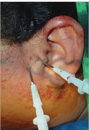
Figure 1. Clinical view of TMJ arthrocenthesis.
Table 1. Epidemiological aspects of participants.
Nº Gender Age Main complaint Diagnosis Side
1 Fem 20 Pain Internal de-rangement Bilateral
2 Fem 34 Pain Internal de-rangement Bilateral
3 Fem 39 Pain Closed lock Bilateral
4 Fem 43 Pain Internal
de-rangement Bilateral
5 Fem 39 Limited mouth
opening Closed lock Bilateral
6 Fem 32 Pain Internal
noises, deviation on maximum mouth opening, and pain on mandibular movements, which was catalogued accor-ding to the pain visual analog scale. Radiography inclu-ded a maxillary panoramic radiogram and a TMJ specific panoramic radiogram to establish anatomical changes of the mandibular condyle and reduced joint space.
The first surgical procedure was TMJ arthrocentesis. All procedures were done by a single surgeon. Procedures were done under local anesthesia and sedation. Nitzan et al.’s (1991) surgical technique was used. A line was drawn from the corner of the eye - tragus and the first mark was made 10mm from the tragus and 0.5mm below the line. The second point was marked 20mm from the tragus and 1mm below the line. A 40x12 needle was placed on each point and the joint was irrigated with 250ml of saline under continuous pressure (Figure 1).
Postoperative follow-up for all patients was 11.5 months (6 to 17 months). Postoperative data were the same preoperative variables. This study was presented to and approved by the Pernambuco University Research Ethics Committee (number 004/04) and participants read and signed a free and informed consent form.
RESULTS
Objective findings
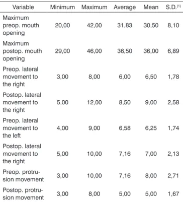
Table 2 summarizes and compares objective pre and postoperative findings. All patients had improved mouth opening following arthrocentesis. Preoperative mouth opening was 31.83mm ±8.10mm and postoperative mouth opening was 36.50mm ±6.89mm. Lateral movements and protrusion were unaltered. Further information is shown on Table 2.
Three patients presented joint noises on the pre-operative clinical exam. At the end of treatment, two no longer had joint noises and one had a reduction in joint clicks.
Subjective findings
Every patient had moderate to severe preoperative pain. Preoperative pain visual analog scale scores averaged 7 ±1.78. On follow-up the pain score fell to an average 4.33 ±1.03. All patients reported an improved general clinical status and a reduction of internal derangement symptoms. (Table 3)
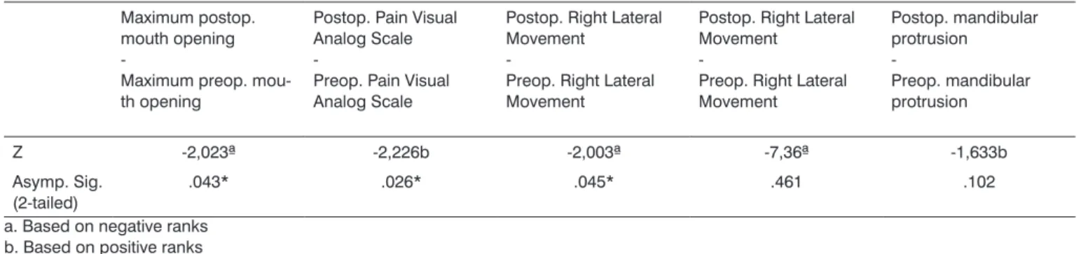
Maximum mouth opening, right lateral movements and the pain visual analog scale were statistically significant (p < 0.05) based on the Wilcoxon test. (Table 4)
Table 2. Measurement of the pre and postoperative mandibular mo-vement amplitude.
Variable Minimum Maximum Average Mean S.D.(1)
Maximum preop. mouth opening
20,00 42,00 31,83 30,50 8,10
Maximum postop. mouth opening
29,00 46,00 36,50 36,00 6,89
Preop. lateral movement to the right
3,00 8,00 6,00 6,50 1,78
Postop. lateral movement to the right
5,00 12,00 8,50 9,00 2,58
Preop. lateral movement to the left
4,00 9,00 6,58 6,25 1,74
Postop. lateral movement to the right
5,00 10,00 7,16 7,00 2,13
Preop.
protru-sion movement 3,00 10,00 7,16 8,00 2,71 Postop.
protru-sion movement 3,00 8,00 5,00 5,00 1,67 (1) - Standard Deviation
Table 3. Pre and postoperative pain scale scores.
Variable Minimum Maximum Average Mean S.D.(1)
Preop. Pain Visual Ana-log Scale
5,00 10,00 7,00 6,50 1,78
Postop. Pain Visual Ana-log Scale
3,00 6,00 4,33 4,00 1,03
(1) - Standard Deviation
DISCUSSION
Table 4. Statistical analysis of objective and subjective findings.
Maximum postop. mouth opening
-Maximum preop. mou-th opening
Postop. Pain Visual Analog Scale
-Preop. Pain Visual Analog Scale
Postop. Right Lateral Movement
-Preop. Right Lateral Movement
Postop. Right Lateral Movement
-Preop. Right Lateral Movement Postop. mandibular protrusion -Preop. mandibular protrusion
Z -2,023ª -2,226b -2,003ª -7,36ª -1,633b
Asymp. Sig. (2-tailed)
.043* .026* .045* .461 .102
a. Based on negative ranks b. Based on positive ranks c. Wilcoxon Signed Ranks Test
of TMJ dysfunction (Spallaccia et al., 2000).
There are no longitudinal studies to compare the success and lack of success of this approach, and further studies are required to scientifically demonstrate the in-dication and predictability of results. Efficacy in various papers are: Murakami et al. (1995) - 70%; Dimitroulis et al. (1995) - 98%; Hosaka et al. (1996) - 79%; Fridrich et al. (1996) - 75%; Nitzan et al. (1997) - 95%. These studies suggest that arthrocentesis is an efficient method with relatively high success rates.
All patients had improvement in symptoms related to the intra-articular derangement and increased mandibu-lar movements. These results are simimandibu-lar to published stu-dies (Frost et al., 1992; Dimitroulis, 1995; Stein, 1995; Nitzan et al., 1997; Carvajal, Laskin, 2000). Internal derangement and closed lock conditions are usually associated with common symptoms such as pain, limited mouth opening and altered mandibular function, which may explain our results. Increased pain leads to reduced mouth opening and eventually to altered movements. By correcting one problem, the remaining two may also improve. (Carvajal; Laskin, 2000).
Lavage of the upper joint space reduces pain by removing inflammation mediators from the joint (Quinn, Bazan, 1990), increasing mandibular mobility by removing intra-articular adhesions (Spallacia et al., 2000), eliminating the negative pressure within the joint, recovering disc and fossa space (Nitzan et al., 1991), and improving disc mobi-lity, which reduces the mechanical obstruction caused by the anterior position of the disc (Moses et al., 1989).
In our study we observed that symptom reduction resulted in improved mandibular function without neces-sarily producing an anatomical relationship between the mandibular head, the articular disc and the glenoid cavity.
Rather, improved mandibular function appeared to be due to the removal of intra-articular adhesions and the resulting increased mobility of the disc.
CONCLUSION
The methodology used in this series showed effective improvement in the treatment by arthrocentesis of patients with mandibular internal derangement and closed lock.
REFERENCES
1. Barkin S, Weinberg S. Internal derangements of the temporomandi-bular joint: the role of arthroscopic surgery and arthrocentesis. J Can Dent Assoc 2000;66:199-202.
2 arvajal WA, Laskin DM. Long-term evaluation of arthrocentesis for the treatment of internal derangements of the temporomandibular joint. J Oral Maxillofac Surg 2000;58:852-7.
3. Dimitroulis G, Dolwick MF, Martinez GA. Temporomandibular joint arthrocentesis and lavage for the treatment of closed lock: A follow up study. Br J Oral Maxillofac Surg 1995;33:23-7.
4. Fridrich KL, Wise JM, Zeitler DL. Prospective comparison of arthros-copy and arthrocentesis for temporomandibular joint disorders. J Oral Maxillofac Surg 1996;54:816-20.
5. Frost DE, Kendell BD. The use of arthrocentesis for treatment of tem-poromandibular joint disorders. J Oral Maxillofac Surg 1999;57:583. 6. Haason O, Levy Y. Arthrocentesis e lavagem da articulação temporo-mandibular: indicações no tratamento da abertura de boca limitada. Rev Paul Odontol 1999;21:4-6.
7. Hosaka H, Murakami K, Goto K, Tadahiko L. Outcome of arthrocen-tesis for temporomandibular joint with closed lock at 3 years follow up. Oral Surg Oral Med Oral Pathol 1996;82:501-4.
8. Moses JJ, Sartoris D, Glass R, Tanaka T, Poker I. The effect of ar-throscopy surgical lysis and lavage of the superior joint space on temporomandibular joint disk position and mobility. J Oral Maxillofac Surg 1989;47:674-8.
therapy and arthroscopy lysis and lavage. Oral Surg Oral Med Oral Pathol 1995;80:253-7.
10. Nitzan DW, Dolwick MF, Heft MW. Arthroscopy lavage and lysis of the temporomandibular joint: a change in perspective. J Oral Maxillofac Surg 1990;48:798-801.
11 Nitzan DW, Dolwick MF. An alternative explanation for the genesis of closed lock symptoms in the internal derangement process. J Oral Maxillofac Surg 1991;49:810-15.