Validity of Klotho, CYR61 and YKL-40 as ideal predictive
biomarkers for acute kidney injury: review study
Validade de Klotho, CYR61 e YKL-40 como biomarcadores preditivos ideais
para lesão renal aguda: estudo de revisão
Osama Mosa
I, Milan Skitek
II, Ales Jerin
IIIHealth Science College at Al-Leith, Umm Al-Qura University, Saudi Arabia
ABSTRACT
CONTEXT AND OBJECTIVE: Acute kidney injury (AKI) is still a headache for clinicians and scientists as a possible reason for increased death among intensive care unit (ICU) patients after invasive cardiac surgery. Furthermore, the diagnostic process for AKI using conventional biomarkers is not suicient to ensure early warning of this condition because of the morbid inluence of non-renal factors that deinitively delay the time for the prognosis. These imposed limitations have led to signiicant amounts of research targeted towards identifying novel biomarkers for AKI with a sustained degree of sensitivity and speciicity. Here, we reviewed previous studies conducted on the Klotho, CYR61 and YKL-40 biomarkers in relation to AKI.
DESIGN AND SETTING: Review of the literature conducted in the Institute of Clinical Chemistry & Bio-chemistry, Ljubljana University Medical Center, Slovenia.
METHODS: The literature was searched in PubMed and the Cochrane Library. From the database of this specialty, we selected 17 references that matched our context for detailed analysis and further investigation.
RESULTS: The studies reviewed showed notable diferences in their results relating to the diagnostic im-pact of Klotho, CYR61 and YKL-40 on early prediction of AKI.
CONCLUSIONS: The results regarding the Klotho, CYR61 and YKL-40 biomarkers showed markedly equivo-cal performance in the previous studies and did not fulill the expectations that these factors would form valid possible biomarkers for AKI.
RESUMO
CONTEXTO E OBJETIVO: A lesão renal aguda (LRA) ainda é uma dor de cabeça para os clínicos e cientistas como possível razão para o aumento da mortalidade entre os pacientes de unidade de terapia intensi-va (UTI) após cirurgia cardíaca invasiva. Além disso, o processo de diagnóstico para LRA usando biomarca-dores convencionais não é suiciente para garantir um alerta precoce desta condição, devido à inluência mórbida de fatores não renais que podem retardar o tempo para o prognóstico. Essas limitações geraram quantidades signiicativas de pesquisas orientadas para identiicar novos biomarcadores para LRA com um grau adequado de sensibilidade e especiicidade. Revisamos estudos anteriores realizados sobre os biomarcadores Klotho, CYR61, YKL-40 para LRA.
TIPO DE ESTUDO E LOCAL: Revisão da literatura realizada no Instituto de Química Clínica e Bioquímica, Centro Médico da Universidade de Ljubljana, Eslovênia.
MÉTODOS: A literatura foi pesquisada no PubMed e Cochrane Library. A partir da base de dados da es-pecialidade, selecionamos 17 referências que combinavam com o contexto para uma análise detalhada e mais investigação.
RESULTADOS: Os estudos revisados mostraram diferenças notáveis nos resultados sobre o impacto diag-nóstico de Klotho, CYR61 e YKL-40 sobre a detecção precoce do LRA.
CONCLUSÃO: Os resultados em relação aos biomarcadores Klotho, CYR61 e YKL-40 mostraram desem-penho marcadamente equívoco nos estudos anteriores e não cumpriram as expectativas de que estes fatores constituam possíveis biomarcadores válidos para LRA.
IPhD. Lecturer of Clinical Biochemistry, Department of Public Health, Health Science College at Al-Leith, Umm Al-Qura University, Saudi Arabia.
IIPhD. Professor and Head of Institute of Clinical Chemistry and Biochemistry, Ljubljana University Medical Center, Ljubljana, Slovenia.
IIIPhD. Associate Professor and Head of Department of Hormones and Tumor Markers, Institute of Clinical Chemistry and Biochemistry, Ljubljana University Medical Center, Ljubljana, Slovenia.
KEY WORDS:
Acute kidney injury. Thoracic surgery. Proteins. Biomarkers. Review.
PALAVRAS-CHAVE:
INTRODUCTION
Acute kidney injury (AKI) is a highly progressive critical prob-lem that oten occurs ater invasive cardiac surgery using cardio-pulmonary bypass (CBP).1,2 It threatens the life of intensive care
unit (ICU) hospitalized patients through accompanying irre-versible adverse outcomes that ultimately contribute to a 60%
increase in mortality rate.3 Deining AKI is dependent on
mea-surement of baseline serum creatinine, the traditional biomarker of kidney function, which remains unchanged until a sudden
50% of kidney function is lost.4 Moreover, AKI has been found
to be strongly afected by dietary status, exercise, protein supple-ments, corticosteroids, age, gender and muscle mass.5,6 herefore,
there is an urgent need for novel biomarkers to predict and diag-nose AKI at its earlier stages, so as to prevent complications and potentiate therapeutic approaches.
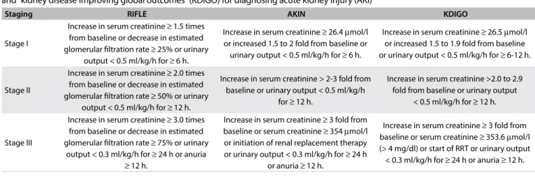
Classiication of AKI
The Acute Dialysis Quality Initiative Group (ADQI) meeting in 2004 gave rise to a new regular criterion for analyzing kid-ney function, termed Risk Injury Failure Loss of function and
End stage (RIFLE).7,8 RIFLE was dependent on serum
creati-nine (SCr) or urinary output (UO) measurements to deter-mine the prognostic severity of deterioration of kidney
func-tion, classified into three stages.8 Many studies mentioned
that the usefulness of RIFLE was affected by the following substantial limitations: [1] calculation of the SCr baseline using the Modification of Diet in Renal Disease (MDRD) equation showed high specificity for chronic kidney disease (CKD) but not AKI; [2] SCr was directly influenced by non-specific factors and hence was unreliable; [3] using UO was a good alternative for SCr, but it was affected by diuretics and could only be measured by using a bladder catheter in an ICU and not among long-stay hospitalized patients; and
[4] SCr was considered to be a marker for renal function, not kidney injury.9
Subsequently, a modiied standard was published in 2007 under the name “Acute Kidney Injury Network (AKIN)”, with the aim of closing gaps generated by RIFLE. AKIN used two values of SCr within two days instead of baseline SCr, regardless of glo-merular iltration rate (GFR) changes. According to AKIN, stage 3 AKI was conirmed when the duration of increased SCr levels did not exceed 48 h and the patient required renal replacement therapy (RRT).10
he failure of both RIFLE and AKIN to fulill precise prog-nostic stratiication of AKI severity and to provide a uniied dei-nition of AKI was the reason for establishing the Kidney Disease Improving Global Outcomes (KDIGO) guidelines. hese novel criteria suggested that AKI should be deined by SCr levels that reached 26.4 μmol/l within 48 h or increased to a level 1.5 times higher than the baseline level within 7 days, which provides a suf-icient timeframe for AKI diagnosis.11 he diferences between all
the diagnostic criteria are summarized in Table 1.
Epidemiology of AKI
he AKI incidence rate worldwide has remained imprecise because of the small number of case report studies, gaps in the data collected from patients and diferences in deinitions of
AKI between developed and developing countries.12-14 Recent
studies conducted in the USA and Spain showed incidences of approximately 23.8 cases per 1000 discharges and 209 cases per million, respectively.15,16 A recent population-based study
con-ducted in the UK reported high incidence of AKI, of 1811 cases
per million in 2003.17 A report from Kuwait indicated an
inci-dence of 4.1 cases per 100,000 population per year.18 In addition,
the annual incidences for AKI in Brazil and northern India were 7.9 and 6.4 cases per 1000 admissions.19,20 Notably, the mortality
Staging RIFLE AKIN KDIGO
Stage I
Increase in serum creatinine ≥ 1.5 times from baseline or decrease in estimated glomerular iltration rate ≥ 25% or urinary
output < 0.5 ml/kg/h for ≥ 6 h.
Increase in serum creatinine ≥ 26.4 μmol/l or increased 1.5 to 2 fold from baseline or
urinary output < 0.5 ml/kg/h for ≥ 6 h.
Increase in serum creatinine ≥ 26.5 μmol/l or increased 1.5 to 1.9 fold from baseline or urinary output < 0.5 ml/kg/h for ≥ 6-12 h.
Stage II
Increase in serum creatinine ≥ 2.0 times from baseline or decrease in estimated glomerular iltration rate ≥ 50% or urinary
output < 0.5 ml/kg/h for ≥ 12 h.
Increase in serum creatinine > 2-3 fold from baseline or urinary output < 0.5 ml/kg/h
for ≥ 12 h.
Increase in serum creatinine >2.0 to 2.9 fold from baseline or urinary output
< 0.5 ml/kg/h for ≥ 12 h.
Stage III
Increase in serum creatinine ≥ 3.0 times from baseline or decrease in estimated glomerular iltration rate ≥ 75% or urinary
output < 0.3 ml/kg/h for ≥ 24 h or anuria
≥ 12 h.
Increase in serum creatinine ≥ 3 fold from baseline or serum creatinine ≥ 354 μmol/l or initiation of renal replacement therapy or urinary output < 0.3 ml/kg/h for ≥ 24 h
or anuria ≥ 12 h.
Increase in serum creatinine ≥ 3 fold from baseline or serum creatinine ≥ 353.6 μmol/l (> 4 mg/dl) or start of RRT or urinary output < 0.3 ml/kg/h for ≥ 24 h or anuria ≥ 12 h.
rates in developed countries were found to be lower than those in developing countries, where young adults and children were very badly afected.21
Prospective biomarkers
Klotho
Klotho (KL) is a novel phosphatonin encoded by the anti-aging KL gene located on chromosome 13q12 as an inactive
single-pass transmembrane protein.22 Upon activation through action
by membrane bound-secretases like ADAM10 and ADAM17, driven by insulin, the extracellular domain is cleaved and its
serum, urine and cerebrospinal luid levels become elevated.23
his ectodomain was termed a soluble Klotho, which would possibly bind directly with FGFR and tend to form an active
complex exhibiting high ainity against FGF,24 thereby
allevi-ating oxidative stress through suppression of growth factors
and stimulation of calcium ion channels(TRPV5 and TRPV6)23
and potassium channels(ROMK)25 but not sodium-phosphate
cotransporters.26 Meanwhile, the remaining membrane Klotho
would function as a coreceptor for bone regulatory hormone
FGF23.27 Normally, Klotho shows greater expression in distal
rather than proximal convoluted tubules in the kidneys, and in the choroid plexus of the brain rather than in the heart and parathyroid gland.28
he pathological importance of Klotho emerged through studies on animal models for AKI that had previously under-gone ischemic reperfusion injury (IRI) or unilateral ure-thral obstruction (UUO). hus, a transient reduction in renal Klotho mRNA expression was shown in response to renal tubu-lar injury.29, 30 Other studies on Klotho applied to humans have
demonstrated that the urinary and plasma levels of Klotho in patients with AKI are notably lower than in healthy individu-als.29 From these observations, it has been proposed that Klotho
has a role in exacerbating renal damage and has potential as a likely biomarker for AKI.
Cysteine-rich protein 61 (CYR61)
CYR61 is a cysteine-rich matricellular protein encoded by the CYR61 gene located on chromosome 1p22.3. It is intercalated with various integrins and heparin sulfate proteoglycans and is associated with extracellular matrix formation, cell adhe-sion, proliferation, diferentiation, angiogenesis, apoptosis and inlammation due to its biochemical features, which resemble
Wnt-1 proto-oncogene, and its number of growth factors.31
Additionally, renal CYR61 mRNA and protein expression, along with urinary levels, have been found to increase in IRI animal models that sufered from signiicant hypoxia, despite
being indistinguishable at renal levels in normal tissues.32,33
his result provides encouragement to study CYR61 levels in humans, in order to elucidate its preventive and/or predictive role against AKI.
Chitinase-3-like protein 1 (YKL-40)
Chitinase-3-like protein 1 (CHI3L1) or YKL-40 is a 40 kDa
gly-coprotein34 that is expressed from the CHI3L1 gene located on
chromosome 1q31-q32.35 It is considered to be a member of
the family of 18 glycoside hydrolases that encompasses chitin-ases but without any enzymatic activity. It is secreted by vari-ous cell types, including macrophages, chondrocytes and some types of cancer cells.34 Furthermore, Johansen et al. revealed that
YKL-40 increased inlammation through activation of the innate
immune response and regulation of tissue remolding.35 In
addi-tion, Maddens et al. collected urine samples from mice that pre-sented sepsis two days ater intrauterine injection of E. coli,and from human patients with sepsis. hey showed similar
quan-titative increases in comparison with controls without AKI.36
herefore, studies on YKL-40 remain a prerequisite for under-standing the pathophysiology of AKI.
OBJECTIVE
he objective of the current review was to focus on the suitabil-ity and validsuitabil-ity of Klotho, CYR61 and YKL-40 as ideal predictive biomarkers for acute kidney injury.
METHODS
We conducted a comprehensive systematic search by using the main known databases: PubMed, SCOPUS, SciELO, Lilacs, ScienceDirect and Google Scholar. The MeSH search terms included: (‘‘Klotho and Acute Kidney Injury’’), (‘‘CYR61 and Acute Kidney Injury’’) AND (Chitinase-3-like protein 1 and Acute Kidney Injury’’). The search strategy was designed for the PubMed database and was altered as needed for use in other databases. Our last search was finished in January 2016. References were written in the English language. The inclu-sion criterion was that all research articles, review articles and observational studies included needed to match our context, i.e. “the propensity of CYR61, Klotho and YKL-40 to be novel biomarkers for AKI”. Additionally, we excluded papers that investigated these biomarkers in relation to chronic kidney disease (CKD) and other diseases as well as AKI.
RESULTS
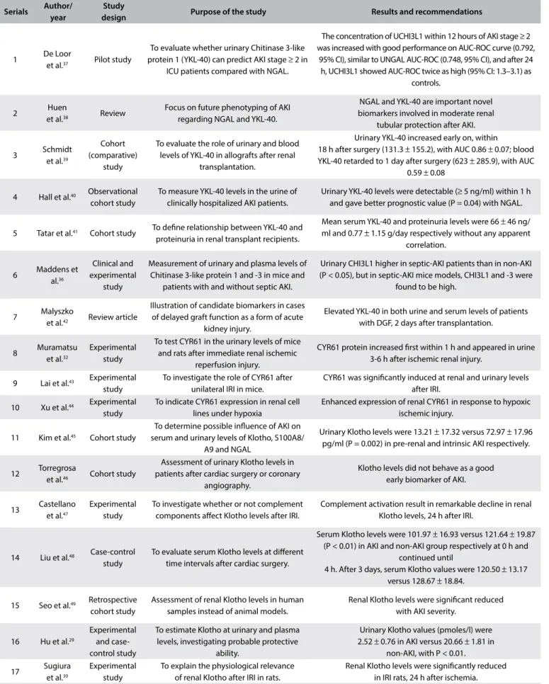
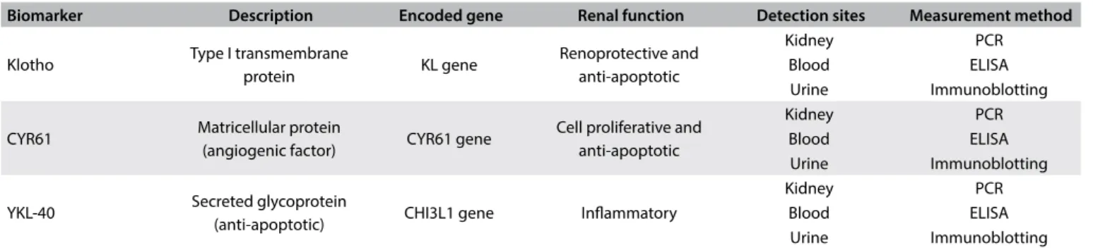
summarized the main results and recommendations for each study in Table 3.29,30,32,36-49 Finally, a synopsis of the biomarkers
studied, showing general descriptions, functions and techniques used for measurements, was produced as Table 4.
DISCUSSION
In this review article, we discuss the propensity of some novel biomarkers for early detection of AKI. Traditional biomarkers have been proven to be unable to satisfactorily distinguish AKI during the irst 24 hours before kidney function is disrupted. his is certain to delay the diagnostic process and gives rise to the pos-sibility that the patient’s condition will worsen. Despite the pau-city of studies on biomarkers and AKI (for reasons mentioned earlier), we conducted a comprehensive review of the literature encompassing all papers relating to our context, focusing on all the results.
Recent papers have inferred that reduced levels of Klotho correlated with emergence of sot tissue calciications, cardiovas-cular diseases, senescence, cancers, chronic hypertension, osteo-porosis, renal failure, diabetes mellitus, oxidative stress and ure-mic parathyroid hyperplasia.50-52 Furthermore, Hu et al. observed
that Klotho levels in both plasma and urine declined immedi-ately in AKI animal models and were detectable within 3 h ater injury. his change preceded any changes in serum creatinine by 1 day and plasma NGAL by 5 h, thus suggesting that Klotho may
be an early biomarker for renal parenchymal injury.29,53 In the
same manner, Kim et al. demonstrated that there were lower uri-nary Klotho levels in patients with pre-renal AKI than those with intrinsic AKI, and that this was not accompanied by any change in NGAL at the serum and urinary levels.45
Sugiura et al. indicated that renal Klotho levels in rats started to fall on the irst day and completely returned to normal within 10 days.30 On the other hand, Seo et al. studied human subjects
and showed that renal Klotho levels were reduced, compared
with high serum creatinine levels, according to AKI severity.49
Likewise, Castellano et al. observed that Klotho levels were sig-niicantly increased in renal biopsies on cadaveric donors before transplantation and markedly reduced in patients with delayed grat function (DGF), in comparison with patients with early grat function. Furthermore, serum Klotho levels showed a sig-niicant decrease in DGF patients two years ater transplantation, thus suggesting that the complement component has a modula-tory role through activation of the nuclear factor kappa B (NF-kB) signaling pathway.47
A clinical study on urinary Klotho levels found that these were lower in AKI patients than in healthy individuals and
rec-ommended that this should be a candidate biomarker for AKI.29
Surprisingly, Torregrosa et al. concluded that there was no difer-ence in urinary Klotho levels measured by means of the ELISA (enzyme-linked immunosorbent assay) technique between AKI
Table 2. Outlines of the search strategies used for each database
Database
used Search strategy
Number of papers yielded per searchable database
Number of inclusions
Number of exclusions
PubMed
Klotho AND “acute kidney injury”[MeSH Terms] 31
Included in the r
eview ar
ticle
Ex
cluded because of duplica
tion or lack of ma
tch
with specializa
tion of pr
oposed descr
iption
Cysteine rich protein 61 and “acute kidney injury” 11 Chitinase-3-like protein 1 and “acute kidney injury” 4
Scopus
Klotho and “ acute kidney injury” 45 CYR61 and “ acute kidney injury” 9 Chitinase-3-like protein 1 and “acute kidney injury” 5
SciELO
Klotho and AKI/“acute kidney injury” 2
CYR61 1
YKL-40 2
Cochrane Library
Klotho 21
CYR61 4
Chitinase-3-like protein 1 6
LILACS
Klotho 8
CYR61 1
YKL-40 2
Science Direct
Klotho biomarker and “acute kidney injury” 256 CYR61 and “acute kidney injury” 108 YKL-40 and “acute kidney injury” 77
Google Scholar
Klotho and AKI 909
CYR61 biomarker and “acute kidney injury” 663 YKL-40 biomarker and acute kidney injury 752
Table 3. Summary of characteristics and main results of the 17 previous studies included in this review
Serials Author/ year
Study
design Purpose of the study Results and recommendations
1 De Loor
et al.37 Pilot study
To evaluate whether urinary Chitinase 3-like protein 1 (YKL-40) can predict AKI stage ≥ 2 in
ICU patients compared with NGAL.
The concentration of UCHI3L1 within 12 hours of AKI stage ≥ 2 was increased with good performance on AUC-ROC curve (0.792,
95% CI), similar to UNGAL AUC-ROC (0.748, 95% CI), and after 24 h, UCHI3L1 showed AUC-ROC twice as high (95% CI: 1.3–3.1) as
controls.
2 Huen
et al.38 Review
Focus on future phenotyping of AKI regarding NGAL and YKL-40.
NGAL and YKL-40 are important novel biomarkers involved in moderate renal
tubular protection after AKI.
3 Schmidt et al.39
Cohort (comparative)
study
To evaluate the role of urinary and blood levels of YKL-40 in allografts after renal
transplantation.
Urinary YKL-40 increased early on, within 18 h after surgery (131.3 ± 155.2), with AUC 0.86 ± 0.07; blood YKL-40 retarded to 1 day after surgery (623 ± 285.9), with AUC
0.59 ± 0.08
4 Hall et al.40 Observational
cohort study
To measure YKL-40 levels in the urine of clinically hospitalized AKI patients.
Urinary YKL-40 levels were detectable (≥ 5 ng/ml) within 1 h and gave better prognostic value (P = 0.04) with NGAL.
5 Tatar et al.41 Cohort study To deine relationship between YKL-40 and
proteinuria in renal transplant recipients.
Mean serum YKL-40 and proteinuria levels were 66 ± 46 ng/ ml and 0.77 ± 1.15 g/day respectively without any apparent
correlation.
6 Maddens et al.36
Clinical and experimental
study
Measurement of urinary and plasma levels of Chitinase 3-like protein 1 and -3 in mice and
patients with and without septic AKI.
Urinary CHI3L1 higher in septic-AKI patients than in non-AKI (P < 0.05), but in septic-AKI mice models, CHI3L1 and -3 were
found to be high.
7 Malyszko
et al.42 Review article
Illustration of candidate biomarkers in cases of delayed graft function as a form of acute
kidney injury.
Elevated YKL-40 in both urine and serum levels of patients with DGF, 2 days after transplantation.
8 Muramatsu et al.32
Experimental study
To test CYR61 in the urinary levels of mice and rats after immediate renal ischemic
reperfusion injury.
CYR61 protein increased irst within 1 h and appeared in urine 3-6 h after ischemic renal injury.
9 Lai et al.43 Experimental
study
To investigate the role of CYR61 after unilateral IRI in mice.
CYR61 was signiicantly induced at renal and urinary levels after IRI.
10 Xu et al.44 Experimental
study
To indicate CYR61 expression in renal cell lines under hypoxia
Enhanced expression of renal CYR61 in response to hypoxic ischemic injury.
11 Kim et al.45 Cohort study
To determine possible inluence of AKI on serum and urinary levels of Klotho, S100A8/
A9 and NGAL
Urinary Klotho levels were 13.21 ± 17.32 versus 72.97 ± 17.96 pg/ml (P = 0.002) in pre-renal and intrinsic AKI respectively.
12 Torregrosa
et al.46 Cohort study
Assessment of urinary Klotho levels in patients after cardiac surgery or coronary
angiography.
Klotho levels did not behave as a good early biomarker of AKI.
13 Castellano et al.47
Experimental study
To investigate whether or not complement components afect Klotho levels after IRI.
Complement activation result in remarkable decline in renal Klotho levels, 24 h after IRI.
14 Liu et al.48 Case-control
study
To evaluate serum Klotho levels at diferent time intervals after cardiac surgery.
Serum Klotho levels were 101.97 ± 16.93 versus 121.64 ± 19.87 (P < 0.01) in AKI and non-AKI group respectively at 0 h and
continued until
4 h. After 3 days, serum Klotho values were 120.50 ± 13.17 versus 128.67 ± 18.84.
15 Seo et al.49 Retrospective
cohort study
Assessment of renal Klotho levels in human samples instead of animal models.
Renal Klotho levels were signiicant reduced with AKI severity.
16 Hu et al.29
Experimental and case-control study
To estimate Klotho at urinary and plasma levels, investigating probable protective
ability.
Urinary Klotho values (pmoles/l) were 2.52 ± 0.76 in AKI versus 20.66 ± 1.81 in
non-AKI, with P < 0.01. 17 Sugiura
et al.30
Experimental study
To explain the physiological relevance of renal Klotho after IRI in rats.
and non-AKI patients ater cardiac surgery or coronary angi-ography, thus dismissing the possibility that Klotho would be a sensitive AKI biomarker.46 Recently, Liu et al. showed that there
was a notable immediate decline in serum Klotho levels in AKI
patients compared with non-AKI (101 ± 16.93 versus 121.64 ±
19.87) ater cardiac valve replacement surgery, although the pre-operative levels had been steady and close together without any signiicant diference. Subsequently, 24 hours ater the operation, the levels exhibited stepwise recovery towards the preoperative (baseline) levels. his observation indicated that serum Klotho might be a sensitive biomarker limited to a short time ater sur-gery. An emerging suggestion to use the SCr/KL ratio instead of serum creatinine or Klotho alone could improve their diagnostic sensitivity for AKI at later times.48
Studies on Klotho were found to exhibit a variety of problem-atic issues: almost all the studies related to animal models rather than humans, with a narrow scale; there were unexplained varia-tions between comparable studies; the mechanism of Klotho in AKI remains unknown, the behavior of Klotho in animal models difered from its behavior in humans; there was a lack of knowl-edge of ideal Klotho timing and normal cutof ranges; and the urinary and plasma levels of Klotho were not indicative for renal Klotho, which might suggest that confounding factors and dis-crepancies in laboratory methodologies were present.
According to Vaidya and Muramatsu et al., CYR-61 was rap-idly stimulated in the proximal renal tubules and was excreted in urine within 3-6 h ater bilateral renal ischemic injury in rats. Its peak was within 6-9 h and it declined ater 24 h.32,54 Consequently,
urinary CYR61 might act as an acceptable biomarker and screen-ing tool for AKI, with follow-up in both preclinical and clini-cal studies.32,55 Moreover, Lai et al. conducted experiments on
mice that proved that proinlammatory TGF-β enhanced renal
CYR61 in mRNA and protein levels within 10 days ater occur-rences of unilateral ureteral obstruction (UUO).56 Subsequently,
CYR61 gave rise to inlammatory sequelae through activation of monocyte chemoattractant protein-1 (MCP-1), thereby leading
to monocyte chemotaxis and macrophage iniltration.57 his
evi-dence revealed that inhibition of CYR61 could prevent adverse consequences that would contribute towards irreversible AKI-CKD transition, through postponing inlammation, tubulointer-stitial ibrosis and apoptosis.43 Furthermore, Xu et al. conducted
experiments on renal cell lines under conditions of hypoxia and found that CYR61 expression prevented apoptosis through phos-phorylation of BAD, which released anti-apoptotic factors (bcl-2, bcl-xl) and enhanced cell proliferation through activation of the
Akt and ERK signaling pathways.44
Other previous papers investigating CYR61 expression found that it was induced by several growth factors, exposure to UV irradiation,58 hypoxic conditions, vigorous exercise,59
bac-terial infections60 and viral infections.61 Likewise, Pendurthi
et al. mentioned that clotting factor VIIa (FVIIa) and throm-bin triggered CYR61 redundancy, forced through blood
coagu-lation.62 his observation matched with Hviid et al., who
indi-cated that CYR61 levels increased at sites of surgical wound closure and that CYR61 was absent from systemic blood, which might explain the mediatory role of platelets in accumulations of CYR61 at sites of tissue injury in AKI patients.63
he diagnostic capacity of urinary CYR61 as a biomarker might be blocked through: 1) its poor speciicity, since it is nor-mally abundant under both physiological and pathological con-ditions; 2) its rapid decline over time in spite of AKI progres-sion; 3) the insensitivity of the immunoblotting technique used in quantiication in urine; and 4) the fact that most studies were con-ducted on animal models because of diiculty in obtaining sam-ples from human patients without prolonged routine registry for clinical trials in accordance with the World Health Organization (WHO) requirements and without prior patient approval.
Hall et al. showed that increased levels of urinary YKL-40 of up to 5 ng/ml were moderately correlated with AKI progression and/or mortality among patients. Moreover, apparent increases in YKL-40 levels in urine were observed in cases of kidney transplantation among patients hospitalized within 24 hours of
PCR = polymerase chain reaction; ELISA = enzyme-linked immunosorbent assay.
Table 4. Description of biomarkers, their functions and measurement methods
Biomarker Description Encoded gene Renal function Detection sites Measurement method
Klotho Type I transmembrane
protein KL gene
Renoprotective and anti-apoptotic
Kidney PCR
Blood ELISA Urine Immunoblotting
CYR61 Matricellular protein
(angiogenic factor) CYR61 gene
Cell proliferative and anti-apoptotic
Kidney PCR
Blood ELISA Urine Immunoblotting
YKL-40 Secreted glycoprotein
(anti-apoptotic) CHI3L1 gene Inlammatory
Kidney PCR
developing AKI.40 Further proof was presented by Maddens et
al., showing that urinary levels of YKL-40 were elevated in sep-tic AKI patients. Taken together, YKL-40 with the best renal tro-ponins (NGAL) might improve stratiication of the risk of AKI among patients without any indications of primary renal damage and strengthen early prediction of sepsis-induced AKI.36, 38
Another study by Schmidt and Malyszko et al. reported that urinary YKL-40 was better than serum YKL-40 levels for distin-guishing between delayed grat function and slow or immediate grat function, within 3 days ater kidney transplantation. Delayed grat function produces greater severity of ischemic kidney injury, while the damage from other types tends to become repaired.39,42
Synergistically, Hall et al. recommended that urinary YKL-40 could be used as an accurate and reliable biomarker to identify patients at risk of AKI following transplantation, rather than urinary or plasma NGAL.40 Conversely, a pilot study by De Loor et al. demonstrated
that the urinary concentrations of YKL-40 and NGAL in ICU patients with AKI stage ≥ 2 measured within 12 h or 24 h exhibited higher convergent diagnostic performance than did serum YKL-40, which did not show any predictive power against AKI.37 Moreover,
Tatar et al. concluded that high levels of serum YKL-40 was accom-panied by increased CRP and proteinuria levels in kidney trans-plant recipients, thus indicating its inlammatory role.41 Although
YKL-40 showed many important beneits, the pathophysiologi-cal mechanism that leads to its expression in cases of AKI remains uncertain and validated cutofs remain largely absent.
CONCLUSION
he results regarding the Klotho, CYR61 and YKL-40 biomarkers showed markedly equivocal performance in the previous studies and did not fulill the expectations that these factors would form valid possible biomarkers for AKI.
REFERENCES
1. Dirkes S. Acute kidney injury: not just acute renal failure anymore? Crit Care Nurse. 2011; 31(1):37-49; quiz 50.
2. Rewa O, Bagshaw SM. Acute kidney injury-epidemiology, outcomes and economics. Nat Rev Nephrol. 2014; 10(4):193-207.
3. Liangos O, Wald R, O’Bell JW, et al. Epidemiology and outcomes of acute renal failure in hospitalized patients: a national survey. Clin J Am Soc Nephrol. 2006; 1(1):43-51.
4. Moran SM, Myers BD. Course of acute renal failure studied by a model of creatinine kinetics. Kidney Int. 1985; 27(6):928-37.
5. Star RA. Treatment of acute renal failure. Kidney Int. 1998; 54(6):1817-31. 6. Stevens LA, Lafayette RA, Perrone RD, Levey AS. Laboratory evaluation of
kidney function. In: Schrier RW, editor. Diseases of the Kidney and Urinary Tract. 8th ed. Philadelphia: Lippincott Williams & Wilkins; 2007. p. 299-336.
7. Bagga A, Bakkaloglu A, Devarajan P, et al. Improving outcomes from acute kidney injury: report of an initiative. Pediatr Nephrol. 2007; 22(10):1655-8.
8. Bellomo R, Ronco C, Kellum JA, et al. Acute renal failure - deinition, outcome measures, animal models, luid therapy and information technology needs: the Second International Consensus Conference of the Acute Dialysis Quality Initiative (ADQI) Group. Crit Care. 2004; 8(4):R204-12.
9. Lopes JA, Jorge S. The RIFLE and AKIN classiications for acute kidney injury: a critical and comprehensive review. Clinical Kidney Journal. 2013; 6(1):8-14. Available from: http://ckj.oxfordjournals.org/ content/6/1/8.full.pdf+html. Accessed in 2016 (Jun 7).
10. Mehta RL, Kellum JA, Shah SV, et al. Acute Kidney Injury Network: report of an initiative to improve outcomes in acute kidney injury. Crit Care. 2007; 11(2):R31.
11. Abstract. Kidney Int Suppl (2011). 2012;2(2):142.
12. Lameire N, Van Biesen W, Vanholder R. The rise of prevalence and the fall of mortality of patients with acute renal failure: what the analysis of two databases does and does not tell us. J Am Soc Nephrol. 2006; 17(4):923-5.
13. Cerdá J, Lameire N, Eggers P, et al. Epidemiology of acute kidney injury. Clin J Am Soc Nephrol. 2008; 3(3):881-6.
14. Lameire N, Van Biesen W, Vanholder R. The changing epidemiology of acute renal failure. Nat Clin Pract Nephrol. 2006; 2(7):364-77. 15. Xue JL, Daniels F, Star RA, et al. Incidence and mortality of acute renal
failure in Medicare beneiciaries, 1992 to 2001. J Am Soc Nephrol. 2006; 17(4):1135-42.
16. Liaño F, Pascual J. Epidemiology of acute renal failure: a prospective, multicenter, community-based study. Madrid Acute Renal Failure Study Group. Kidney Int. 1996; 50(3):811-8.
17. Ali T, Khan I, Simpson W, et al. Incidence and outcomes in acute kidney injury: a comprehensive population-based study. J Am Soc Nephrol. 2007; 18(4):1292-8.
18. Abraham G, Gupta RK, Senthilselvan A, van der Meulen J, Johny KV. Cause and prognosis of acute renal failure in Kuwait: a 2-year prospective study. J Trop Med Hyg. 1989; 92(5):325-9.
19. Noronha IL, Schor N, Coelho SN, et al. Nephrology, dialysis and transplantation in Brazil. Nephrol Dial Transplant. 1997; 12(11):2234-43. 20. Srivastava RN, Bagga A, Moudgil A. Acute renal failure in north Indian
children. Indian J Med Res. 1990; 92:404-8.
21. Arora P, Kher V, Rai PK, et al. Prognosis of acute renal failure in children: a multivariate analysis. Pediatr Nephrol. 1997; 11(2):153-5.
22. Matsumura Y, Aizawa H, Shiraki-Iida T, et al. Identiication of the human klotho gene and its two transcripts encoding membrane and secreted klotho protein. Biochem Biophys Res Commun. 1998; 242(3):626-30.
23. Chen CD, Podvin S, Gillespie E, Leeman SE, Abraham CR. Insulin stimulates the cleavage and release of the extracellular domain of Klotho by ADAM10 and ADAM17. Proc Natl Acad Sci U S A. 2007; 104(50):19796-801.
25. Cha SK, Hu MC, Kurosu H, et al. Regulation of renal outer medullary potassium channel and renal K(+) excretion by Klotho. Mol Pharmacol. 2009; 76(1):38-46.
26. Hu MC, Moe OW. Klotho as a potential biomarker and therapy for acute kidney injury. Nat Rev Nephrol. 2012; 8(7):423-9.
27. Hu MC, Kuro-o M, Moe OW. Klotho and chronic kidney disease. Contrib Nephrol. 2013; 180:47-63.
28. Kuro-o M. Overview of the FGF23-Klotho axis. Pediatr Nephrol. 2010; 25(4):583-90.
29. u MC, Shi M, Zhang J, et al. Klotho deiciency is an early biomarker of renal ischemia-reperfusion injury and its replacement is protective. Kidney Int. 2010; 78(12):1240-51.
30. ugiura H, Yoshida T, Tsuchiya K, et al. Klotho reduces apoptosis in experimental ischaemic acute renal failure. Nephrol Dial Transplant. 2005; 20(12):2636-45.
31. Perbal B. CCN proteins: multifunctional signaling regulators. Lancet. 2004; 363(9402): 62-4.
32. Muramatsu Y, Tsujie M, Kohda Y, et al. Early detection of cysteine rich protein 61 (CYR61, CCN1) in urine following renal ischemic reperfusion injury. Kidney Int. 2002; 62(5):1601-10.
33. Kolesnikova TV, Lau LF. Human CYR61-mediated enhancement of bFGF-induced DNA synthesis in human umbilical vein endothelial cells. Oncogene. 1998; 16(6):747-54.
34. Hakala BE, White C, Recklies AD. Human cartilage gp-39, a major secretory product of articular chondrocytes and synovial cells, is a mammalian member of a chitinase protein family. J Biol Chem. 1993; 268(34):25803-10. 35. Johansen JS, Jensen BV, Roslind A, Nielsen D, Price PA. Serum YKL-40,
a new prognostic biomarker in cancer patients? Cancer Epidemiol Biomarkers Prev. 2006; 15(2):194-202.
36. Maddens B, Ghesquière B, Vanholder R, et al. Chitinase-like proteins are candidate biomarkers for sepsis-induced acute kidney injury. Mol Cell Proteomics. 2012; 11(6):M111.013094.
37. De Loor J, Decruyenaere J, Demeyere K, et al. Urinary chitinase 3-like protein 1 for early diagnosis of acute kidney injury: a prospective cohort study in adult critically ill patients. Crit Care. 2016; 20(1):38. 38. Huen SC, Parikh CR. Molecular phenotyping of clinical AKI with novel
urinary biomarkers. Am J Physiol Renal Physiol. 2015; 309(5):F406-13. 39. Schmidt IM, Hall IE, Kale S, et al. Chitinase-like protein Brp-39/YKL-40 modulates the renal response to ischemic injury and predicts delayed allograft function. J Am Soc Nephrol. 2013; 24(2):309-19. 40. Hall IE, Stern EP, Cantley LG, Elias JA, Parikh CR. Urine YKL-40
is associated with progressive acute kidney injury or death in hospitalized patients. BMC Nephrol. 2014; 15:133.
41. Tatar E, Gungor O, Celtik A, et al. Correlation between serum YKL-40 (chitinase-3-like protein) level and proteinuria in renal transplant recipients. Ann Transplant. 2013; 18:95-100.
42. Malyszko J, Lukaszyk E, Glowinska I, Durlik M. Biomarkers of delayed graft function as a form of acute kidney injury in kidney transplantation. Sci Rep. 2015;5:11684.
43. Lai CF, Lin SL, Chiang WC, et al. Blockade of cysteine-rich protein 61 attenuates renal inlammation and ibrosis after ischemic kidney injury. Am J Physiol Renal Physiol. 2014; 307(5):F581-92.
44. Xu Y, Shen X, Ma R, Jiang W, Zhang W. Protection of renal tubular epithelial cells from apoptosis by Cyr61 expression under hypoxia. Cell Biology International Reports. 2014; 21(2):47-52. Available from: http://onlinelibrary.wiley.com/doi/10.1002/cbi3.10016/full. Accessed in 2016 (Jun 7).
45. Kim AJ, Ro H, Kim H, et al. Klotho and S100A8/A9 as Discriminative Markers between Pre-Renal and Intrinsic Acute Kidney Injury. PLoS One. 2016; 11(1):e0147255.
46. Torregrosa I, Montoliu C, Urios A, et al. Urinary Klotho measured by ELISA as an early biomarker of acute kidney injury in patients after cardiac surgery or coronary angiography. Nefrología. 2015; 35(2):172-8. 47. Castellano G, Intini A, Stasi A, et al. Complement Modulation of Anti-Aging Factor Klotho in Ischemia/Reperfusion Injury and Delayed Graft Function. Am J Transplant. 2016; 16(1):325-33.
48. Liu YJ, Sun HD, Chen J, et al. Klotho: a novel and early biomarker of acute kidney injury after cardiac valve replacement surgery in adults. Int J Clin Exp Med. 2015; 8(5):7351-8.
49. Seo MY, Yang J, Lee JY, et al. Renal Klotho expression in patients with acute kidney injury is associated with the severity of the injury. Korean J Intern Med. 2015;30(4):489-95.
50. Bian A, Neyra JA, Zhan M, Hu MC. Klotho, stem cells, and aging. Clin Interv Aging. 2015; 10:1233-43.
51. Mitobe M, Yoshida T, Sugiura H, et al. Oxidative stress decreases klotho expression in a mouse kidney cell line. Nephron Exp Nephrol. 2005; 101(2):e67-74.
52. Canalejo R, Canalejo A, Martinez-Moreno JM, et al. FGF23 fails to inhibit uremic parathyroid glands. J Am Soc Nephrol. 2010; 21(7):1125-35. 53. Hu MC, Kuro-o M, Moe OW. The emerging role of Klotho in clinical
nephrology. Nephrol Dial Transplant. 2012;27(7):2650-7.
54. Vaidya VS, Ferguson MA, Bonventre JV. Biomarkers of acute kidney injury. Annu Rev Pharmacol Toxicol. 2008; 48:463-93.
55. Trof RJ, Di Maggio F, Leemreis J, Groeneveld AB. Biomarkers of acute renal injury and renal failure. Shock. 2006; 26(3):245-53.
56. Lai CF, Chen YM, Chiang WC, et al. Cysteine-rich protein 61 plays a proinlammatory role in obstructive kidney ibrosis. PLoS One. 2013; 8(2):e56481.
57. Deshmane SL, Kremlev S, Amini S, Sawaya BE. Monocyte chemoattractant protein-1 (MCP-1): an overview. J Interferon Cytokine Res. 2009; 29(6):313-26.
58. Quan T, He T, Shao Y, et al. Elevated cysteine-rich 61 mediates aberrant collagen homeostasis in chronologically aged and photoaged human skin. Am J Pathol. 2006;169(2):482-90.
60. Wiedmaier N, Müller S, Köberle M, et al. Bacteria induce CTGF and CYR61 expression in epithelial cells in a lysophosphatidic acid receptor-dependent manner. Int J Med Microbiol. 2008; 298(3-4):231-43.
61. Kim SM, Park JH, Chung SK, et al. Coxsackievirus B3 infection induces cyr61 activation via JNK to mediate cell death. J Virol. 2004; 78(24):13479-88.
62. Pendurthi UR, Ngyuen M, Andrade-Gordon P, Petersen LC, Rao LV. Plasmin induces Cyr61 gene expression in ibroblasts via protease-activated receptor-1 and p44/42 mitogen-protease-activated protein kinase-dependent signaling pathway. Arterioscler Thromb Vasc Biol. 2002; 22(9):1421-6.
63. Hviid CVB, Pripp AH, Aasen OA, Danckert-Krohn C. Postoperative Accumulation of Cyr61/CCN1 in Surgical Wound Fluid Precedes Cytokine Activation and is Disparate from Systemic Alterations. Journal of Infectious Diseases & Therapy. 2014; 2(6):181. Available from: http:// www.esciencecentral.org/journals/postoperative-accumulation-of-cyrccn-in-surgical-2332-0877.1000181.pdf. Accessed in 2016 (Jun 7).
Sources of funding: No funding sources were available
Conlict of interest: The authors declare that they did not have any competing interests
Date of irst submission: February 10, 2016
Last received: April 18, 2016
Accepted: May 22, 2016
Address for correspondence:
Osama Mosa
Umm Al Qura University Health Science College at Al-Leith P.O. Box 127, Ekremaa St. Al-Leith, Saudi Arabia Tel. +966541485058