Full Length Research Paper
The medicinal species Myracrodruon urundeuva
Fr. All. presents antidiabetic and hypolipidemic
properties, probably related to its antioxidant and
anti-inflammatory actions
Alisson Cordeiro Moreira
1, Ítalo Cordeiro Moreira
1, Viviane de Jesus Alves
1, José
Bégue Moreira Carvalho
1, Maria Janice Pereira Lopes
1, Pedro Everson Alexandre
de Aquino
2, Luzia Kalyne Almeida Moreira Leal
2, Emmanuel Vinicius Oliveira
Araújo
2, Nayara Coriolano de Aquino
3, Antônio Marcos Esmeraldo Bezerra
4,
Janaína Serra Azul Monteiro Evangelista
5, Edilberto Rocha Silveira
3, Glauce
Socorro de Barros Viana
1,21
Faculty of Medicine Estácio of Juazeiro do Norte (Estácio/FMJ)
2
Faculty of Medicine of the Federal University of Ceará (UFC)
3
Department of Organic and Inorganic Chemistry (UFC)
4
Department of Plant Science (UFC)
5
Faculty of Veterinary Medicine of the State University of Ceará (UECE), Brazil
*Corresponding author. E-mail: gbviana@live.com, Phone: 55(85) 3242-3064
Received 25 February, 2018, Accepted 5 March, 2018
The Type 2 diabetes (T2D) is a global epidemics and considered as an inflammation related disease. Myracrodruon urundeuva (Anacardiaceae) is used in Northeast Brazil, because of its
anti-inflammatory properties. The species is known to present tannins and chalcones among its active compounds. The objectives of the work were to investigate the antidiabetic and hypolipidemic activities of the stem bark decoction (MUSB) from a cultivated specimen of M. urundeuva, in the alloxan-induced diabetes model. For that, male Wistar rats were divided into
normal controls, untreated and treated (MUSB, 50 and 100 mg/kg, 7 days) diabetic groups. Afterwards, the animals were subjected to biochemical measurements (glycemia, cholesterol, triglycerides and ALT and AST liver transaminases) before (48 h, post-alloxan) and after treatments, then euthanized for histopathological studies. The data were analyzed by ANOVA
and Tukey’s test and considered significant for p<0.05. We observed significant decreases in glycemia, cholesterol and triglycerides values. ALT and AST values were within normal ranges. The histopathological examination revealed changes of somehow less intensity in treated diabetic, relatively to diabetic animals without treatment. An interesting finding was the beta cell proliferation observed in diabetic pancreas after MUSB treatments. The antidiabetic and hypolipidemic effects are possibly related to the anti-inflammatory and antioxidant actions of MUSB. The regulation of beta cell mass, as a strategic target for T2D treatment, should estimulate translational studies dealing with the effects of M. urundeuva on beta cell
proliferation.
Key words: Myracrodruon urundeuva, antidiabetic and hypolipidemic effects, beta cell proliferation
Vol. 4 (1), pp. 342-351, March, 2018
Copyright ©2017 Global Journal of Medicinal Plants Research Author(s) retain the copyright of this article.
Glob. J. Med. Plant Res. 343
INTRODUCTION
Type 2 diabetes (T2D) is a metabolic disorder characterized by the presence of hyperglycemia, due to defective insulin secretion and/or defective insulin action. The chronic hyperglycemia is associated with long-term microvascular complications, as well as an increased risk of cardiovascular disease. According to the Global Report on Diabetes, from the World Health Organization (WHO), 2016 [1], the number of cases and the prevalence of T2D have been increasing over the past few decades. An estimated 422 million adults were living with diabetes, in 2014, and the global prevalence, since 1980, rised from 4.7% to 8.5% in the adult population. This reflects an increase in associated risk factors, such as overweight or obesity. Diabetes prevalence has risen faster in low- and middle-income countries than in high-income countries.
The causes of increasing T2D are embedded in a very complex group of genetic and epigenetic systems that act within an equally complex framework determining behavior and also with environmental influences [2]. Furthermore, T2D presents significant social and economic burdens related to complications that account for increased morbidity and mortality. Inflammatory response likely contributes to T2D occurrence by causing insulin resistance and is, in turn, intensified in the presence of hyperglycemia to promote long-term complications. Thus, targeting inflammatory pathways could possibly be a component of strategies to prevent and control diabetes and related complications.
Furthermore, insufficient insulin secretion by pancreatic beta cells, in order to compensate for insulin resistance, is a fundamental cause of diabetic hyperglycemia [3,4], making beta cells a central player in the pathogenesis of diabetes [5]. Thus, beta cell dysfunction and decreased beta cell mass are crucial to diabetes development, shifting the goal for diabetes treatment from merely reducing glucose concentrations to preventing decline in beta cell function [6]. Although obesity, dyslipidemia and insulin resistance are important risk factors for T2D development, the major factor is beta cell failure [7].
Myracrodruon urundeuva Fr. All. belongs to the Anacardiaceae family and is widely used in popular medicine, in the Northeastern region of Brazil (where it is known as “aroeira-do-sertão”), mainly in the treatment of inflammation-related conditions. Importantly, M. urundeuva is one of the medicinal species most used in that semi-arid region of Brazil [8,9,10]. Previously [11,12,13,14] we showed that the M. urundeuva anti-inflammatory activities are probably due to the presence of phenol compounds, as tannins and chalcones, among others.
Lately, we demonstrated that this species also protects mesencephalic cells against 6-OHDA-induced neurotoxicity and reduces dopaminergic loss, in a model
of Parkinson’s disease in rats [15,16]. This effect, in turn,
emphasizes the potential importance of M. urundeuva as a therapeutic strategy in the treatment of neurological conditions, as neurodegenerative disorders where inflammation is also involved.
Furthermore, a subclinical inflammatory reaction has been shown to precede the onset of T2D and to have an important role in the destruction of pancreatic beta cells, leading to T1D [17]. In addition, evidences indicate that the pattern of circulating inflammatory cytokines modifies the risk for T2D. Thus, the elevation of IL-1β and IL-6 alone or combined increases the risk for this disorder [18]. Moreover, a better understanding of inflammatory mechanisms involved with T2D may provide newer therapeutic strategies for the treatment of this disease.
Thus, considering the association of T2D with inflammation and the potent anti-inflammatory properties of M. urundeuva, the objectives of the present work were to evaluate the possible antidiabetic effects of this species. For that, we measured blood biochemical parameters (glycemia, total cholesterol and triglycerides) of diabetic rats subjected to the alloxan-induced diabetes. Besides, histological analyses of pancreas, liver and kidney were also carried out.
MATERIAL AND METHODS
2.1. Drugs. Alloxan was purchased from Sigma-Aldrich (MO, USA). All the kits for measurements of biochemical parameters were from Labtest Diagnóstica S.A. (Lagoa Santa, MG, Brazil). Antibodies for immunohistochemistry assays were from Santa Cruz Biotechnology (Dallas, TX, USA) or Merck-Millipore (Darmstadt, Germany). All other reagents were of analytical grade.
2.2. Animals. Male Wistar rats (200-250 g) from the Animal House of the Faculty of Medicine Estácio of Juazeiro do Norte (Estácio/FMJ), Ceará, Brazil, were divided into the following groups: normal controls (administered with distilled water), untreated diabetic (administered with distilled water) and diabetic treated with the M.urundeuva stem bark decoction (MUSB), at the doses of 50 and 100 mg/kg, p.o., daily for 7 days. The project was approved by the Institutional Ethics Committee on Animal Experimentation of the Estácio/FMJ (2014.1-004). The experiments were performed according to the Guide for the Care and Use of Laboratory Animals (NIH, USA, 2011).
2.3. Plant material. The seeds from wild specimens of
M. urundeuva were cultivatedin the horticulture sector of the Federal University of Ceará (UFC, Fortaleza, Brazil), in February 2012, and the exsiccatae are deposited at the UFC Prisco Bezerra Herbarium, under the number 48,904. The stem bark of a cultivated specimen of the E
Moreira et al., 344
this M. urundeuva stem bark were triturated after drying at room temperature and, then, submitted to decoction for 15 min, in 900 mL water (3x), yielding the decoction liquid that was lyophilized to yield 210.5 g of a solid residue designated MUSB. The analysis was made with an Acquity UPLC system coupled to a quadrupole/time-of-flight system (both: Waters, Milford, MA, USA). MUSB
was chromatographed on an Acquity BEH C18 (1.7 μm,
2.1 × 150 mm; also: Waters, Milford, MA, USA) column at 40°C. The identification of the compounds present in the stem bark of the decoction was carried out by comparison of the mass spectrometric data, obtained
under both negative and positive electrospray ionization conditions, with the data available in the literature. The correspondent peak of each compound was numbered after its elution order, identified and classified into seven groups: non-proteic amino acids (one compound: 1), carboxylic acids (three compounds: 2, 4 & 5), catechins (two compounds: 6 & 18), chlorogenic and cinnamic acid derivatives (seven compounds: 8, 12, 13, 15, 17, 20 &
22), hydrolysables tannins (six compounds: 3, 10, 14,
16, 21 & 25), condensed tannins (eight compounds: 7, 9,
11, 19, 23, 24, 26 & 27) and dimeric chalcones (four compounds: 28, 29, 30 & 31).
UPLC-ESI-QTOF-MS chromatogram of the decoction from the stem bark of a cultivated specimen of Myracrodruon urundeuva
Glob. J. Med. Plant Res. 345
Proposed structures for the identified compounds of the decoction from the stem bark of a cultivated specimen of Myracrodruon urundeuva.
2.4. Alloxan-induced diabetes model. The cytotoxic action of alloxan is mediated byreactive oxygen species (ROS) which enters the beta cells, via a glucose transporter (GLUT-2). The action of ROS and the massive increase in cytosolic calcium concentration cause beta cells destruction [20, 21]. Moreover, the excess generation of ROS due to hyperglycemia causes oxidative stress, further exacerbating the development and progression of diabetes and its complications [22]. For the diabetic groups, the animals were intravenously injected (penile vein) with the alloxan solution (40-50 mg/kg). The animals had their blood collected from the retroorbital plexus, 48 h after the alloxan injection (when treatments started) and 7 days later, for biochemical measurements.
2.5. Measurements of biochemical parameters. For the determination of sera glucose, cholesterol, triglycerides and liver transaminases (AST and ALT), the
animals from untreated and treated diabetic groups were injected with alloxan and, 48 h later, these animals as well as nondiabetic ones had their blood collected (1 mL) from the retro-orbital plexus, under light anesthesia, for biochemical measurements. This was the experimental zero-day (T0). Then, the treatments started, lasting for 7 days. At the 7th day (T7), the animals had their blood collected again for a new biochemical determination. All conventional laboratory measurements were done
according to the manufacturer’s instructions (Labtest
Diagnóstica S.A.).
2.6. Histological analyses. After 7-day treatments, the animals were euthanized and pancreas, liver and kidneys from all groups were dissected, fixed in 10% buffered formalin, followed by immersion in a 70% alcohol solution and embedded in paraffin. These
paraffin blocks were used for the preparation of 6 μm
Moreira et al., 346
examined by optic microscopy, with x100 and x400 magnifications.
2.7. Immunohistochemical assays. After 7-day treatments, the animals were euthanized and the pancreases from all groups were dissected, fixed in 10% buffered formalin, followed by immersion in a 70% alcohol solution for 24 h and embedded in paraffin. The
paraffin blocks were used for the preparation of 6 μm
sections and immunohistochemical assays. For that, the sections were deparaffinized, dehydrated in xylol and ethanol, and immersed in 0.1 M citrate buffer (pH 6) under microwave heating for 18 min, for antigen recovery. After cooling at RT for 20 min, the sections were washed in PBS, followed by a 15 min blockade of the endogenous peroxidase with a 3% H2O2 solution. The sections were incubated overnight at 4°C with rabbit primary antibodies (anti-iNOS or anti-COX-2) diluted in
PBS-BSA, according to the manufacturers’ instructions.
At the next day, the sections were washed in PBS and incubated for 30 min with the secondary biotinylated rabbit antibody (anti-IgG, 1:200 dilution) in PBS-BSA. After washing in PBS, the sections were incubated for 30 min with the conjugated streptavidin peroxidase complex (ABC Vectastain® complex, Vector Laboratories, Burlingame, CA, USA). After another washing with PBS,
the sections were stained with 3,3’diaminobenzidine
-peroxide (DAB) chromophore, counter-stained with Mayer hematoxylin, dehydrated and mounted in microscope slides for analyses.
2.8. Statistical analyses. The data were presented as means±SEM. The immunohistochemistry data were semi-quantified by the Image J software (NIH, USA) and all data were analyzed by One-way ANOVA, followed by Tukey as the post hoc test and considered significant for p<0.05.
RESULTS
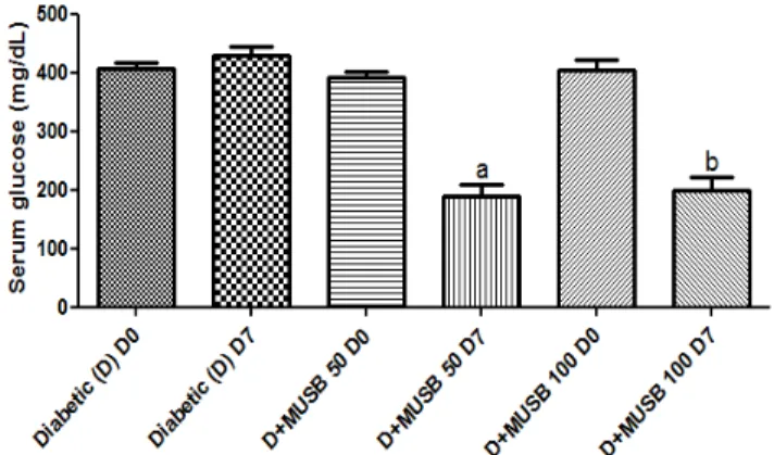
3.1. Blood glycemia. While no changes in blood glucose were observed in diabetic animals, at T0 in relation to T7, 44 and 38% reductions were demonstrated in diabetic animals after a 7-day treatment with MUSB 50 and 100 mg/kg, respectively, related to each group at T0 (Figure 1).
3.2. Blood cholesterol and triglycerides. Similar decreases (around 35%) were observed in cholesterol values, after 7-day treatments with MUSB, at both doses used (Figure 2). Higher decreases (74 and 75%) were observed in triglycerides (TG) levels from diabetic animals, after MUSB treatments with the doses of 50 and 100 mg/kg, respectively, in relation to T0 of each group (Figure 3).
3.3. Blood liver transaminases. Although, for all groups tested (diabetic rats untreated and treated with
MUSB, 50 and 100 mg/kg) there was an increase in concentrations of both AST and ALT, these changes were in the range of reference values for the animal species used in the present work (Figures 4 and 5).
Figure 1: Myracrodruon urundeuvadecoction (MUSB) treatments, for 7 days, decreases blood glucose levels in alloxan-induced diabetic rats (D). a. vs. D+MUSB-50 T0, q=11.49, p<0.001; b. vs. D+MUSB-100 T0, q=9.702, p<0.001. T0= 48 h after alloxan-induced diabetes (One way ANOVA and Tukey as the post hoc test).
Figure 2: Myracrodruon urundeuvadecoction (MUSB) treatments, for 7 days,decreases blood cholesterol levels in alloxan-induced diabetic rats (D). a. vs. D+MUSB-50 T0, q=6.865, p<0.001; b. vs. D+MUSB-100 T0, q=5.477, p<0.01. T0=48 h after alloxan-induced diabetes (One way ANOVA and Tukey as the post hoc test).
Glob. J. Med. Plant Res. 347
Figure 4: Myracrodruon urundeuvadecoction (MUSB) treatments, for 7 days, did not reverse the increase in AST levels observed in the alloxan-induced diabetic rats (D). a. vs. Diabetic (D) T0, q= 4.121, p<0.05; b. vs. D+MUSB-100 T0, q=4.308. T0=48 h after alloxan-induced diabetes (One way ANOVA and Tukey as the post hoc test).
Figure 5: Myracrodruon urundeuva decoction (MUSB-100) treatments, for 7 days, did not alter ALT levels observed in the alloxan-induced diabetic rats, D (One way ANOVA and Tukey as the
post hoc test).
3.4. Histopathological analyses (HE staining).
Pancreas from controls showed preserved acini and normal islets, while diabetic rats presented pancreatic islets with atresia and reduced size. Interestingly, the diabetic animals after treatment with MUSB, at both doses (50 and 100 mg/kg), showed pancreatic islets with a proliferative aspect (Figure 6). The liver microscopic analyses of the control group showed, in general, a normal histological pattern, but also some perivascular inflammatory infiltrate and congested vessels, while the diabetic group showed congested vessels, interstitial edema, sinusoidal dilatation and inflammatory infiltration. The diabetic animals after treatment with MUSB, at the lower dose (50 mg/kg), sometimes showed congested vessels, sinusoidal dilatation, intersticial edema and inflammatory infiltrate. However, normal lobuli and septa were usually observed, as well as a normal portal triad and normal hepatocytes. The 100 mg/kg-treated group showed congested vessels and interstitial hemorrhage, sinusoidal dilatation and inflammatory infiltrate. All these
liver alterations after the MUSB treatments were however of less intensity, in relation to the diabetic liver
Figure 6: Representative photomicrographs (HE, x400) of normal and alloxan-induced diabetic rat pancreas, untreated (D) or treated with MUSB (50 and 100 mg/kg, p.o., 7 days). The untreated diabetic pancreas (D) shows atresic and congested islets with reduced size. In diabetic rat pancreas after MUSB treatments, islets of Langerhans appear with proliferative characteristics. T0=48 h after diabetes induction and T7=7 days latter (Nikon Eclipse Nis, Software Nis 4.0, scale=100 and 200 μm).
without treatment (Figure 7). While kidneys from the control group showed normal features, the diabetic group presented tubular tumefaction, interstitial and intratubular hemorrhage, congested interstitial capillary, vacuolar degeneration and tubular cells degeneration. The diabetic group treated with MUSB, at the dose of 50 mg/kg, showed interstitial hemorrhage, congested glomerular capilaries and vacuolized cells of the dense macula detaching from the basal membrane, small cytoplasmic vacuoles in the proximal contorted tubules, discrete inflammatory infiltrate and a discontinuous tubular epithelium. A similar pattern, although with less changes, was observed in diabetic rats after MUSB treatment, at the dose of 100 mg/kg. Thus, the kidneys showed vacuolar and glomerular degeneration with discontinuous Bowman capsule cells, intratubular proteic material deposition, interstitial hemorrhage, edema and vacuole and tubular degeneration (Figures 8A and 8B).
Moreira et al., 348
from the normal controls, after MUSB treatments with both doses (Figure 9B).
Figure 7: Representative photomicrographs (HE, x400) of normal and alloxan-induceddiabetic rat liver, untreated (D) or treated with MUSB (50 and 100 mg/kg, p.o., 7 days). In the untreated diabetic liver, there are inflammatory cells in the space of Disse and hepatocytes with pyknotic nuclei. After MUSB-50 and MUSB-100 treatments inflammatory infiltrates with mast cells and lymphocytes were still observed (Nikon Eclipse Nis, Software Nis 4.0, scale=20 μm).
Figure 8: Representative photomicrographs (HE, x400) of normal and alloxan-induced diabetic rat kidney, untreated (D) or treated with MUSB (50 and 100 mg/kg, p.o., 7 days). A. renal medulla: In the untreated diabetic kidney, there are hemorrhage, distal convoluted tubule with tumefaction and vacuolar degeneration of epithelial cells. After MUSB treatments, intersticial hemorrhage and hydropic degeneration of renal tubular epithelium are shown. B. renal cortex: In the untreated diabetic kidney, there are intersticial congested vessels. After MUSB treatments, there are glomeruli tumefaction, vacuolar degeneration and intraglomerular hemorrhage (Nikon Eclipse Nis, Software Nis 4.0, scale=20 μm).
Figure 9: Myracrodruon urundeuva(MUSB-100) treatments, for 7 days, decreases iNOS and COX-2 immunoreactivity in pancreas from alloxan-induced diabetic rats (D). iNOS: a. vs. normal controls, q=18.08, p<0.001; b. vs. D+MUSB 50, q=9.379;p<0.001; c. vs. D+MUSB 100, q=15.31, p<0.001; d. vs. normal controls, q=7.363; p<0.01; e. vs. D+MUSB 100, q=5.553, p<0.05. COX-2: a. vs. normal controls, q=22.66, p<0.001; b. vs. D+MUSB 50, q=26.28, p<0.001; c. vs. D+MUSB 100, q=26.13, p<0.001 (One way ANOVA and Tukey as the post hoc test).
DISCUSSION
Type 2 diabetes (T2D), the most common form of the disease, is characterized by insufficient secretion of insulin from pancreatic beta-cells, coupled with impaired insulin action in muscles, liver and adipose tissue, named as insulin resistance. The impaired insulin secretion could be due to a decline in beta-cells secretory rate or to a decrease in beta-cell mass or both [23]. The increasing prevalence of T2D throughout the world has had a major impact on the development of diabetic kidney disease, one of the most frequent complications of diabetes [24]. Besides, renal complications are an important contributor to mortality in patients with T2D [25].
Glob. J. Med. Plant Res. 349
Diabetes is mainly due to oxidative stress increasing reactive oxygen species that can have major effects. Many plants contain different natural antioxidants, in particular tannins, flavonoids, C and E vitamins, that
have the ability to maintain β-cells performance and to
decrease glucose levels in the blood. Both experimental and clinical studies suggest that oxidative stress plays a major role in the pathogenesis of both types of diabetes
mellitus. This oxidative stress leads to β-cell destruction
by apoptosis. The antidiabetic properties of several medicinal species are mediated through their antioxidant and/or antiapoptotic properties in a streptozotocin induced stress model [28]. Furthermore, diabetes induces oxidative stress in the liver, that is characterized by increased concentration of reactive oxygen species (ROS) in the tissue and significant reduction in its antioxidant defenses. Such oxidative unbalance in liver cells may play a relevant role in the genesis of the chronic liver disease associated with diabetes [29].
In the present work, we showed for the first time the hypoglycemic effect of M. urundeuva in the model of alloxan-induced diabetes in rats. The aqueous extract (decoction, MUSB) was administered orally for 7 days, at the doses of 50 and 100 mg/kg. This medicinal species is well know for its anti-inflammatory properties, as evaluated in acute models of inflammation [11,12] and these effects seem to be, at least partly, related to inhibition of pro-inflammatory cytokines, as TNF-alpha [16]. Interestingly, M. urundeuva belongs to the Anacardiaceae family, as Anacardium occidentale and
Mangira indica, medicinal species known for their antidiabetic activity [30, 31, 32, 33].
Furthermore, M. urundeuva, at the present study both doses, significantly decreased total cholesterol and triglyceride contents in the serum of diabetic animals, after a 7-day treatment. Cholesterol is important to health, but can be harmful when its levels are too high, contributing to the narrowing of arteries. Unfortunately, people with diabetes are more prone to having unhealthy high cholesterol levels, what contributes to cardiovascular disease. In addition, triglycerides are the most common type of fat in the body. A high triglyceride level, combined with low HDL cholesterol or high LDL cholesterol, is associated with atherosclerosis and the build up of fatty deposits in artery walls that increase the risk for heart attack, peripheral artery disease and stroke [34].
Diabetes is associated with a high risk of atherosclerosis and cardiovascular disease and, thus, it is the leading cause of death among patients with type 2 diabetes. T2D lipid abnormalities are defined by a high concentration of TG and a low concentration of HDL cholesterol [34]. Abnormalities in lipid metabolism in T2D are among the major factors contributing to an increased cardiovascular risk. The primary quantitative lipoprotein abnormalities are increased TG levels and decreased HDL-cholesterol levels [35]. Thus, the treatment of dyslipidemia in insulin resistant patients with T2D has
been successful in reducing cardiovascular disease (CVD). CVD is a major cause of morbidity and mortality in T2D patients, with two- to four-fold increases, when compared with non-diabetic individuals. Thus, LDL cholesterol, TG, and HDL cholesterol are all appropriate targets for therapy [36], pointing out the importance of
new andsafer therapeutic strategies.
In the present study, the histopathological changes observed in pancreas, liver and kidneys from diabetic rats, after MUSB treatments, were in general of less intensity, in relation to those of diabetic groups without
treatment. Interestingly, while diabetic rats presented
pancreatic islets with atresia and reduced size, diabetic animals after MUSB treatments showed beta-cells from pancreatic islets in a proliferative state.
The regulation of beta-cell mass represents a critical issue for understanding diabetes, a disease characterized by a near-absolute (T1D) or relative (T2D) deficiency in the number of pancreatic beta-cells. The low capacity for self-replication in the adult is limited and does not result in significant regeneration, following extensive tissue injury. Thus, chronically increased metabolic demands also can lead to beta-cell failure. Furthermore, the stimulation of beta-cell replication, inhibition of cell apoptosis, and induction of beta-cell neogenesis are probably the best options for pharmacological restoration of a functional beta-cell mass, in diabetes. Pharmacological regulation of the beta-cell mass, therefore, represents another interesting target for the treatment of T2D patients [37].
A growing body of evidence [38- 43] points out to the importance of beta-cell mass to T2D development and progression. Beta-cell dysfunction in T2D patients might be induced by beta-cell loss and/or its functional defects. A very important point is that most people with insulin resistance will never develop T2D, which only results if the patients beta-cells fail to provide sufficient insulin [42]. Furthermore, the production of interleukin-1beta by beta-cells have been linked to beta-cells death in T2D [44].
Moreira et al., 350
COX-2 is upregulated in a variety of pathological conditions, including diabetes, and this event is associated with activation of downstream inflammatory reactions [48, 49]. Earlier [50], the administration of a selective COX-2 inhibitor was shown to prevent the onset of diabetes in mice. We observed an almost 3-times increase in COX-2 immunoreactivity in the diabetic pancreas, in relation to normal controls, and this increase was completely reversed after MUSB treatments. This could be justified by the anti-inflammatory effects of M. urundeuva, as earlier demonstrated by us [11, 12].
Diabetes is a global health problem and an economic burden. Although several antidiabetic drugs are available, the need for novel therapeutic agents with improved efficacy and few side-effects remains. Drugs derived from natural compounds are more attractive than synthetic drugs, because of their diversity and minimal side-effects. Published data suggest that natural compounds directly enhance insulin secretion, prevent pancreatic beta-cell apoptosis and modulate pancreatic beta-cell differentiation and proliferation [51]. Thus, it is essential to continuously investigate natural compounds as sources of novel pharmaceuticals, and to investigate their mechanisms of action, seeking the development of potential and safer antidiabetics.
Many plants are natural antioxidants and effective herbal medicines, in part due to their antidiabetic compounds, such as flavonoids, tannins, phenolics, and alkaloids, that improve the performance of pancreatic tissues by increasing the insulin secretion or decreasing the intestinal absorption of glucose and tannins. Phenolic compounds are present in M. urundeuva stem bark. Furthermore, natural and synthetic chalcones, as well, show antidiabetic and hypolipidemic effects [52-55]. In the present study, we showed that, by causing beta-cell proliferation and presenting an anti-inflammatory activity, M. urundeuva is a potential candidate to be considered in translational studies for the adjuvant treatment of T2D.
Conflict of interests
The authors declare no conflict of interests.
ACKNOWLEDGMENTS
The authors thank the financial support from the Brazilian National Research Council (CNPq) and the Foundation for the Support of Technological and Scientific Development of the State of Ceará (FUNCAP). The authors are also grateful to Prof. M.O.L. Viana for the ortographic revision of the manuscript.
REFERENCES
1. WHO. Global report on diabetes (2016).
2. Chen L, Magliano DJ, Zimmet PZ (2012). The worldwide epidemiology of type 2 diabetes mellitus - present and future perspectives. Nat Rev Endocrinol. 8:228-236.
3. Prentki M, Nolan CJ (2006). Islet beta cell failure in type 2 diabetes. J. Clin. Invest. 116: 1802–1812.
4. Thorens B (2013). The required beta cell research for improving treatment of type 2 diabetes. J. Intern. Med. 274:203-214. 5. Vetere, A, Choudhary,A, Burns, SM, Wagner, BK (2014).
Targeting the pancreatic Β-cell to treat diabetes. Nature Rev.
Drug Disc. 13:278–289.
6. Jung KY, Kim KM, Lim S (2014). Therapeutic approaches for preserving or restoring pancreatic β-cell function and mass. Diabetes Metab. J. 38:426-436.
7. Song I, Muller C, Louw J, Bouwens L (2015). Regulating the beta cell mass as a strategy fot ype-2 diabetes treatment. Curr. Drug Targets. 16:516-524.
8. Albuquerque UP (2006). Re-examining hypotheses concerning the use and knowledge of medicinal plants: a study in the Caatinga vegetation of the NE Brazil. J. Ethnobiol. Ethnomed. 2006; 2:30. Doi:10.1186/1746-4269-2-30.
9. Coutinho PC, Soares ZA, Ferreira EC, Souza D, Oliveira RS, Lucena RFPL (2015). Knowledge and use of medicinal plants in the semi-arid region of Brazil. Braz. J. Biol. Sci. 3: 51-74. 10. Penido AB, Morais SM, Ribeiro AB, Silva AZ (2016).
Ethnobotanical study of medicinal plants in Imperatriz, State of Maranhão, Northeast Brazil. Acta Amazonica. 46: 345-354. 11. Viana GSB, Bandeira MAM, Moura LC, Souza-Filho MVP, Matos
FJA, Ribeiro RA (1997). Analgesic and anti-inflammatory effects of the tannin fraction from Myracrodruon urundeuva Fr. All. Phytother. Res. 11: 118-122.
12. Viana GS, Bandeira MA, Matos FJ (2003). Analgesic and anti-inflammatory effects of chalcones isolated from Myracrodruon urundeuva Allemão. Phytomedicine. 10:189-95.
13. Souza SM, Aquino LC, Milach AC Jr., Bandeira MA, Nobre ME, Viana GS (2007). Anti-inflammatory and antiulcer properties of tannins from Myracrodruon urundeuva Allemão (Anacardiaceae) in rodents. Phytother. Res. 21:220-225. 14. Albuquerque RJM, Leal LKAM, Bandeira MA, Viana GSB,
Rodrigues LV (2011). Chalcones from Myracrodruon urundeuva
are efficacious in guinea pig ovalbumin-induced allergic conjunctivitis. Braz. J. Pharmacog. 21: 953-962.
15. Nobre-Júnior HV, Oliveira RA, Maia FD, Nogueira MAS, Moraes MO, Bandeira MAM, Andrade GM, Viana GSB (2009). Neuroprotective effects of chalcones from Myracrodruon urundeuva on 6-hydroxydopamine-induced cytotoxicity in rat mesencephalic cells. Neurochem. Res. 34:1066-1075.
16. Calou IBF, Bandeira MA, Aguiar-Galvão W, Cerqueira GS, Siqueira R, Neves KRT, Brito GAC, Viana GSB (2014). Neuroprotective Properties of a standardized extract from
Myracrodruon urundeuva Fr. All. (Aroeira-Do-Sertão), as evaluated by a Parkinson’s disease model in rats. Parkinson’s
Disease Volume 2014, Article ID
519615.http://dx.doi.org/10.1155/2014/519615.
17. Hohmeier HE, Tran VV, Chen G, Gasa R, Newgard CB (2003). Inflammatory mechanism in diabetes: lessons from β-cell. Int J Obes Relat. Metabol. Disord. Suppl. 3S12-16.
18. Spranger J, Kroke A, Möhlig M, Hoffmann K, Bergmann MM, Ristow M, Boeing H, Pfeiffer AFH (2003). Inflammatory cytokines and the risk to develop type 2 diabetes: results of the prospective population-based European Prospective Investigation into Cancer and Nutrition (EPIC)–Potsdam Study. Diabetes. 52, 812-817.
19. Aquino F G, Walter BMT, Ribeiro JF (2007). Espécies vegetais de uso múltiplo em reservas legais de cerrado - Balsas, MA. Rev. Bras. Biociênc. 5: 147-149.
20. Szkudelski T (2001). The mechanism of alloxan and streptozotocin action in β-cells of the rat pancreas. Physiol. Res. 50:536-546.
21. Lenzen S (2008). The mechanisms of alloxan- and streptozotocin-induced diabetes. Diabetologia. 51:216-226.
Glob. J. Med. Plant Res. 351
science to clinical practice. Cardiovasc. Diabetology. 2005, 4:5 DOI: 10.1186/1475-2840-4-5
23. Cantley J, Ashcroft FM (2015). Q&A: insulin secretion and type 2
diabetes: why do β-cells fail? BMC Biology. 15;13:33 DOI:
10.1186/s12915-015-0140-6
24. Tuttle KR, Bakris GL, Bilous RW, Chiang JL, de Boer IH, Goldstein-Fuchs J, Hirsch IB, Kalantar-Zadeh K, Narva AS, Navaneethan SD, Neumiller JJ, Patel UD, Ratner RE, Whaley-Connell AT, Molitch ME (2014). Diabetic kidney disease: a report from an ADA Consensus Conference. Diabetes Care. 37:2864-83.
25. Carney EF (2016). Renal complications and excess mortality in type 2 diabetes mellitus. Nature Rev. Nephrol. 12, 2.
26. Lucchesi AN, Cassettari LL, Spadella CT (2015). Alloxan-induced diabetes causes morphological and ultrastructural changes in rat liver that resemble the natural history of chronic fatty liver disease in humans. J. Diabetes Res. 2015, Art. ID 494578, 11 pages. http://dx.doi.org/10.1155/2015/494578.
27. Harris EH (2005). Elevated liver function tests in Type 2 diabetes. Clin. Diabetes 23:3.
28. Kalekar SA, Munshi RP, Thatte UM (2013). Do plants mediate their anti-diabetic effects through anti-oxidant and anti-apoptotic actions? an in vitro assay of 3 Indian medicinal plants. BMC Complement. Altern. Med. 13: 257.
29. Lucchesi AN, Freitas NT, Cassettari LL, Marques SFG, Spadella ST (2013). Diabetes mellitus triggers oxidative stress in the liver of alloxan-treated rats: a mechanism for diabetic chronic liver disease. Acta Cirúrgica Brasileira. 28: 502-508.
30.Ojewole JAO (2005). Antiinflammatory, analgesic and hypoglycemic effects of Mangifera indica Linn. (Anacardiaceae) stem‐bark aqueous extract. Methods Find.Exp. Clin. Pharmacol. 27:547–554.
31. Shah KA, Patel MB, Patel RJ, Parmar PK (2010). Mangifera Indica (Mango) Pharmacogn. Rev. 4: 42–48.
32. Patel DK, Prasad SK, Kumar R, Hemalatha S (2012). An overview on antidiabetic medicinal plants having insulin mimetic property Asian Pac. J. Trop. Biomed. 2: 320–330.
33. Jaiswal YS, Tatke PA, Gabhe SY, Vaidya AB (2016). Antidiabetic activity of extracts of Anacardium occidentale Linn. Leaves on n-streptozotocin diabetic rats. J. Tradit. Complem. Med. 2016. http://dx.doi.org/10.1016/j.jtcme.2016.11.007.
34. Bitzur R, Chen H, Kamari Y, Shaish A, Harats D (2009). Triglycerides and HDL cholesterol. Diabetes Care. 32(suppl 2): S373-S377.
35. Vergés B (2015). Pathophysiology of diabetic dyslipidaemia: where are we? Diabetologia. 58:886–899
36. Ginsberg HN, Zhang Y-L, Hernandez-Ono A (2005). Regulation of plasma triglycerides in insulin resistance and diabetes. Arch. Med. Res. 36: 232–240.
37. Bouwens L, Rooman I (2005). Regulation of pancreatic beta-cell mass. Physiol. Rev. 85: 255-70.
38. Ahrén B (2005). Type 2 diabetes, insulin secretion and beta-cell mass. Curr Mol Med. 5: 275-86.
39. Baggio LL, Drucker DJ (2006). Therapeutic approaches to preserve islet mass in type 2 diabetes. Ann. Rev. Med. 57:265-81.
40. Matveyenko AV, Veldhuis JD, Butler PC (2008). Adaptations in pulsatile insulin secretion, hepatic insulin clearance, and beta-cell mass to age-related insulin resistance in rats. Am. J. Physiol. Endocrinol. Metab. 295(4):E832-41.
41. Cho J-H, Kim J-W, Shin J-A, Shin J, Yoon K-H (2011). β‐cell mass in people with type 2. J. Diabetes Investig. 24: 2(1): 6–17. 42. Weir GC, Bonner-Weir S (2013). Islet β cell mass in diabetes and
how it relates to function, birth, and death. Ann. N.Y. Acad. Sci. 1281:92-105.
43. Meier JJ, Bonadonna RC (2013). Role of reduced β-cell mass versus impaired β-cell function in the pathogenesis of type 2 diabetes. Diabetes Care. 36 (Suppl 2): S113–S119.
44. Donath MY, Shoelson SE (2011). Type 2 diabetes as an inflammatory disease. Nat. Rev. Immunol. 11:98–107.
45. Fujimoto M, Shimizu N, Kunii K, Martyn JA, Ueki K, Kaneki M (2005). A role for iNOS in fasting hyperglycemia and impaired insulin signaling in the liver of obese diabetic mice. Diabetes. 54:1340-1348.
46. Soskié S, Dobutovié BD, Sudar EM, Obradovié MM, Nikolié DM, Djordjevic JD, Radak DJ, Mikhailidis DP, Isenovié ER (2011). Regulation of inducible nitric oxide synthase (iNOS) and its potential role in insulin resistance, diabetes and heart failure. The Open Cardiovasc Med J. 5:153-163.
47. Keklikoglu N, Akinci S (2013). The role of iNOS in beta cell destruction in diabetes. Oxid Antioxid Med Sci. 2:251-254. 48. Bagi Z, Erdei N, Papp Z, Edes I, Koller A (2006). Up-regulation of
vascular cyclooxygenase-2 in diabetes mellitus. Pharmacol Rep. 58: Suppl:52-56.
49. Kellog AP, Cheng HT, Pop-Busui R (2008). Cyclooxygenase-2 pathway as a potential therapeutic target in diabetic peripheral neuropathy. Curr Drug Targets. 9:68-76.
50. Tabatabaie T, Waldon AM, Jacob JM, Floyd RA, Kotake Y (2000). COX-2 inhibition prevents insulin-dependent diabetes in low-dose streptozotocin-treated mice, Biochem. Biophys. Res. Commun. 273:699-704.
51. Oh YS (2015). Plant-derived compounds targeting pancreatic beta cells for the treatment of diabetes. Evidence-Based Complementary and Alternative Medicine. Article ID 629863, 12 pages.http://dx.doi.org/10.1155/2015/629863
52. Enoki T, Ohnogi H, Nagamine K, Kudo Y, Sugiyama K, Tanabe M, Kobayashi E, Sagawa H, Kato I (2007). Antidiabetic activities of chalcones isolated from a Japanese herb, Angelica keiskei. J. Agric. Food Chem. 55:6013-17.
53. LI Xiang-hua, ZOU Han-jun, WU, An-hui, YE, Yang-liang, SHEN, Jian-hua (2007). Structure-based drug design of a novel family of
chalcones as PPARα agonists: virtual screening, synthesis,
and biological activities in vitro. Acta Pharmacol Sin. 28: 2040–2052.
54. Hsieh CT, Hsieh TJ, El-Shazly M, Chuang DW, Tsai YH, Yen CT, Wu SF, Wu YC, Chang FR (2012). Synthesis of chalcone derivatives as potential anti-diabetic agents. Biorg. Med. Chem. Lett. 22: 3012-3915.