REVISÃO / REVIEW
INTRODUCTION
The prevalence of early cancers (invading no deeper than the submucosa) has increased, especially in Asian countries, where upper digestive screening is common(14,31). Similarly, in the United States, there has been an increased diagnosis of early lesions starting in the Barrett’s esophagus(1,16,29).
Disease conined to the esophagus or local disease is present in 22% of the cases, and a minority of the patients have disease that is limited to the mucosa or submucosa (T1N0M0). An esophagectomy is the irst line of therapy for patients with stage T1N0M0. For patients with a localized and potentially resectable form of the disease, the median survival strongly correlates with disease stage, which is where the goal of surgery is the curative resection R0(2,5,14,31). Advanced age and comorbidities increase the risk of postoperative cardiorespiratory complications, anastomotic leakage, reoperation rates, wound infection, and death(15). Endoscopic resection (ER)
SURGERY VERSUS ENDOSCOPIC
THERAPIES FOR EARLY CANCER AND
HIGH-GRADE DYSPLASIA IN THE
ESOPHAGUS: a systematic review
Fabio Alberto Castillo
BUSTAMANTE
1, Eduardo Guimarães
HOURNEAUX DE MOURA
1,
Wanderley
BERNARDO
2, Rubens Antonio Aissar
SALLUM
3, Edson
IDE
1and Elisa
BABA
1Received 17/8/2015 Accepted 19/9/2015
ABSTRACT - Background - Esophageal cancer occurs as a local disease in 22% of cases, and a minority of this disease is limited to the mucosa
or submucosa (early lesions). Endoscopic mucosal resection, endoscopic submucosal dissection, photodynamic therapy, laser therapy, and argon plasma coagulation have emerged as alternatives to surgical resection for early lesions. Objective - The aim of this systematic review is to identify studies that statistically compare survival, disease-free survival, morbidity and mortality associated with the procedure, and mor-tality associated with cancer in the endoscopic versus surgical therapies. Data sources - A systematic review using MEDLINE, COCHRANE, EMBASE, EBSCO, LILACS, Library University of Sao Paulo, BVS, and SCOPE. Study selection - Randomized controlled trial, controlled clinical trial, clinical trial, and cohort study. Criteria - Studies that statistically compare survival, disease-free survival, morbidity and mortality associated with the procedure, and mortality associated with cancer in patients who underwent endoscopic and surgical therapy for early lesions of esophageal cancer. Data extraction - Independent extraction of the articles by two authors using predeined data ields, including study quality indicators. Limitation - Only retrosprospective cohort studies comparing the endoscopic and surgical therapies were recovered. Results - The survival rates after 3 and 5 years were different and exhibited superiority with the surgical therapies over time. Endoscopy is superior in the control of mortality related to cancer with a high rate of disease recurrence. With regard to the comorbidity and the mortality associated with the procedure, endoscopy is superior. Conclusion - There is no evidence from clinical trials. In this systematic review, surgical therapies showed superiority for survival, and endoscopic therapies showed superiority in the control of mortality related to cancer with a high rate of disease recurrence; also, for the comorbidity and the mortality associated with the procedure, endoscopy is superior. Prospective, controlled trials with large sample sizes are necessary to conirm the results of the current analysis.
HEADINGS- Esophageal neoplasms, surgery. Gastrointestinal endoscopy. Morbidity. Review.
Declared conflict of interest of all authors: none Disclosure of funding: no funding received
1 Serviço de Endoscopia Gastrointestinal, Departamento de Gastroenterologia, Hospital das Clínicas, Faculdade de Medicina Universidade de São Paulo, São Paulo, SP,
Brasil; 2 Projeto Diretrizes, Associação Médica Brasileira, São Paulo, SP, Brasil; 3 Divisão de Cirurgia Esofágica, Departamento de Gastroenterologia, Hospital das Clinicas
Universidade de São Paulo, São Paulo, SP, Brasil.
Correspondence: Fabio Alberto Castillo Bustamante. Serviço de Endoscopia Gastrointestinal, Departamento de Gastroenterologia, Hospital das Clinicas, Faculdade de Medicina, Universidade de São Paulo (FM-USP). Av. Dr. Enéas de Carvalho Aguiar, 255, 6º andar, Bloco 3 – CEP: 05403-000 – São Paulo, SP, Brasil. E-mail: facastillobu@gmail.com
is an alternative to surgical resection of the mucosal and submucosal neoplastic lesions and has the beneit of not having major surgical complications. Endoscopic mucosal resection (EMR), endoscopic submucosal dissection (ESD), photodynamic therapy (PDT), laser therapy, and argon plasma coagulation (APC) have been developed as alternatives to surgical resection for early lesions. The benefit of these therapies is obvious and has been supported by multiple case study publications, as well as systematic reviews, with no statistical data analysis. Therefore, the beneits of these therapies compared with surgery are still not clear(3,4,11,17,27).
OBJECTIVES
METHODS
PROSPERO 2014: CRD42014013170(33).
Eligibility criteria
Types of studies: (Randomized Controlled Trial, Con-trolled Clinical Trial, Comparative Study).
Types of participants: Early esophageal cancers that are classiied as Tis (high-grade dysplasia, which includes all noninvasive neoplastic epithelial that was formerly called carcinoma in situ) and T1 tumours, which are split into T1a and T1b subcategories depending on the depth of the invasion(9).
• M1 - Limited to the epithelial layer • M2 - Invades the lamina propria
• M3 - Invades into but not through the muscularis mucosa
M1 tumours correspond to the Tis stage in the AJCC stage deinition, while both M2 and M3 tumours would be considered T1a lesions.
Tumours invading the submucosa are subclassiied as follows(28):
• SM1 - Penetrates the shallowest one-third of the sub-mucosa
• SM2 - Penetrates into the intermediate one-third of the submucosa
• SM3 - Penetrates the deepest one-third of the submucosa All of these subcategories would be considered T1b disease according to the AJCC stage deinitions(8). Early esophageal cancer is considered to be T1b SM1 (200 µ inva-sion to the submucosa).
Types of intervention: esophagectomy or any approach compared with endoscopic mucosal resection (EMR), endoscopic submucosal dissection (ESD), photodynamic therapy (PDT), laser therapy, and argon plasma coagulation (APC).
Types of outcome measures: statistically compare survival, disease-free survival, the complications associated with the procedure (morbidity and mortality), and the mortality associated with cancer.
In this search, we do not limit ourselves by year of pub-lication or by language.
Search MEDLINE
P: (Esophageal Neoplasms OR Esophageal Neoplasm OR Neoplasm Esophageal OR Middle third submucosal layer OR Deep third submucosal layer OR Submucosal esophageal cancer AND ESOPHAG*) AND I: (Surgi-cal Procedures, Operative OR OREsophagectomy OR Esophagectomies) AND C: (Endoscopy OR Surgical Procedures, Endoscopic OR Gastrointestinal Surgery, En-doscopic OR Esophagogastroduodenoscopies).
EBSCO, EMBASE, LILACS, COCHRANE, LIBRARY UNIVERSITY OF SAO PAULO, BVS, SCOPE
P: Esophagus cancer AND early AND I: surgery AND C: endoscopy.
Study selection
Two reviewers performed the eligibility assessment, which was performed independently in an unblinded standardized manner. Each reviewer completed a thorough reading of the abstract to identify studies comparing endoscopic with surgical techniques. The studies that were selected were read in their entirety and were excluded according with
criteria from JADAD(13) NEWCASTLE OTTAWA(10) and
Methodology Check List SIGN(26). We excluded studies that did not report the results in absolute numbers. Disagreements between reviewers were resolved by consensus.
Data collection process
We extracted the data through a detailed reading of the results of each study, including the information on mortality at 3 and 5 years, disease-free survival at 5 years, complications associated with the procedure (mortality and morbidity), and mortality associated with cancer. We only included the absolute numbers reported in the text of the article or with the analysis of the graphs. We excluded an article for not reporting the results in absolute numbers. One review author extracted the data described below from the included studies, and the second author checked the extracted data. Disagree-ments were resolved by discussion between the two review authors; we did not contact authors for further information.
Data items
We select items from populations with early adenocar-cinoma or squamous cell cancer of the esophagus (EAC ESCC), and we understood these cancers as mucosal and submucosal M1, M2, M3, SM1, SM2, and SM3.
We compared endoscopic submucosal dissection, endoscopic mucosal resection, photodynamic therapy, and ablative therapies (EMR, ESD, PDT, TA) with esophagectomies by a transhiatal or a transthoracic approach and minimally invasive techniques (TH, TT, MI).
Risk of bias in the individual studies
The first approach to identify the risk of bias was undertaken using the NEWCASTLE OTTAWA (11) and Methodology Check List: Cohort studies SIGN(26).
Synthesis of results
We performed an analysis with the available software RevMan5(7) using the following characteristics: dichotomous data type, statistical method of Mantel-Haenszel, fixed effect model analysis, effect measure risk difference, study conidence interval 95%, total conidence interval 95%, and organized by year of study.
Risk of bias across the studies
RESULTS
Study selections
We identiied 9859 studies through the PUBMED search and through the COCHRANE, EMBASE, EBSCO, LILACS, Library University of Sao Paulo, BVS and SCOPE we found 9527 studies. After eliminating the repeat studies, we identi-ied a total of 16699 studies. We excluded studies that did not have information concerning early esophagus cancer. NO PROSPECTIVE CLINICAL TRIAL WAS FOUND. We chose 20 studies that we reviewed with the NEWCASTLE OTTAWA and the Methodology Check List: Cohort studies SIGN and excluded 7 of these studies. We included 13 studies for the qualitative analysis and 12 studies for the quantitative analysis (Figure 1).
Study characteristics
The characteristics of the studies are summarized in Table 1 and describe the oncologic diagnosis analysed. The number of interventions for each treatment group and the report in absolute numbers gives each outcome of interest extracted.
TABLE 1. Study characteristics
Study Population Intervention (MoAP)*MAP* year (MAC)*Survival 3 Survival 5 year (MAC)* DFS*
Merkow RP, 2014 (18)
M1-2-3 SM1-2-3 EAC ESCC 04-10
E 1428 (EMR ESD TA) 7 1092
S 3962 (NS)* 139 3470
Wani S, 2014 (32)
M1-2-3 SM1-2-3 EAC ESCC 98-09
E 430 (NS)* 318 266
S 1586 (NS)* 1237 1110
Ngamruengphong, S 2013 (20)
M1-2-3 SM1-2-3 EAC 98-09
E 306 (NS)* 223 177 (56) 248 5y
S 1312 (NS)* 1023 918 (289) 1023 5y
Tian J, 2011 (30)
SM1-2-3 EAC 95-10
E29 (EMR) 19 17
S39 (TT TH) 35 31
Pech O, 2011 (22)
M1-2-3 EAC 96-09
E 76 (NS)* 0 73 68 69-5y
S 38 (TT) 1 35 35 38-5y
Zehetner J, 2011 (34)
M1-2-3 EAC 01/out
E 40 (EMR PDT) 0 (0) 38 (0) 0
S 61 (TH TT MI VSR) 0 (16) 57 (0) 7
Prasad GA, 2009 (24)
M1-2-3 EAC 98-07
E 132 (EMR PDT) 0(18) 109 (1) 105-5y
S 46 (TH TT) 1(17) 43 (1) 45-5y
Das A, 2008 (6)
M1-2-3 SM1-2-3 EAC ESCC 98-03
E 99 (EMR PDT TA) 76 76
S 643 (NS)* 521 502
Schembre DB, 2008 (25)
M1-2-3 EAC 98-05
E 62 (EMR APC PDT) 1 55 (0)
S 32 (NS)* 0 30 (0)
Prasad GA, 2007 (23)
M1 EAC 94-04
E 129 (EMR PDT) 0 122 118 (0) 110-5y
S 70 (TT TH) 1 (27) 67 64 (0) 66-5y
Paciico RJ, 2003 (21)
M1-2-3 SM1-2-3 EAC 96-01
E 24 (EMR PDT) 0(4) 20-1y
S 64 (NS)* 1(34) 64-1y
Fujita H, 2001 (9)
M1-2-3 ESCC 81-97
E 37 (EMR) 0 (2) 26(0) 22 (0)
S 35 (TT TH) 5 (24) 26(0) 24 (0)
M: mucosa; SM: submucosa; EAC: early adenocarcinoma; ESCC: squamous cell carcinoma; EMR: endoscopic mucosal resection; ESD: endoscopic submucosal dissection; TA: trans abdominal; TH: trans hiatal; TT: trans thoracic; MI: minimally invasive; MAP: mortality associate to procedure; MoAP: morbidity associated to procedure; MAC: mortality associated to cancer; DFS: disease free survival; NS not speciied.
FIGURE 1. Flow diagram studies selection(19)
Records identified through PUBMED
(n = 9859)
Additional records identified through Cochrane Controlled Trials Register, EMBASE, EBSCO, LILIACS, Library University of São Paulo,
Research website BVS and SCOPE Science Direct.
Records in total excluding repeated (n = 16699)
Records excluded (n = 16625) Not early esophageal
Records excluded (n = 55) Not comparing surgery with
endoscopic therapy
Full-text articles excluded (n = 7) Poor rating in NEWCASTLE OTTAWA and Methodology Check List: Cohort
studies SIGN
Full-text articles excluded (n = 1) No results in absolute numbers Records screened
(n = 74)
Full-text articles assessed for eligibility
(n = 20)
Studies included in qualitative synthesis
(n = 13)
Studies included in quantitative synthesis
(n = 12)
Risk of bias within the studies
The irst approach to identify risk of bias was undertaken
using the NEWCASTLE OTTAWA(10) and Methodology
Check List: Cohort studies SIGN(26). Because all of the studies were retrospective cohorts from hospital care data networks, the rate was generally good.
RESULTS OF THE INDIVIDUAL STUDIES AND THE SYNTHESIS OF THE RESULTS
3-year survival
Risk Difference in 3-year survival for the endoscopy and surgery therapies.
Nine studies reported the 3-year survival, and the heterogeneity test indicated χ2=9.35 and I2=14%, which demonstrated a insigniicant heterogeneity. The ixed-effects model was adopted, and the RD was -0.04 (95% CI: -0.07, –0.01) (Figure 2). An analysis of the pooled data revealed differences between the two therapies. There was a statistically signiicant increase in survival with the surgical therapy.
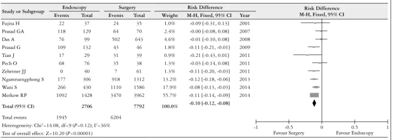
5-year survival
Risk Difference in 5-year survival for the endoscopy and surgery therapies.
Ten studies reported the 5-year survival, and the heteroge-neity test indicated χ2=14.08 and I2=36%, which demonstrat-ed a low heterogeneity. The ixdemonstrat-ed-effects model was adoptdemonstrat-ed, and the RD was -0.10 (95% CI: -0.12, –0.08) (Figure 3). An analysis of the pooled data revealed differences between the two therapies. There was a statistically signiicant increase in survival with surgical therapy.
Disease-free survival after 5 years
Risk Difference in disease-free survival after 5 years for the endoscopy and surgery therapies.
Five studies reported disease-free survival after 5 years, and the heterogeneity test indicated χ2 =29.28 and I2 =86%, which demonstrated a high heterogeneity. The ixed-effects model was adopted, and the RD was -0.02 (95% CI: -0.06, –0.02) (Figure 4). In the funnel plot analysis, the study by Ngamruengphong S. (2013) was identified as a source of
FIGURE 2. Risk difference in 3 year survival, for endoscopy and surgery therapies
Study or Subgroup
Experimental Control Risk Difference
Risk Difference M-H, Fixed, 95% CI
Events Total Events Total Weight M-H, Fixed, 95% CI Year
Fujita H 26 37 26 35 2.2% -0.04 [-0.25, 0.17] 2001
Prasad GA 122 129 67 70 5.5% -0.01 [-0.07, 0.05] 2007
Schembre DB 55 62 30 32 2.6% -0.05 [-0.17, 0.06] 2008
Das A 76 99 521 643 10.4% -0.04 [-0.13, 0.05] 2008
Zehetner JJ 38 40 57 61 2.9% 0.02 [-0.08, 0.11] 2011
Tian J 19 29 35 39 2.0% -0.24 [-0.44, -0.04] 2011
Pech O 73 76 35 38 3.1% 0.04 [-0.06, 0.14] 2011
Ngamruengphong S 223 306 1023 1312 30.2% -0.05 [-0.11, 0.00] 2013
Wani S 318 430 1237 1586 41.1% -0.04 [-0.09, 0.01] 2014
Total (95% CI) 1208 3816 100.0% -0.04 [-0.07, -0.01]
Total events 950 3031
Heterogeneity: Chi2=9.35, df=8 (P=0.31); I2=14%
Test of overall effect: Z=2.96 (P=0.003) Favour Surgery Favour Endoscopy
-1 -0.5 0 0.5 1
FIGURE 3. Risk difference in 5 year survival, for endoscopy an surgery therapies
Study or Subgroup Endoscopy Surgery Risk Difference Risk Difference
M-H, Fixed, 95% CI
Events Total Events Total Weight M-H, Fixed, 95% CI Year
Fujita H 22 37 24 35 1.0% -0.09 [-0.31, 0.13] 2001
Prasad GA 118 129 64 70 2.4% -0.00 [-0.08, 0.08] 2007
Das A 76 99 502 643 4.6% -0.01 [-0.10, 0.08] 2008
Prasad G 109 132 43 46 1.8% -0.11 [-0.21, -0.01] 2009
Tian J 17 29 31 39 0.9% -0.21 [-0.43, 0.01] 2011
Pech O 68 76 35 38 1.3% -0.03 [-0.14, 0.08] 2011
Zehetner JJ 0 40 7 61 1.3% -0.11 [-0.20, -0.03] 2011
Ngamruengphong S 177 306 918 1312 13.2% -0.12 [-0.18, -0.06] 2013
Wani S 266 430 1110 1586 17.9% -0.08 [-0.13, -0.03] 2014
Merkow RP 1092 1428 3470 3962 55.7% -0.11 [-0.14, -0.09] 2014
Total (95% CI) 2706 7792 100.0% -0.10 [-0.12, -0.08]
Total events 1945 6204
Heterogeneity: Chi2=14.08, df=9 (P=0.12); I2=36%
heterogeneity; by consensus, the reviewers opted to withdraw this work. Four studies reported disease-free survival after 5 years, and the heterogeneity test indicated χ2=3.71 and I2=19%, which demonstrated a low level of heterogeneity. The ixed-effects model was adopted, and the RD was -0.13 (95% CI: -0.17, –0.08) (Figure 4). An analysis of the pooled data revealed differences in the disease-free survival in the ifth year and showed better outcomes for the surgical therapy.
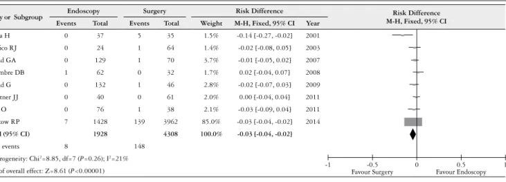
Mortality associated with the procedure
Risk Difference in the mortality associated with the pro-cedure for the endoscopy and surgery therapies.
Eight studies reported mortality associated with the procedure, and the heterogeneity test indicated χ2=8.85 and I2=21%, which demonstrated an insigniicant heterogeneity. The ixed-effects model was adopted, and the RD was -0.03 (95% CI: -0.04, –0.02) (Figure 5). An analysis of the pooled
data revealed differences between the two therapies. There was a statistically signiicant increase in mortality with the surgical therapy.
Morbidity associated with the procedure
Risk ratio in the morbidity associated with the procedure for the endoscopy and surgery therapies.
Four studies reported morbidity associated with the procedure, and the heterogeneity test indicated χ2=6.38 and I2=53%, which demonstrated a moderately high level of heterogeneity. The ixed-effects model was adopted, and the risk difference was -0.24 (95% CI: -0.31, -0.16) (Figure 6). In the funnel plot analysis, we identiied true heterogeneity, and the reviewers opted to perform a Risk Ratio analysis. Four studies reported morbidity associated with the procedure, and the analysis of the heterogeneity test indicated χ2=2.61 and I2=0% low heterogeneity. Therefore, the
ixed-FIGURE 4. Risk difference in disease-free survival 5 years, for endoscopy and surgery therapies
0.02
0.04
0.06
0.08
0.1
-1 -0.5 0 0.5 1
0 SE (RD)
Study or Subgroup Endoscopy Surgery Risk Difference Risk Difference
M-H, Fixed, 95% CI
Events Total Events Total Weight M-H, Fixed, 95% CI Year
Paciico RJ 20 24 64 64 14.3% -0.17 [-0.32, -0.01] 2003
Prasad GA 110 129 66 70 37.1% -0.09 [-0.17, -0.01] 2007
Prasad G 105 132 45 46 27.9% -0.18 [-0.26, -0.10] 2009
Pech O 69 76 38 38 20.7% -0.09 [-0.17, -0.02] 2011
Ngamruengphong S 248 306 1023 1312 0.0% 0.03 [-0.02, 0.08] 2013
Total (95% CI) 361 218 100.0% -0.13 [-0.17, -0.08]
Total events 304 213
Heterogeneity: Chi2=3.71, df=3 (P=0.29); I2=19%
Test of overall effect: Z=5.34 (P<0.00001) Favour Surgery Favour Endoscopy
-1 -0.5 0 0.5 1
Study or Subgroup Endoscopy Surgery Risk Difference Risk Difference
M-H, Fixed, 95% CI
Events Total Events Total Weight M-H, Fixed, 95% CI Year
Paciico RJ 20 24 64 64 4.7% -0.17 [-0.32, -0.01] 2003
Prasad GA 110 129 66 70 12.3% -0.09 [-0.17, -0.01] 2007
Prasad G 105 132 45 46 9.2% -0.18 [-0.26, -0.10] 2009
Pech O 69 76 38 38 6.8% -0.09 [-0.17, -0.02] 2011
Ngamruengphong S 248 306 1023 1312 67.0% 0.03 [-0.02, 0.08] 2013
Total (95% CI) 667 1530 100.0% -0.02 [-0.06, 0.02]
Total events 552 1236
Heterogeneity: Chi2=29.28, df=4 (P<0.00001); I2=86%
Test of overall effect: Z=1.15 (P=0.25) Favour Surgery Favour Endoscopy
-1 -0.5 0 0.5 1
effects model was adopted, and the Risk Ratio was 0.28 (95% CI: 0.18, 0.46) (Figure 6). An analysis of the pooled data revealed differences between the two therapies. There was a statistically significant increase in the morbidity associated with the surgical therapy procedure.
Associated cancer mortality at 5 years
Risk difference associated with cancer mortality at 5 years for the endoscopy and surgery therapies.
Five studies reported an associated cancer mortality at 5 years, and the heterogeneity test indicated χ2=16.57 and
FIGURE 5. Risk difference in mortality associated with the procedure, for endoscopy and surgery therapies
Study or Subgroup Endoscopy Surgery Risk Difference Risk Difference
M-H, Fixed, 95% CI
Events Total Events Total Weight M-H, Fixed, 95% CI Year
Fujita H 0 37 5 35 1.5% -0.14 [-0.27, -0.02] 2001
Paciico RJ 0 24 1 64 1.4% -0.02 [-0.08, 0.05] 2003
Prasad GA 0 129 1 70 3.7% -0.01 [-0.05, 0.02] 2007
Schembre DB 1 62 0 32 1.7% 0.02 [-0.04, 0.07] 2008
Prasad G 0 132 1 46 2.8% -0.02 [-0.07, 0.03] 2009
Zehetner JJ 0 40 0 61 2.0% 0.00 [-0.04, 0.04] 2011
Pech O 0 76 1 38 2.1% -0.03 [-0.09, 0.04] 2011
Merkow RP 7 1428 139 3962 85.0% -0.03 [-0.04, -0.02] 2014
Total (95% CI) 1928 4308 100.0% -0.03 [-0.04, -0.02]
Total events 8 148
Heterogeneity: Chi2=8.85, df=7 (P=0.26); I2=21%
Test of overall effect: Z=8.61 (P<0.00001) -1 Favour Surgery-0.5 0 Favour Endoscopy0.5 1
FIGURE 6. Risk ratio in morbidity associated with the procedure for endoscopy and surgery therapies
0.02
0.04
0.06
0.08
0.1
-1 -0.5 0 0.5 1
0 SE (RD)
Study or Subgroup Endoscopy Surgery Risk Difference Risk Difference
M-H, Fixed, 95% CI
Events Total Events Total Weight M-H, Fixed, 95% CI Year
Fujita H 2 37 5 35 8.3% 0.38 [0.08, 1.82] 2001
Paciico RJ 4 24 34 64 29.9% 0.31 [0.12, 0.79] 2003
Prasad G 18 132 17 46 40.6% 0.37 [0.21, 0.65] 2009
Zehetner JJ 0 40 16 61 21.2% 0.05 [0.00, 0.74] 2011
Total (95% CI) 233 206 100.0% 0.28 [0.18, 0.46]
Total events 24 72
Heterogeneity: Chi2=2.61 df=3 (P=0.46); I2=0%
Test of overall effect: Z=5.11 (P<0.00001) -1 Favour Surgery-0.5 0 Favour Endoscopy0.5 1
Study or Subgroup Endoscopy Surgery Risk Difference Risk Difference
M-H, Fixed, 95% CI
Events Total Events Total Weight M-H, Fixed, 95% CI Year
Fujita H 2 37 5 35 19.2% -0.09 [-0.23, 0.05] 2001
Paciico RJ 4 24 34 64 18.6% -0.36 [-0.56, -0.17] 2003
Prasad G 18 132 17 46 36.4% -0.23 [-0.38, -0.08] 2009
Zehetner JJ 0 40 16 61 25.8% -0.26 [-0.38, -0.15] 2011
Total (95% CI) 233 206 100.0% -0.24 [-0.31, -0.16]
Total events 24 72
Heterogeneity: Chi2=6.38 df=3 (P=0.09); I2=53%
Test of overall effect: Z=6.04 (P<0.00001) -1 Favour Surgery-0.5 0 Favour Endoscopy0.5 1
I2=76%, which demonstrated a high level of heterogeneity. Therefore, the fixed-effects model was adopted, and the RD was -0.04 (95% CI: -0.06, -0.02, P=0.0004) (Figure 7). In the funnel plot analysis, the study by Prasad GA (2007) was identiied as a source of heterogeneity. By consensus, the reviewers opted to withdraw this work. Four studies reported an associated cancer mortality at 5 years, and the heterogeneity test indicated χ2=4.57 and I2=34%, which demonstrated a low level of heterogeneity. Therefore, the fixed-effects model was adopted, and the RD was -0.04 (95% CI: -0.07, –0.02) (Figure 7). An analysis of the pooled data revealed differences between the two therapies. There was a statistically signiicant increase associated with cancer mortality at 5 years in the surgical therapy procedure.
Risk of bias across the studies
In relation to the heterogeneity across the studies because both the endoscopic and surgical treatment protocols are dif-ferent in various parts of the world, even more of the therapies described in each study were different. It is typical to identify combination therapies, including EMR, ESD, PDT, or ablative therapies, in one or more sessions. Additionally, the surgical techniques described are different in each study. There are predominantly open approaches (TT, TH) in the older studies and minimally invasive approaches in the recent studies (MI). In the analysis of the disease-free survival at 5 years, in the funnel plot and I2 analysis identiied publication bias in the study by Ngamruengphong S. (2013). After excluding this study, we observed a frank tendency to obtain better results
FIGURE 7. Risk difference in 5 years associated with câncer mortality for endoscopy and surgery therapies
0.02
0.04
0.06
0.08
0.1
-1 -0.5 0 0.5 1
0 SE (RD)
Study or Subgroup Experimental Control Risk Difference Risk Difference
M-H, Fixed, 95% CI
Events Total Events Total Weight M-H, Fixed, 95% CI Year
Fujita H 0 37 0 35 2.8% 0.00 [-0.05, 0.05] 2001
Prasad GA 0 129 0 70 0.0% 0.00 [-0.02, 0.02] 2007
Prasad G 1 132 1 46 5.3% -0.01 [-0.06, 0.03] 2009
Ngamruengphong S 56 306 289 1312 38.9% -0.04 [-0.09, 0.01] 2013
Wani S 21 430 159 1586 53.0% -0.05 [-0.08, -0.03] 2014
Total (95% CI) 905 2979 100.0% -0.04 [-0.07, -0.02]
Total events 78 449
Heterogeneity: Chi2=4.57 df=3 (P=0.21); I2=34%
Test of overall effect: Z=3.57 (P=0.0004) Favour Surgery Favour Endoscopy
-1 -0.5 0 0.5 1
Study or Subgroup Experimental Control Risk Difference Risk Difference
M-H, Fixed, 95% CI
Events Total Events Total Weight M-H, Fixed, 95% CI Year
Fujita H 0 37 0 35 2.6% 0.00 [-0.05, 0.05] 2001
Prasad GA 0 129 0 70 6.6% 0.00 [-0.02, 0.02] 2007
Prasad G 1 132 1 46 5.0% -0.01 [-0.06, 0.03] 2009
Ngamruengphong S 56 306 289 1312 36.3% -0.04 [-0.09, 0.01] 2013
Wani S 21 430 159 1586 49.5% -0.05 [-0.08, -0.03] 2014
Total (95% CI) 1034 3049 100.0% -0.04 [-0.06, -0.02]
Total events 78 449
Heterogeneity: Chi2=16.57 df=4 (P=0.002); I2=76%
Test of overall effect: Z=3.56 (P=0.0004) Favour Surgery Favour Endoscopy
-1 -0.5 0 0.5 1
with surgical therapies regarding disease-free survival at 5 years. However, it is important to consider that the excluded study was the most recent study and that it also had the largest population sample; therefore, it had more population homogeneity. This inding could explain an improvement in the results of the disease-free survival in this study.
In the analysis associated cancer mortality at 5 years, the funnel plot and I2 analysis identiied publication bias in the study by Prasad GA (2007). After excluding this study, we observed a frank tendency to obtain better results with endoscopy therapies and observed an increased mortality associated with cancer at 5 years with surgical therapies. More recent publications by the same author have shown more homogeneous results in relation to the other studies. Additionally, the bias was apparently given by population differences and may have been determined by the time lapse in which the study was conducted.
In the analysis of the morbidity associated with proce-dure, the funnel plot and I2 analysis identiied true hetero-geneity. By consensus, the reviewers opted to calculate the risk ratio with the low heterogeneity. This true heteroge-neity is determined by population differences and mainly interventions.
DISCUSSION
Esophagectomy is the traditional treatment for early neoplasia in the esophagus. It is associated with a substantial morbidity and mortality rate, and endoscopic management is an important modality in these patients. We reviewed the literature to compare the two therapies.
In this analysis, we included early cancer of the esophagus, adenocarcinoma, and squamous cell cancer. We categorized these as M1, M2, M3, or SM1 according to the classiication of Paris. Several endoscopic treatments were used in this analysis, including endoscopic mucosal resection (EMR), endoscopic submucosal dissection (ESD), photody-namic therapy (PDT), ratio frequency ablation (RFA), and argon plasma coagulation (APC). Several technical surgeries were also used in this analysis.
Esophagectomy is associated with more frequent major adverse events, which include bleeding, stenosis, anastomotic leakage, and others that cause extended hospitalization, high expenditure, and death. For the procedure-related morbidity
and the procedure-related mortality, the differences were signiicant and favoured the endoscopy therapy.
The survival rates after 3 and 5 years were not similar and showed superiority in the surgical therapies over time. The difference in esophageal neoplasia–related death between the two treatments was signiicant, and the endoscopic therapies were superior in the analysis of the mortality associated with cancer, excluding the population selection bias. Although the recurrence rate is higher than the endoscopic therapies, as demonstrated by the analysis of the 5-year disease-free survivals, apparently disease control can be achieved with monitoring, identiication, and effective treatment of these recurrences.
Several limitations of the present study need to be considered. All of the studies included in this analysis were retrospective co-horts. Additionally, the characteristics of the patients were not comparable in a number of the studies, and the patients treated endoscopically were older and had more medical comorbidities than those treated surgically, which may lead to a signiicant bias in the results. Several endotherapies, including ESD, EMR, PDT, RFA, and APC, were used. The treatment eficacy of each modality was different. The operation type of esophagec-tomy and the level of experience of the operators were also different in all studies.
CONCLUSION
This analysis is limited by the availability of only retrospective studies. In this analysis, the superiority of the surgical therapies on survival is evident, which increases over time and is apparently inluenced by biases in the selection of the population. When this bias is removed, endoscopy exhibits superiority in controlling the mortality related to cancer, but it has a high rate of disease recurrence. With regard to the comorbidity and the mortality associated with the procedure, endoscopy is superior. Prospective, controlled trials with large sample sizes are required to conirm the results of the current analysis.
Systematic review registration number: CRD42014013170.
Authors’ contributions
REFERENCES
1. Alexandrou A Davis PA, Law S, Murthy S, Whooley BP. Squamous cell carcinoma and adenocarcinoma of the lower third of the esophagus and gastric cardia: simi-larities and differences. Dis Esophagus. 2002;15:290.
2. Bedenne L, Michel P, BouchéO, Milan C, Mariette C, Conroy T, et al. Chemoradia-tion followed by surgery compared with chemoradiaChemoradia-tion alone in squamous cancer of the esophagus: FFCD 9102. J Clin Oncol. 2007;25:1160.
3. Bennett C, Green S, Barr H, Bhandari P, Decaestecker J, Ragunath K, et al. Surgery versus radical endotherapies for early cancer and high grade dysplasia in Barrett’s oesophagus. Cochrane Database Syst Rev. 2010:CD007334.
4. Bennett C, Green S, Decaestecker J, Almond M, Barr H, Bhandari P, et al. Surgery versus radical endotherapies for early cancer and high-grade dysplasia in Barrett’s oesophagus. Cochrane Database Syst Rev. 2012:11:CD007334.
5. D’Amico TA. Outcomes after surgery for esophageal cancer. Gastrointest Cancer Res. 2007;1:188-96.
6. Das A, Singh V, Fleischer DE, Sharma VK. A comparison of endoscopic treatment and surgery in early esophageal cancer: an analysis of surveillance epidemiology and end results data. Am J Gastroenterol. 2008;103:1340-5.
7. Department Cochrane Informatics and Knowledge Management. Cochrane Infor-matics and Knowledge Manegement Department. 2014. [Internet]. [cited 2014 Oct 5]. Available from: http://tech.cochrane.org/revman/download.
8. Edge SB, Byrd DR, Compton CC, et al (Eds), American Joint Committee on Cancer Staging Manual, 7th. Springer, New York. 2010. p. 103.
9. Fujita H, Sueyoshi S, Yamana H, Shinozaki K, Toh U, Tanaka Y, Mine T, et al. Optimum treatment strategy for supericial esophageal cancer: endoscopic mucosal resection versus radical esophagectomy. World J Surg. 2001;25:424-31.
10. GA Wells, B Shea, D O’Connell, J Peterson, V Welch, M Losos, P Tugwell. The Newcastle-Ottawa Scale (NOS) for assessing the quality of nonrandomised studies in meta-analyses OTTAWA HOSPITAL 2014. [Internet]. [cited 2015 Aug 03]. Available from: http://www.ohri.ca/programs/clinical_epidemiology/oxford.asp.
11. Green S, Tawil A, Barr H, Bennett C, Bhandari P, Decaestecker J, et al. Surgery versus radical endotherapies for early cancer and high grade dysplasia in Barrett’s oesophagus. Cochrane Database Syst Rev. 2009:CD007334.
12. Hirst J Smithers BM, Gotley DC, Thomas J, Barbour A. Deining cure for esopha-geal cancer: analysis of actual 5-year survivors following esophagectomy. Ann Surg Oncol. 2011;18:1766-74.
13. Jadad AR, Moore RA, Carroll D, et al. Assessing the quality of reports of ran-domized clinical trials: is blinding necessary? Controlled Clin. Trials. 1996;17:1-12. 14. Kanamoto A, Yamaguchi H, Nakanishi Y, Tachimori Y, Kato H, Watanabe H.
Clinicopathological study of multiple supericial oesophageal carcinoma. Br J Surg. 2000;87:1712.
15. Lightdale CJ. Esophageal cancer. American College of Gastroenterology. Am J Gastroenterol. 1999;94:20-9.
16. Mariette C, Finzi L, Piessen G, Van Seuningen I, Triboulet JP. Esophageal carcino-ma: prognostic differences between squamous cell carcinoma and adenocarcinoma. World J Surg. 2005;29:39.
17. Menon D, Stainski T, Wu H, Lau D, Wong C. Endoscopic treatments for Barrett’s esophagus: a systematic review of safety and effectiveness compared to esophagec-tomy. BMC Gastroenterol. 2010;10:111.
18. Merkow RP, Bilimoria KY, Keswani RN, Chung J, Sherman KL, Knab LM, et al. Treatment trends, risk of lymph node metastasis, and outcomes for localized esophageal cancer. J Natl Cancer Inst. 2014;106.
19. Moher D Liberati A, Tetzlaff J, Altman DG. The PRISMA Group (2009). Preferred Reporting Items for Systematic Reviews and Meta-Analyses: The PRISMA State-ment. 2009. [cited 2014 Mar 8]. [Internet]. Available from: http://www.prisma-state-ment.org/statement.htm.
20. Ngamruengphong S, Wolfsen HC, Wallace MB. Survival of patients with supericial esophageal adenocarcinoma after endoscopic treatment vs surgery. Clin Gastroen-terol Hepatol. 2013;11:1424-9.
21. Paciico RJ, Wang KK, Wongkeesong LM, Buttar NS, Lutzke LS. Combined en-doscopic mucosal resection and photodynamic therapy versus esophagectomy for management of early adenocarcinoma in Barrett’s esophagus. Clin Gastroenterol Hepatol. 2003;1:252-7.
22. Pech O, Bollschweiler E, Manner H, Leers J, Ell C, Hölscher AH. Comparison between endoscopic and surgical resection of mucosal esophageal adenocarcinoma in Barrett’s esophagus at two high-volume centers. Ann Surg.2011;254:67-72. 23. Prasad GA, Wang KK, Buttar NS, Wongkeesong LM, Krishnadath KK, Nichols
FC 3rd, Lutzke LS, Borkenhagen LS. Long-term survival following endoscopic and surgical treatment of high-grade dysplasia in Barrett’s esophagus. Gastroenterology. 2007;132:1226-33.
24. Prasad GA, Wu TT, Wigle DA, Buttar NS, Wongkeesong LM, Dunagan KT, En-doscopic and surgical treatment of mucosal (T1a) esophageal adenocarcinoma in Barrett’s esophagus. Gastroenterology. 2009;137:815-23.
Bustamante FAC, Hourneaux de Moura EG, Bernardo W, Sallum RAA, Ide E, Baba E. Terapias cirúrgicas versus endoscópicas para câncer precoce e
displasia de alto grau no esôfago: uma revisão sistemática. Arq Gastroenterol. 2016,53(1):10-9.
RESUMO - Contexto- Cerca de 22% dos casos de câncer esofágico ocorrem como uma doença local e uma minoria é considerada lesão precoce, isto é,
está limitada à mucosa ou submucosa. A ressecção endoscópica da mucosa, dissecção endoscópica da submucosa, a terapia fotodinâmica, a terapia
laser e coagulação com plasma de argônio se desenvolveram como alternativas à ressecção cirúrgica para lesões precoces. Objetivo- O objetivo desta
revisão sistemática é identiicar estudos que comparam terapia endoscópica com terapia cirúrgica, quanto à sobrevivência, à sobrevivência livre de
doença, à morbidade e a mortalidade associada ao procedimento e a mortalidade associada ao câncer. Fontes de dados- Revisão sistemática utilizando
MEDLINE, COCHRANE, EMBASE, EBSCO, LILACS, Biblioteca da Universidade de São Paulo, BVS e ESCOPE. Seleção de estudo- Estudo
randomizado controlado, ensaio clínico e estudo de coorte. Critérios- Estudos que comparam a sobrevivência, a sobrevivência livre de doença, a
morbidade e a mortalidade associadas ao procedimento e mortalidade associada ao câncer na endoscópica e terapia cirúrgica para lesões precoces
de câncer de esôfago. Extração de dados- Extração independente de artigos com dois autores usando campos de dados pré-deinidos, incluindo
indicadores de qualidade do estudo. Limitação- Somente estudos de coorte retrospectivos comparando endoscopia e a cirurgia foram recuperados.
Resultados- As taxas de sobrevida após 3 e 5 anos foram diferentes e mostrou-se superioridade das terapias cirúrgicas em relação às endoscópicas
ao longo do tempo. A endoscopia é superior no controle da mortalidade relacionada ao câncer com alta taxa de recorrência da doença. Em relação
à morbidade e mortalidade associadas ao procedimento, a endoscopia é superior. Conclusão- Não há evidências de ensaios clínicos. Esta revisão
sistemática mostrou superioridade na sobrevivência das terapias cirúrgicas. As terapias endoscópicas evidenciam superioridade no controle da
mortalidade relacionada ao câncer com uma alta taxa de recorrência da doença. Além disso, a endoscopia correlaciona-se com menor morbidade
e mortalidade associadas à intervenção. Ensaios controlados com grandes amostras são necessários para conirmar os resultados da análise atual.
25. Schembre DB, Huang JL, Lin OS, Cantone N, Low DE. Treatment of Barrett’s esophagus with early neoplasia: a comparison of endoscopic therapy and esophagec-tomy. [Journal] // Gastrointest Endosc. 2008;67:595-601.
26. Scotland Healtcare improvement. 2014. [Internet]. [cited 2015 Aug 4]. Available from: http://www.sign.ac.uk/guidelines/index.html.
27. Sgourakis G, Gockel I, Lang H. Endoscopic and surgical resection of T1a/T1b esophageal neoplasms: a systematic review. World J Gastroenterol. 2013;19:1424-37. 28. Shimada H, Nabeya Y, Matsubara H, Okazumi S, Shiratori T, Shimizu T, et al.
Prediction of lymph node status in patients with supericial esophageal carcinoma: analysis of 160 surgically resected cancers. Am J Surg. 2006;191:250.
29. Siewert JR, Ott K Are squamous and adenocarcinomas of the esophagus the same disease? Semin Radiat Oncol. 2007;17:38.
30. Tian J Prasad GA, Lutzke LS, Lewis JT, Wang KK. Outcomes of T1b esophageal adenocarcinoma patients. Gastrointest Endosc.2011;74:1201-6.
31. Wang GQ, Jiao GG, Chang FB, Fang WH, Song JX, Lu N, Lin DM, Xie YQ, Yang L Long-term results of operation for 420 patients with early squamous cell esophageal carcinoma discovered by screening. Ann Thorac Surg. 2004;77:1740. 32. Wani S, Drahos J, Cook MB, Rastogi A, Bansal A, Yen R, Sharma P, Das A
Comparison of endoscopic therapies and surgical resection in patients with early esophageal cancer: a population-based study. Gastrointest Endosc. 2014;79:224-32.
33. York University of PROSPERO - International prospective register of systematic reviews. 2014. [2014 Aug 11]. [Internet] Available from: http://www.crd.york.ac.uk/ PROSPERO/display_record.asp?ID=CRD42014013170.