R E S E A R C H
Open Access
Molecular detection of
Leishmania
spp. in
road-killed wild mammals in the Central Western
area of the State of São Paulo, Brazil
Virginia Bodelão Richini-Pereira, Pamela Merlo Marson, Enio Yoshinori Hayasaka, Cassiano Victoria,
Rodrigo Costa da Silva and Hélio Langoni
*Abstract
Background:Road-killed wild animals have been classified as sentinels for detecting such zoonotic pathogens as Leishmaniaspp., offering new opportunities for epidemiological studies of this infection.
Methods:This study aimed to evaluate the presence ofLeishmaniaspp. andLeishmania chagasiDNA by PCR in tissue samples (lung, liver, spleen, kidney, heart, mesenteric lymph node and adrenal gland) from 70 road-killed wild animals.
Results:DNA was detected in tissues of oneCavia aperea(Brazilian guinea pig), fiveCerdocyon thous(crab-eating fox), oneDasypus septemcinctus(seven-banded armadillo), twoDidelphis albiventris(white-eared opossum), one Hydrochoerus hydrochoeris(capybara), twoMyrmecophaga tridactyla(giant anteater), oneProcyon cancrivorus (crab-eating raccoon), twoSphiggurus spinosus(porcupine) and oneTamandua tetradactyla(lesser anteater) from different locations in the Central Western part of São Paulo state. TheLeishmania chagasiDNA were confirmed in mesenteric lymph node of oneCerdocyon thous. Results indicated common infection in wild animals.
Conclusions:The approach employed herein proved useful for detecting the environmental occurrence of Leishmaniaspp. andL. chagasi, as well as determining natural wild reservoirs and contributing to understand the host-parasite interaction.
Keywords:Road-killed animal,Leishmaniaspp,Leishmania chagasi, PCR, Zoonosis
Background
Leishmaniosis is a zoonotic, parasitic disease caused by kinetoplastid flagellate protozoan parasites of the genus Leishmania that infects several mammal species, inclu-ding humans, and is transmitted by the phlebotomine sandfly. Leishmania species include visceral, cutaneous and mucocutaneous forms of the disease in both the Old and New Worlds [1,2].
Great concern has been sparked by the contribution that global warming might be making to the recent in-crease in the number of reported cases and geographical areas [3]. Environmental, demographic and human be-havioral factors contribute to the changing landscape of
leishmaniasis, which includes increased risk factors for zoonotic cutaneous leishmaniasis and new scenarios as-sociated with the zoonotic visceral leishmaniasis [4].
Studies on Leishmania spp. in wild animals have be-come more numerous in Brazil due to the importance of these species in the life cycle of leishmaniasis [5]. Studies involving road-killed instead of laboratory research ani-mals have become more frequent in helminthological, epidemiological, morphological and genetic areas [6-12]. However, the use of molecular techniques for detection of microorganisms in these samples is recent [13-17].
Despite the difficulty of culturing and histopathologic-ally analyzing tissue samples from road-killed animals, molecular techniques can be used in the identification and typing of pathogens by polymerase chain reaction (PCR), which presents high specificity and sensitivity to a certain fragment of the pathogen’s specific DNA [18]. * Correspondence:hlangoni@fmvz.unesp.br
Departamento de Higiene Veterinária e Saúde Pública, Faculdade de Medicina Veterinária e Zootecnia, Universidade Estadual Paulista (UNESP), Distrito de Rubião Júnior, s/n, Botucatu, SP, Brasil
Leishmania kinetoplastic DNA (kDNA)-specific probes have been used for the detection and identification of this protozoan, and demonstrated to be useful for epi-demiological field studies because a large number of samples can be handled simultaneously [19]. A minicircle of kDNA (0.8 to 1 kb in length) is an ideal target, since it is present in 10,000 copies per cell and because its se-quences are known for mostLeishmaniaspecies [20].
The present work aimed to describe possible new hosts for leishmaniasis by using molecular tools to detect Leish-maniaspp. in tissues of road-killed wild mammals. Thus, research into new hosts by molecular techniques is dis-tinctive in epidemiological studies of pathogens and repre-sents suitable indicators of environmental contamination byLeishmaniaspp. [21].
Methods
Animals and studied area
Seventy road-killed wild animals were studied: one Cal-lithrix penicillata (black-tufted marmoset), four Cavia aperea (Brazilian guinea pig), one Cebus apella (capu-chin monkey), 13Cerdocyon thous(crab-eating fox), three Dasypus novemcinctus(nine-banded armadillo), one Dasy-pus septemcinctus (seven-banded armadillo), nine Didel-phis albiventris (white-eared opossum), oneEira barbara (tayra), oneEuphractus sexcinctus(six-banded armadillo), two Gallictis vittata (grison), two Hydrochoerus hydro-chaeris (capybara), one Leopardus tigrinus (leopard cat), fiveLepus europaeus(brown hare), threeLutreolina crassi-caudata (latrine opossum), two Mazama gouazoubira (brown brocket deer), one Myocastor coypus(coypu), six Myrmecophaga tridactyla (giant anteater), three Procyon cancrivorus (crab-eating raccoon), two Puma concolor (cougar), twoRattus rattus(black rat), fiveSphiggurus spi-nosus(porcupine) and twoTamandua tetradactyla(lesser anteater). Only recently killed animals (1–7 hours) and those with no exposed viscera were collected. This study is in accordance with the Brazilian Institute of Environ-ment and Renewable Natural Resources’ (IBAMA) nor-mative statement n. 119 of October 11, 2006, chapter VI, art .26, which authorizes the collection and transport of animals that were found dead for scientific or didactic purposes. This work was also approved by the Ethics Committee for Animal Experimentation at our Institution (CEEA/FMVZ n.211/2008).
The geographic positions of the road-killed animals, established through global positioning system (GPS), were plotted on a digital map using a geographic data-base by the TerraView 3.6.0 [22].
Molecular detection
DNA extraction from the animals’tissue samples (lung, spleen, liver, kidney, heart, mesenteric lymph node and adrenal gland) was carried out by using the kit Illustra™
Tissue & Cells Genomic Prep Mini Spin (GE Healthcare, USA). PCR reactions were performed by employing the primers LinR4 (5’-GGGTTGGTGTAAAATAGGG-3’) and Lin19 (5’-CAGAACGCCCCTACCCG-3’), described by Aransayet al. [20], to amplify a 720 bp fragment. Samples positive for Leishmania spp. PCR were also assayed for Leishmania braziliensis complex and Leishmania mexi-cana complex [23,24]. Genus-specific primers for Leish-maniaspp. were used in order to identify the DNA of all possibleLeishmaniaspecies that cause visceral or cutane-ous leishmaniasis. The cycling profile consisted of an ini-tial denaturation at 95°C for three minutes, followed by 30 cycles at 95°C for 30 seconds, 63°C for 30 seconds and 72°C for one minute, and a final extension at 72°C for seven minutes. Positive controls were included in each as-say and consisted of 10 ng of DNA extracted from Leish-mania major(MHOM/SU/1973/5-ASKH) andL. chagasi (MHOM/BR/2002/LPC-RPV). Negative controls were: ul-trapure water and DNA from T. cruzi (ColTryp 0032/ MCAN/BR/2008/CAO) that were added to the mix-PCR. The PCR mixture was composed of 10 mM Tris HCl pH 8.0, 50 mM KCl, 1.5 mM MgCl2, 0.2 mM dNTP, 10ρmol of each primer, 0.2 units ofTaqDNA polymerase,
and 10 ng DNA template.
The amplification ofLeishmania chagasiDNA was per-formed utilizing primers Lc14 (5’-CGCACGTTATATC TACAGGTTGAG-3’) and Lc15 (5’- TGTTTGGGATT GAGGTAATAGTGA-3’) on a 190 bp fragment, by using the following cycling profile: initial denaturation at 94°C for four minutes, 40 cycles of 94°C for 30 seconds, 59°C for 30 seconds, 72°C for 30 seconds, and 70°C for ten mi-nutes. Positive controls were included in each assay and consisted of 10 ng of DNA extracted from L. chagasi (MHOM/BR/2002/LPC-RPV). Negative controls were: ul-trapure water and DNA from T. cruzi (ColTryp 0032/ MCAN/BR/2008/CAO) that were added to the mix-PCR. The PCR mixture was composed of 10 mM Tris HCl pH 8.0, 50 mM KCl, 1.5 mM MgCl2, 0.2 mM dNTP, 10
ρmol of each primer, 0.5 units ofTaq DNA polymerase,
and 10 ng DNA template.
Results and discussion
The present results draw attention to a very important source of research and emphasize the importance of using this biological resource in an epidemiological stu-dy of zoonotic infection.
One of the main problems in elucidating leishmaniasis epidemiology is to identify and confirm that a vertebrate host is a natural reservoir. The natural reservoirs are widely unknown because of the difficulties in capturing a sufficient number of wild animals and due to the tech-niques used in isolating and identifying the parasite.
This approach of using road-killed wild animals for the molecular detection of Leishmania spp. may rep-resent a useful alternative to the utilization of captured ones in research studies, as indicated by animal research ethics committees. In the present study, a great diversity of road-killed wild mammal species was found. Culture analysis and histopathology are difficult and laborious. Sensitive and specific molecular tools allow pathogens to be identified without the need of culturing.
In this paper, molecular detection of Leishmania spp. and Leishmania chagasi was attempted from several wildlife species using PCR. Several studies have reported the presence of this parasite in mammalian species, in-cluding rodents, carnivores, primates and marsupials [5,26-29].
Table 1 contains the results of the PCR and identifies percentages of amplicon obtained in road-killed wild animals positive for Leishmania spp. and Leishmania chagasi from deposited homologue DNA sequences, as
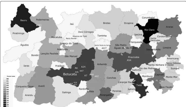
determined by BLASTn analysis. Figure 1 illustrates the human cutaneous leishmaniasis data corresponding to the cases seen in the central western area of the state of São Paulo, Brazil, from 1998 to 2010 [30]. Figure 2 displays the human visceral leishmaniasis data corresponding to the cases seen in the central western area of the state of São Paulo, Brazil, from 1998 to 2010 and geographic location of the positive road-killed animals evaluated [31].
Leishmania DNA was detected in 5/12 (41.67%; CI95% 19.22-68.42%) samples fromCerdocyon thous (crab-eating fox). A previous report indicates seropositivity in wild non-captive Cerdocyon thous [5,28]. The importance of these animals as reservoirs depends on their ability to transmit the infection to sandflies rather than on their infection rate; it is also a function of their capability to (re)introduce the pathogen intoLeishmania-free dog populations [32].
In the current study, Leishmania spp. DNA was de-tected in 1/3 Procyon cancrivorus (crab-eating raccoon). Voltarelliet al.[33] reported the presence ofLeishmania antibodies inProcyon cancrivorusin Northwestern Paraná. These findings suggest that these species can act as a re-servoir forLeishmaniaspp.
The members of Didelphidae, represented by Didel-phis albiventris specimens (white-eared opossums), are habitat generalists and currently occur in areas near dwel-lings, including farms, yards and urban centers [34]. This species is already proven to be a leishmaniasis reservoir and, for its synanthropic habits, it plays an important role in the peridomestic-forest traffic of degraded areas [35-37]. The present study confirms that molecular detection of
Table 1 Data on road-killed wild animals, including the sex, tissue, PCR and sequencing results for molecular detection onLeishmaniaspp. andLeishmania chagasi
Species Animal Sex Tissue (PCR positive) % identity/GenBank access
Procyon cancrivorus A1 Male kidney 99%/AJ270142.1Leishmaniaspp.
Cerdocyon thous A4 Male heart, mesenteric lymph node 100%/AJ270142.1Leishmaniaspp.
Cerdocyon thous A6 Male spleen, heart 100%/AJ270142.1Leishmaniaspp.
Cerdocyon thous A7 Male heart 100%/AJ270141.1Leishmaniaspp.
Cavia aperea A17 Male heart 99%/AJ270141.1Leishmaniaspp.
Dasypus septemcinctus A18 Male liver 100%/AJ270141.1Leishmaniaspp.
Sphiggurus spinosus A22 # liver, spleen 100%/AJ270142.1Leishmaniaspp.
Tamandua tetradactyla A23 Male lung, liver, mesenteric lymph node 100%/AJ270142.1Leishmaniaspp.
Shiggurus spinosus A24 Female spleen, kidney, heart 100%/AJ270142.1Leishmaniaspp.
Cerdocyon thous A41 Male liver, mesenteric lymph node 100%/AF308682.1Leishmania chagasi
Myrmecophaga. tridactyla A42 Male lung, kidney, heart, mesenteric lymph node 100%/AJ270142.1Leishmaniaspp.
Didelphis albiventris A46 Male liver, spleen, kidney 100%/AJ270142.1Leishmaniaspp.
Didelphis albiventris A47 Male lung 100%/AJ270142.1Leishmaniaspp.
Hydrochoerus hydrochaeris A50 Female lung 100%/AJ270142.1Leishmaniaspp.
Cerdocyon thous A54 Male lung, spleen 100%/AJ270142.1Leishmaniaspp.
Myrmecophaga tridactyla A60 Male lung 100%/AJ270142.1Leishmaniaspp.
Figure 1Geographic location of road-killed animals employed forLeishmaniaspp. molecular detection, correlating to the occurrence of cases of human cutaneous leishmaniasis.
Leishmaniaspp. in 2/8 group members may be com-mon, as was already described in several regions of Brazil: Manaus, Amazonas state; in Barra de Guarituba, Rio de Janeiro state; in Amaraji, Pernambuco; and Bauru, São Paulo state [38-41].
Rodents were represented by five S. spinosus (porcu-pine), four C. aperea (Brazilian guinea pig), two Rattus rattus(black rat), two Hydrochoerus hydrochaeris (capy-bara) and one M. coypus (coypu). Current data show that 8/14 (57.14%; CI95% 35.14-82.34%) specimens of wild rodents were positive, a finding that corroborates the literature that considered some rodent as reservoirs ofLeishmaniaspp.
The superorder Xenarthra was represented by 13 spec-imens: three D. novemcinctus (nine-banded armadillo), oneE. sexcinctus(six-banded armadillo), oneD. septem-cinctus (seven-banded armadillo), two T. tetradactyla (lesser anteater) and six M. tridactyla (giant anteater). These animals present some peculiar physiological and ecological characteristics including a weak immune sys-tem and low body sys-temperature, besides the fact that they live literally immersed in soil and organic matter, mainly in tropical and subtropical regions, under biotic and abiotic conditions that promote multiple encoun-ters with a diverse group of pathogens and vectors.
The present study confirms the occurrence of Leish-mania spp. DNA in armadillos (one D. septemcintus) and anteaters (one T. tetradactyla and two M. tridac-tyla). Casadeval and Pirofski [42] clarified many points on virulence and pathogenicity regarding host immune response and pathogen activity. According to the authors, there are classes of pathogenic microorganisms varying from those that provoke damage in hosts that present an extremely weak immune response to others that cause dis-ease only in a situation of very strong immune response. Therefore, it seems reasonable to consider Leishmania spp. to be a pathogen whose ability to provoke disease also depends on host immune response. Since the cellular im-mune response is weak in armadillos and anteaters, it is possible to detect yeast cells in many of their organs; how-ever, this is not sufficient to cause disease as observed in human hosts. Taken together, these factors make xenarthrans suitable models for studying host-pathogen interaction [43].
These animals are assumed to be sources of infection since the agent’s DNA was found in internal organs; in addition, parasitism may occur in internal and cutaneous organs, facilitating transmission from the blood meal by the vector that inoculates promastigote forms of the agent into the man while sucking.
The identities of the amplicon were confirmed by direct double-strand sequencing which showed 100% similarity with L. chagasi sequence deposited at GenBank (access number AF308682.1) (Table 1).
Even without the DNA detection of the cutaneous leish-maniasis agents, the positive results for Leishmania spp. are interesting. Of the 20 species described in the New World, five have never been reported to have caused vis-ceral human leishmaniasis: Leishmania enriettii, Leish-mania hertigi, Leishmania deanei, Leishmania aristidesi and Leishmania forattinii [44-48]. The L. forattinii was isolated from pooled liver and spleen of opossum Didel-phis marsupialiscaptured in Conchas, SP, Brazil [48,49].
It is suggested that the species isLeishmania forattinii and that the evaluated site is close to that where the parasite was first isolated, since the species nucleotide sequence deposited at the GenBank was not found. Con-sidering the occurrence of both the cutaneous and vis-ceral form, in the studied municipalities, it must be emphasized that the sandfly vector may be present and serve as transmitter of Leishmania to these animals and humans.
These findings corroborate the worldwide distribution of Leishmania spp., considering the wide variety of in-termediate hosts that contribute to the epidemiological transmission chain of this infection.
It is important to emphasize that Bauru, SP, is endemic for leishmaniasis; therefore, our results indicate the need for epidemiological molecular biology research on envir-onmental contamination byL. chagasi.
It was possible to evaluate 22 different wild species, without the necessity of exerting a laborious sampling effort. In fact, the numbers and diversity of road-killed animals are considerably higher and, in general, they are killed after their own natural habitats had been invaded by roads [50]. In this manner, the geographic coor-dinates of the locations of the infected animals are well-integrated in databases that use the geographical information systems (GIS), thus contributing to a bet-ter understanding of pathogen distribution.
These results show risk factors such as free movement of the circulating parasite and vectors, as well as the im-portance of road-killed animals as possible reservoirs for the transmission of Leishmania spp. in addition to the significance of the environment and ecology of these positive mammals in the interaction of Leishmania with different Leishmania species that may be pathogenic to humans.
Conclusions
The presented results focus that road-killed animals may serve as an important reservoir for transmission of Leish-maniaspp. and L. chagasi, as well as contributing to un-derstand the host-parasite interaction.
Competing interests
Authors’contributions
VBRP participated in the design of the study, data collection, laboratory tests, analysis and interpretation of data, writing and editing of the manuscript. PMM and EYH participated in the data collection, laboratory tests and took part in the writing. CV participated in geographical location of animals, analysis and interpretation of data, writing and revision of the manuscript. RCS participated in the analysis and interpretation of data, writing and revision. HL was responsible for the coordination, study design, analysis and interpretation of data, writing and editing of the manuscript. All authors read and approved the final manuscript.
Acknowledgments
The authors would like to thank the State of São Paulo Research Foundation (FAPESP, grants n. 08/09378-8 and n. 08/08291-6) for their funding of this research.
Ethics committee approval
This study is in accordance with the Brazilian Institute of Environment and Renewable Natural Resources’(IBAMA) normative statement n. 119 of October 11, 2006, chapter VI, art .26, which authorizes the collection and transport of animals that were found dead for scientific or didactic purposes. This work was also approved by the Ethics Committee for Animal
Experimentation of our Institution (CEEA/FMVZ n.211/2008).
Received: 6 January 2014 Accepted: 4 June 2014 Published: 16 June 2014
References
1. Gontijo B, Carvalho MLR:Leishmaniose tegumentar americana.Rev Soc Bras Med Trop2003,36(1):71–80.
2. Gontijo CMF, Melo MN:Leishmaniose visceral no Brazil: quadro atual, desafios e perspectivas.Rev Bras Epidemiol2004,7(3):338–349. 3. Desjeux P:Leishmaniasis: currents situation and new perspectives.
Comp Immunol Microbiol Infect Dis2004,27(5):305–318.
4. Gramiccia M, Gradoni L:The current status of zoonotic leishmaniases and approaches to disease control.Int J Parasitol2005,35(11–12):1169–1180. 5. Luppi MM, Malta MC, Silva TM, Silva FL, Motta RO, Miranda I, Ecco R, Santos
RL:Visceral leishmaniasis in captive wild canids in Brazil.Vet Parasitol 2008,155(1–2):146–151.
6. Coyner DF, Wooding JB, Forrester DJ:A comparison of parasitic helminths and arthropods from two subspecies of fox squirrels (Sciurus niger) in Florida.J Wildl Dis1996,32(3):492–497.
7. Cheadle MA, Tanhauser SM, Dame JB, Sellon DC, Hines M, Ginn PE, Mackay RJ, Greiner EC:The nine-banded armadillo (Dasypus novemcinctus) is an intermediate host forSarcocystis neurona.Int J Parasitol2001,
31(4):330–335.
8. Foster GW, Main MB, Kinsella JM, Dixon LM, Terrell SP, Forrester DJ:Parasitic helminths and arthropods of coyotes (Canis latrans) from Florida.
USA Comp Parasitol2003,70(2):162–166.
9. Nelder MP, Reeves WK:Ectoparasites of road-killed vertebrates in northwestern South Carolina, USA.Vet Parasitol2005,129(3–4):313–322. 10. Ferroglio E, Ragagli C, Trisciuoglio A:Physaloptera sibirica in foxes
and badgers from the Western Alps (Italy).Vet Parasitol2009,
163(1–2):164–166.
11. Hoppe EG, Araújo de Lima RC, Tebaldi JH, Athayde AC, Nascimento AA:
Helminthological records of six-banded armadillosEuphractus sexcinctus
(Linnaeus, 1758) from the Brazilian semi-arid region, Patos county, Paraíba state, including new morphological data onTrichohelix tuberculata(Parona and Stossich, 1901) Ortlepp, 1922 and proposal of
Hadrostrongylus ransominov. comb.Braz J Biol2009,69(2):423–428. 12. Miquel J, Foronda P, Torres J, Swiderski Z, Feliu C:Ultrastructural study of
the spermatozoon ofTaenia taeniaeformis(Batsch, 1786) (Cestoda, Cyclophyllidea, Taeniidae), an intestinal parasite ofFelis catusfrom La Palma (Canary Islands, Spain).Parasitol Res2009,104(6):1477–1483. 13. Richini-Pereira VB, Bosco SMG, Griese J, Theodoro RC, Macoris SA, da Silva
RJ, Barrozo L, Tavares PM, Zancopé-Oliveira RM, Bagagli E:Molecular detection ofParacoccidioides brasiliensisin road-killed wild animals.
Med Mycol2008,46(1):35–40.
14. Richini-Pereira VB, Bosco SMG, Theodoro RC, Barrozo L, Pedrini SCB, Rosa PS, Bagagli E:Importance of xenarthrans in the eco-epidemiology of
Paracoccidioides brasiliensis.BMC Res Notes2009,2:1–6.
15. Richini-Pereira VB, Bosco SMG, Theodoro RC, Barrozo L, Bagagli E:
Road-killed wild animals: a preservation problem useful for eco-epidemiological studies of pathogens.J Venom Anim Toxins incl Trop Dis 2010,16(4):607613. http://www.scielo.br/scielo.php?script=sci_arttext&pid= S1678-91992010000400011.
16. Zhao C, Onuma M, Asakawa M, Nagamine T, Kuwana T:Preliminary studies on developing a nested PCR assay for molecular diagnosis and identification of nematode (Heterakis isolonche) and trematode (Glaphyrostomumsp.) in Okinawa rail (Gallirallus okinawae).Vet Parasitol 2009,163(1–2):156–160.
17. Pedrini SC, Rosa PS, Medri IM, Mourão G, Bagagli E, Lopes CA:Search for
Mycobacterium lepraein wild mammals.Braz J Infect Dis2010,14(1):47–53. 18. Persing DH, Smith TF, Tenover FC, White TJ:Diagnostic Molecular
Microbiology: Principles and Applications.Washington, D.C: American Society for Microbiology; 1993:423–430.
19. Esseghir SA, Ftaiti A, Ready PB, Khdraoui B, Zaafouri K, Dellagi K, Ben Ismail R:The squash blot technique and the detection ofLeishmania majorinPhlebotomus papatasiin Tunisia.Arch Inst Pasteur Tunis1993,
70(3–4):493–496.
20. Aransay AM, Scoulica E, Tselentis Y:Detection and identification of
LeishmaniaDNA within naturally infected sand flies by seminested PCR on minicircle kinetoplastic DNA.Appl Environ Microbiol2000,
66(55):1933–1938.
21. Gennari SM, Canón-Franco WA, Yai LE, de Souza SL, Santos LC, Farias NA, Ruas J, Rossi FW, Gomes AB:Seroprevalence ofToxoplasma gondiiantibodies from wild canids from Brazil.Vet Parasitol2004,
121(3–4):337–340.
22. INPE:Instituto Nacional de Pesquisas Espaciais.TerraView: Software; v.3.6.0 [http://www.dpi.inpe.br/terraview/index.php]
23. de Bruijn MH, Barker DC:Diagnosis of New World leishmaniasis: specific detection of species of theLeishmania braziliensiscomplex by amplification of kinetoplast DNA.Acta Trop1992,52(1):45–58. 24. Eresh S, McCallum SM, Barker DC:Identification and diagnosis of
Leishmania mexicanacomplex isolates by polymerase chain reaction.
Parasitololy1994,109(Pt 4):423–433.
25. Tamura K, Dudley J, Nei M, Kumar S:MEGA4: Molecular Evolutionary Genetics Analysis (MEGA) software version 4.0.Mol Biol Evol2007,
24(8):1596–1599.
26. Alexander B, Lozano C, Barker DC, McCann SH, Adler G:Detection of
Leishmania(Viannia)braziliensiscomplex in wild mammals from Colombian coffee plantations by PCR and DNA hybridization.Acta Trop 1998,69(1):41–50.
27. Travi BL, Osorio Y, Becerra MT, Adler GH:Dynamics ofLeishmania chagasi
infection in small mammals of the undisturbed and degraded tropical dry forests of northern Colombia.Trans R Soc Trop Med Hyg1998,
92(3):275–278.
28. Curi NHA, Miranda I, Talamoni AS:Serologic evidence ofLeishmania
infection in free-ranging wild and domestic canids around a Brazilian National Park.Mem Inst Oswaldo Cruz2006,101(1):99–101.
29. Papadogiannakis E, Spanakos G, Kontos V, Menounos PG, Tegos N, Vakalis N:
Molecular detection ofLeishmania infantumin wild rodents (Rattus norvegicus) in Greece.Zoonoses Public Health2010,57(7–8):23–25. 30. Centro de Vigilância Epidemiológica:Leishmaniose tegumentar americana.
Distribuição do número de casos de Leishmaniose Tegumentar por município provável de infecção. Estado de São Paulo, 1998–2012.
http://www.cve.saude.sp.gov.br/htm/zoo/lta_lpi.htm.
31. Centro de Vigilância Epidemiológica:Leishmaniose visceral americana. Distribuição do número de casos e óbitos de LVA segundo município e GVE de infecção. Estado de São Paulo 1998–2011.http://www.cve.saude.
sp.gov.br/htm/zoo/lvah_lpi.htm.
32. Dressen DW:Toxoplasma gondiiinfections in wildlife.J Am Vet Med Assoc 1990,196:274–276.
33. Voltarelli EM, Arraes SMAA, Perles TF, Lonardoni MVC, Teodoro U, Silveira TGV:Serological survey forLeishmaniasp. infection in wild animals from the municipality of Maringá, Paraná State, Brazil.J Venom Anim Toxins incl Trop Dis2009,15(4):732–744. http://www.scielo.br/scielo.php?script=sci_ arttext&pid=S1678-91992009000400011.
34. Câmara T, Murta R:Mamíferos da Serra do Cipó.PUC, Minas, Museu de Ciências Naturais: Belo Horizonte; 2003.
36. Guerra JAO, Ribeiro JAS, Coelho LIARC, Barbosa MGV, Paes MG:
Epidemiologia da leishmaniose tegumentar na comunidade São João, Manaus, Amazonas.Brasil Cad Saúde Pública2006,22(11):2319–2327. 37. Quintal AP, Ribeiro Ede S, Rodrigues FP, Rocha FS, Floeter-Winter LM,
Nunes CM:Leishmaniaspp. inDidelphis albiventrisandMicoureus paraguayanus(Didelphimorphia: Didelphidae) of Brazil.Vet Parasitol2011,
176(2–3):112–119.
38. Arias JR, Naiff RD:The principal reservoir host of cutaneous leishmaniasis in the urban areas of Manaus, Central Amazon of Brazil.Mem Inst Oswaldo Cruz1981,76(3):279–286.
39. Cabrera MAA, Paula AA, Camacho LAB, Marzochi MCA, Xavier SC, da Silva AVM, Jansen AM:Canine visceral leishmaniasis in Barra de Guaratiba, Rio de Janeiro, Brazil: assessment of risk factors.Rev Inst Med Trop Sao Paulo 2003,45(2):79–83.
40. Brandão-Filho SP, Brito ME, Carvalho FG, Ishikawa EA, Cupolillo E, Floeter-Winter L, Shaw JJ:Wild and synanthropic host ofLeishmania(Viannia)
braziliensisin the endemic cutaneous leismaniasis locality of Amaraji, Pernambuco State, Brazil.Trans R Soc Trop Med Hyg2003,97(3):291–296. 41. Santiago ME, Vasconcelos RO, Fattori KR, Munari DP, Michelin Ade F, Lima VM:An investigation ofLeishmaniaspp. inDidelphisspp. from urban and peri-urban areas in Bauru (São Paulo, Brazil).Vet Parasitol2007,
150(4):283–290.
42. Casadevall A, Pirofski LA:Host-pathogen interactions: redefining the basic concepts of virulence and pathogenicity.Infect Immun1999,
67(8):3703–3713.
43. Bagagli E, Bosco SMG:Armadillos and dimorphic pathogenic fungi: ecological and evolutionary aspects.InThe Biology of the Xenarthra.Firstth edition. Edited by Viscaino SF, Loughry WJ. Gainesville: University Press of Florida; 2008:103–110.
44. Muniz J, Medina HSG:Leishmaniose tegumentar do cobaio(Leishmania enriettii).Arq Biol Tecnol1948,3(2):7–25.
45. Herrer A:Leishmania hertigisp. n., from the tropical porcupine,Coendou rothschildi Thomas.J Parasitol1971,57(3):626–629.
46. Lainson R, Shaw JJ:Leishmanias of neotropical porcupines:Leishmania hertigi deaneinov. subsp.Acta Amaz1977,7(1):51–57.
47. Lainson R, Shaw JJ:The role of animals in the epidemiology of South American leishmaniasis.InThe Biology of the Kinetoplastida, Volume 2. Edited by Lumsden WHR, Evans DA. London, New York, San Francisco: Academic Press; 1979:1–116.
48. Yoshida ELA, Silva R, Cortez LS, Corrêa FMA:Encontro de espécie do gêneroLeishmaniaemDidelphis marsupialis auritano Estado de São Paulo, Brasil.Rev Inst Med Trop Sao Paulo1979,21:110–113.
49. Yoshida ELA, Cuba CA, Pacheco RS, Cupolillo E, Tavares CC, Machado GMC, Momen H, Grimaldi JG:Description ofLeishmania (Leishmania) forattinii
sp. n., a new parasite infecting opossums and rodents in Brazil.Mem Inst Oswaldo Cruz1993,88(3):397–406.
50. Laurance WF, Goosem M, Laurance SG:Impacts of roads and linear clearings on tropical forests.Trends Ecol Evol2009,24(12):659–669.
doi:10.1186/1678-9199-20-27
Cite this article as:Richini-Pereiraet al.:Molecular detection of Leishmaniaspp. in road-killed wild mammals in the Central Western area of the State of São Paulo, Brazil.Journal of Venomous Animals and Toxins including Tropical Diseases201420:27.
Submit your next manuscript to BioMed Central and take full advantage of:
• Convenient online submission
• Thorough peer review
• No space constraints or color figure charges
• Immediate publication on acceptance
• Inclusion in PubMed, CAS, Scopus and Google Scholar
• Research which is freely available for redistribution