Printed version ISSN 0001-3765 / Online version ISSN 1678-2690
www.scielo.br/aabc
http://dx.doi.org/10.1590/0001-3765201520130141
Abdominal macrochaetae of female Hylesia oratex Dyar,
1913 (Insecta: Lepidoptera: Saturniidae):
external morphology and medical significance
ROSÂNGELA BRITO1, ALEXANDRE SPECHT2, WILSON S.A. FILHO3, EDEGAR FRONZA4 and CARLOS G.C. MIELKE5
1
Laboratório de Estudos de Lepidoptera Neotropical, Setor de Ciências Biológicas, Departamento de Zoologia, Centro Politécnico, Universidade Federal do Paraná,
Jardim das Américas, Caixa Postal 19020, 81531-980 Curitiba, PR, Brasil
2Laboratório de Entomologia, Embrapa-Cerrados, BR 020, Km 18,
Caixa Postal 08223, 73310-970 Planaltina, DF, Brasil
3
Laboratório de Entomologia, Centro de Ciências Exatas, da Natureza e de Tecnologia, Campus Universitário da Região dos Vinhedos, Universidade de Caxias do Sul,
Caixa Postal 32, 95700-000 Bento Gonçalves, RS, Brasil
4Laboratório de Controle de Pragas, Instituto de Biotecnologia, Universidade de Caxias do Sul,
Rua Francisco Getulio Vargas, Caixa Postal 1352, 95070-560 Caxias do Sul, RS, Brasil
5
Caixa Postal 1206, 84145-000 Carambeí, PR, Brasil
Manuscript received on April 12, 2013; accepted for publication on January 19, 2015
ABSTRACT
The representatives of the genus Hylesia Hübner, [1820] are significant among the medically important Lepidoptera. Adult females use abdominal setae to wrap and protect the eggs that remain for months in
nature. These setae, in contact with human skin, may cause allergic reactions including swelling, itching and local erythema, known as lepidopterism. The morphology of the abdominal scales and setae from
the female H. oratex Dyar, 1913 is herein described and aspects related to their medical significance are discussed. Portions of each abdominal segment were examined through a scanning electron microscope.
Two types of scales without medical importance, and two types of setae with medical importance, classified as "true setae" and "modified setae" were found. The true setae, which are slightly fusiform and have radially arranged lateral projections, are responsible for the allergic reactions caused by skin penetration. The modified setae, which are larger, curved, with the median enlarged and serrated margins, can be
responsible for the release of chemical substances. This information provides a better understanding of the structure of the urticating setae, which are responsible for lepidopterism outbreaks in humans, and
contributes towards the identification of the moth species involved.
Key words: allergic outbreaks, lepidopterism, morphology, urticating moths.
Correspondence to: Rosângela Brito E-mail: rosangela.bri@gmail.com
INTRODUCTION
Butterflies and moths belong to one of the largest
orders of insects (Lepidoptera), known and admired
Hylesia
Hübner [1820], receive significant medical
attention due to their females which bear urticating
setae that come off the abdomen, causing several
outbreaks of dermatitis (Haddad and Cardoso 2003,
Hossler 2009, 2010a, b, Polar et al. 2010, Battisti
et al. 2011). Once an urticating seta penetrates into
the tissue, it may cause skin reactions, persisting
for hours or days and resulting in swelling, itching
and local erythema, known as lepidopterism, and
ultimately forms pruritic plaques, causing fever and
malaise (Battisti et al. 2011). Epidemic outbreaks
of dermatitis have been reported in Mexico,
Venezuela, Guyana, Peru, Argentina, and in some
Brazilian states such as Amapá, Bahia, Minas
Gerais, São Paulo and Rio Grande do Sul. However,
there are very few publications that deal with this
phenomenon (Gusmão et al. 1961, Mascarenhas et
al. 1980, Glasser et al. 1993, Iserhard et al. 2007,
Moreira et al. 2007).
Battisti et al. (2011) classified the "urticating
hairs" of arthropods into three categories: true setae,
modified setae and spines. The first is characterized
by the loss of its neural connection and by the
detachment of its integument at the proximal end.
The second is characterized by its robust base and
connection with the integument, while its neural
connection is lacking. However, another type of
cell is connected to the scale and hypothetically has
a secretory function. The latter has a more complex
structure and can be considered as a structure formed
from the integument, involving a large number of
specialized cells with a sensory function. True setae
have been recorded in
Hylesia
females (Battisti
et al. 2011). Kristensen and Simonsen (1998)
attributed the term "macrochaetes" for bristles or
setae and scales present in Lepidoptera. Another
possible classification for the macrochaetes was
proposed by Rodriguez et al
.
(2004), who, based on
H
.
metabus
(Cramer, 1775), classified them as S1,
S2, S3, S4, with only the last two being urticating.
Hylesia
oratex
Dyar, 1913 occurs within Brazil,
in the states of Goiás, Rio de Janeiro, São Paulo,
Paraná, (D'Abrera 1995, Lemaire 2002, Borges et al.
2003), Santa Catarina (Lima 1947), and Rio Grande
do Sul (Corseuil et al. 2002, Specht et al. 2005), in
addition to being found in Paraguay and Argentina
(Köhler 1931). It is known that its larvae feed on
"erva mate" leaves (
Ilex paraguariensis
St. Hill. -
Aquifoliaceae) (Lima 1947) and, as in
H
.
nigricans
(Berg 1876), form collective silk shelters by joining
leaves (Specht et al. 2006). Female
H
.
oratex
, as well
as in other species of the genus, also bear urticating
setae used to surround their eggs for protection,
unlike males that lack these structures in their bodies
(Rodriguez et al. 2004). Lamy and Lemaire (1983)
reported the setae as corresponding to the type S3
described by Rodriguez et al. (2004), indicating the
possibility of using their morphology for species
level identification of
Hylesia
representatives.
However,
H
.
oratex
´s setae were not included in the
Lamy and Lemaire (1983) study, so the knowledge
of this species is limited, especially with respect the
types and morphology of macrochaetes including
their clinical significance, which lack detailed
information regarding. Thus, the main goal of this
article is to describe the external morphology of
the macrochaetes found on the abdomen of female
H
.
oratex
, relative to their medical significance in
relation to humans.
MATERIALS AND METHODS
H
.
oratex
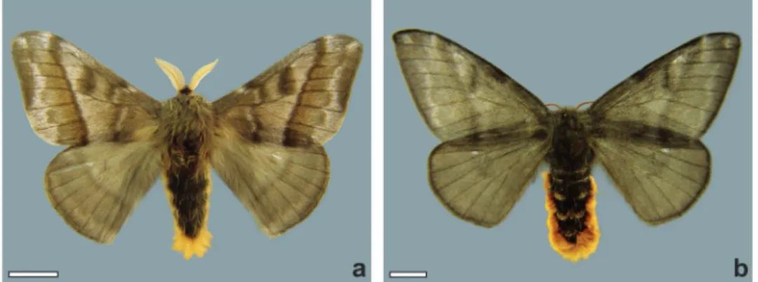
(Figs. 1a and 1b) were obtained from
an egg mass collected in nature at Anta Gorda,
Rio Grande do Sul, on a branch of "erva-mate",
and taken to the laboratory in order to obtain
adults. The larvae were reared at the Laboratório
de Entomologia in the Campus Universitário da
Região dos Vinhedos, Universidade de Caxias do
Sul, in a room at 25 ± 1 °C, 80 ± 10% RH, and 14h
photo phase, and fed on “erva-mate” leaves until
Jesus Santiago Moure, in the Departmento de
Zoologia at the Universidade Federal do Paraná,
Curitiba, Paraná as voucher specimens. Abdomens
were dissected to obtain samples of macrochaetes
from the dorsal, ventral and lateral regions. Scales
and setae of each region were removed and prepared
for histological analysis using scanning electron
microscopy (SEM) at the Centro de Microscopia
Eletrônica of the Universidade Federal do Rio
Grande do Sul (CME-UFRGS/RS). All procedures
were carried out according to the international
practices for animals use and care under the control
of an internal committee of the Universidade de
Caxias do Sul, Brazil.
Figure 1 - Hylesia oratex. (a) male; (b) female (scale = 5 mm, respectively).
RESULTS
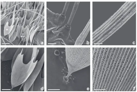
Only non-urticating scales are found on the dorsal
region of the abdomen which can be divided
into two types: fi liform and spatulate scales. The
fi liform scales, corresponding to the seta S1 of
Rodriguez et al.
(2004), are fi liform, cylindrical
at the base, connected to the integument cuticle,
and have a porous surface, with a length from
1000 to 2000µm (Figs. 2a-c). The spatulate scales,
corresponding to the seta S2 of Rodriguez et al
.
(2004), have a narrow base which is connected to
the integument, widening toward the apex with an
ondulated distal end with three, four or slightly
more pointed projections, and, as the previous
type, is porous and also shorter, ranging from 200
to 220µm (Figs. 2d-f).
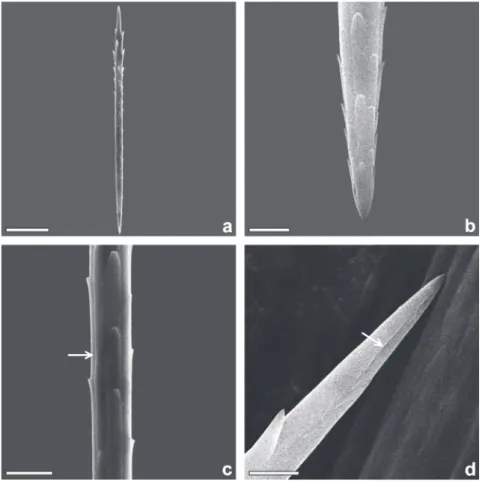
The urticating setae can be divided into two
types: the fi rst, corresponding to the true setae of
Battisti et al
.
(2011) and to the seta S3 of Rodriguez
et al. (2004), are found on the ventral and lateral
regions inserted into a socket. These setae are more
abundant in the central and posterior portions.
They are slightly fusiform and smooth with four
longitudinal grooves (Figs. 3a and 3c) measuring
120-130µm in length with a few lateral projections
arising near the base, extending radially towards
the apex (Fig. 3b). These projections are rounded
and barb-like, varying in numbers from 20 to 25. The
distal end of these setae are ornamented with a discrete
groove, lacking pores or orifi ces (Fig. 3d). The second
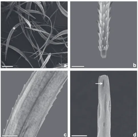
type of setae, corresponding to the modifi ed setae of
Battisti et al
.
(2011) and to the S4 setae of Rodriguez
et al. (2004), are found bordering the ventral side,
together with some fi liform and spatulate scales.
The central portion is wider than the base and apex,
with a length of 1200 to 1400µm (Fig. 4a). The base
contains conspicuous pointed projections directed
towards the distal region (Fig. 4b), being carinated
with serrated edges (Fig. 4c). The apex has a tubular
and rounded shape with small orifi ces which may be
used for secretion (Fig. 4d). In egg masses collected
in the fi eld, the presence of intermixed true setae and
modifi ed setae forming only one layer covering the
egg mass (Figs. 5a and 5b) was observed. However,
due to their larger size, the modifi ed setae are more
DISCUSSION
The fi liform and spatulate scales (Fig. 2), especially
those located in the dorsal portion of the abdomen
of female
H
.
oratex,
were not considered urticating,
due to their high porosity and lack of barb-like
structures. Morphologically, these scales are
very similar to the ones which cover other parts
of
H
.
oratex
´s female body and have no medical
importance, corresponding to the scales described
by Rodriguez e t al
.
(2004) for
H
.
metabus
.
The true setae found in
Hylesia
(Rodriguez et
al. 2004, Specht et al
.
2005, 2007, 2008, Hossler
2009, 2010a, b) correspond to those found in
arthropods, such as spiders, and in larvae and
adults of Lepidoptera (Battisti et al. 2011).
Environmental or human factors are responsible
for the release and/or detachment of the setae from
the abdomen of the female, as they easily detach
by mechanical stimulation (Novak et al. 1987).
They are more likely to induce allergic reactions,
which may cause recurrent dermatitis related to the
outbreaks of insects, affecting various parts of the
body simultaneously (Hossler 2009). Consisting
exclusively of chitin, wax and proteins, when the
setae completely and abundantly penetrate the skin,
they can trigger systemic symptoms and cause
great discomfort, such as local itching, redness
and swelling, while in rare cases they can lead to
anaphylactic reactions (Battisti et al. 2011).
Upon comparing the true seta of
H
.
oratex
with
other
Hylesia
species (Lamy and Lemaire 1983,
Rodriguez et al
.
2004), it was observed that the
number of grooves are similar to the ones of
H
.
iola
Dyar, 1913,
H
.
lineata
Druce, 1886,
H
.
metapyrrha
(Walker, 1855),
H
.
annulata
Schaus, 1911,
H
.
ebalus
(Cramer, 1775),
H
.
gyrex
Dyar, 1913 and
H
.
tapareba
(Dyar, 1913). However, in
H
.
iola
and
H
.
lineata
, this seta does not have a fusiform shape.
The number of barb-like structures on the true seta
of
H
.
oratex
is similar to
H
.
metapyrrha
, while an
apical groove was observed in all species studied
by Lamy and Lemaire (1983). Differences and
Figure 2 - Abdominal scales of the female H. oratex. (a) fi liform and spatulate scales, an arrow indicates fi liform and an asterisk indicates spatulate; (b) base of fi liform scale; (c) median portion
of fi liform scale; (d) general view of spatulate scale; (e) base of spatulate scale; (f) medial portion
similarities among different true setae corroborate
the idea of Lamy and Lemaire (1983) that this
characteristic can be used for species identifi cation
based on the morphology of the true seta, which
causes the dermatitis.
The modifi ed seta is rigid and bears barb-like
structures of various sizes around its base. This
modifi ed seta is present on the abdomen of female
H
.
oratex
at a much lower density than the true seta
which agrees with the description by Battisti et al.
(2011), reporting that its distribution is restricted to
the edges of the "setae fi elds", covering the true setae.
Comparing the modifi ed setae of
H
.
metabus
with
H
.
oratex
, it is possible to fi nd similarities
regarding their size and shape, especially with
respect to the carinate margin and the base and apex
being narrower than the middle portion. However,
a few slight differences can be identifi ed among the
species (
H
.
metabus
and
H
.
oratex
), such as the shape
of the barb-like structures, where in
H
.
metabus
the
apex of these barbs have a triangular shape while in
H
.
oratex
the apex of the barbs are rounded and their
points are directed toward its distal end. Furthermore,
the former bears an apex, which is thin and
tube-like, and has a single opening, whereas the latter
species bears a rounded tube-like apex, with several
openings. The presence of these openings in both
species indicates a probable excretion of chemicals
for protective function. There is a possibility, as
described by Rodriguez et al. (2004), that there is
a cell with secretory function which connects these
setae to the integument (Lundberg et al. 2007).
Figure 3 - True setae of the female H. oratex. (a) general view; (b) base; (c) medialportion, arrow indicates a longitudinal groove; (d) apex, arrow indicates the apical groove
Figure 5 - Egg mass of H. oratex. (a) egg mass on a branch of the host plant; (b) detail of the layer composed of true and modifi ed setae (scales = 5 mm; 200 µm, respectively).
Figure 4 - Modifi ed setae of the female H. oratex. (a) general view (asterisk); (b) pointed
projections at base; (c) medial portion; (d) apex, arrow indicates apical orifi ces (scales = 200, 25, 10, 05 µm, respectively).
True setae and scales were found in the lateral
region of
H
.
oratex,
beyond the modifi ed setae,
while in
H
.
metabus
the modifi ed setae were
predominant (Rodriguez et al. 2004). Observing
the setae layer over the egg mass in
H
.
oratex
,
the true setae and modifi ed setae are mixed into
a single layer, whereas in
H
.
metabus,
according
are predominant. The way the true setae and the
modified setae are used in the preparation of the egg
masses, indicates a complex protective behavior
against possible natural enemies (Lemaire 2002,
Rodriguez et al. 2004, Specht et al. 2005, 2007).
Lepidopterism, as caused by
Hylesia
moths,
does not involve direct contact with the moths, but
rather the presence of setae in the environment (i.e.
Salomon et al. 2005). In this case, most lepidopterism
episodes should be attributed to contact with the
true setae that are released in large quantities in
anthropogenic environments, especially when
the moths are flying abundantly towards light (i.e.
Gusmão et al. 1961, Mascarenhas et al. 1980, Glasser
et al. 1993, Salomon et al. 2005, Iserhard et al. 2007,
Moreira et al. 2007, Polar et al. 2010). Because the
modified setae are heavier and do not easily stand
out from females abdomens, their participation
as a lepidopterism agent is restricted to events in
which patients come in contact with egg masses
during manual disinfection of trees (Casalá et al.
1967). In this case, lepidopterism occurs by physical
contact with the true setae and modified setae, and
the possible contact with the irritating substance
can be released by the modified setae (Battisti et al.
2011). The coexistence of both setae may lead to the
true setae becoming somewhat “coated” with the
secretion that may be released from the modified
setae, thus resulting in the venomous action in
addition to the mechanical one.
For both types of urticating setae described
in this article, it is noteworthy that the treatment
of lepidopterism should be done according to the
symptoms encountered in the patient. However,
the first step should be the complete removal of the
setae present on the skin, using adhesive tape or fine
forceps, and an immediate cleaning with water. The
shape and structure of the true and modified setae
which affect humans may be used to assist in the
indication of
Hylesia
representatives associated with
the dermatitis or the egg masses collected in the
field, as suggested by Lamy and Lemaire (1983).
ACKNOWLEDGMENTS
The authors are grateful to the Electron Microscopy
Center of the Universidade Federal do Rio Grande
do Sul, Denis Santos da Silva and Maurício
Moraes Zenker for his cooperation in preparing
the images.
RESUMO
Os representantes do gênero Hylesia Hübner, [1820] estão entre os principais lepidópteros de importância médica. As fêmeas adultas usam cerdas abdominais para
envolver e proteger seus ovos que podem permanecer
por meses na natureza. Essas cerdas, em contato com a pele humana podem causar reações alérgicas incluindo inchaço local, coceira e eritema conhecido como lepidopterismo. A morfologia das escamas e das cerdas abdominais das fêmeas de H. oratex Dyar,
1913 é aqui descrita e aspectos relacionados à sua
importância médica são discutidos. Porções de cada segmento abdominal foram examinadas através de um microscópio eletrônico de varredura. Dois tipos de escamas sem importância médica, e dois tipos de cerdas
com importância médica, classificadas como "cerdas verdadeiras" e "cerdas modificadas" foram encontradas. As cerdas verdadeiras que são levemente fusiformes e têm projeções laterais dispostas radialmente, são
responsáveis por reações alérgicas provocadas pela
penetração na pele. As cerdas modificadas que são
maiores, curvadas, com as margens medianas alargadas e serrilhadas podem ser responsáveis pela liberação de
substâncias químicas. Essa informação fornece melhor compreensão da estrutura das cerdas urticantes, que
são responsáveis por surtos de lepidopterismo em
humanos, e contribui para a identificação das espécies
de mariposas envolvidas.
Palavras-chave: surtos alérgicos, lepidopterismo, morfologia, mariposas urticantes.
REFERENCES
BATTISTI A, HOLM G, FAGRELL B AND LARSSON S. 2011. Urticating Hairs in Arthropods: Their Nature and Medical
BORGES LR, LÁZZARI SMN AND LÁZZARI FA. 2003. Comparação dos sistemas de cultivo nativo e adensado de erva mate, Ilex paraguariensis St. Hil., quanto à
ocorrência e flutuação populacional de insetos. Rev Bras
Entomol 47(4): 563-568.
CASALÁ A, BIANCHI C, SÁNCHEZ NAVARRO JV, BIANCHI O AND BALSA R. 1967.Granuloma de las manos por nidos de lepidópteros (Hylesia nigricans). Arch Argent Dermat 17(4): 307-313.
CORSEUIL E, SPECHT A AND LANG C. 2002. Saturniídeos (Lepidoptera, Saturniidae) registrados para o Rio Grande do Sul, Brasil. I. Hemileucinae. Biociências 10(2): 147-155. D’ABRERA B. 1995.Saturniidae Mundi. Saturniid moths of the
World – Part 1. Keltern, Automeris Press, 177 p.
GLASSER CM, CARDOSO JL, CARRÉRI-BRUNO GC, DOMINGOS MF, MORAES RHP AND CIARAVOLO RMC. 1993. Surtos epidêmicos de dermatite causada por mariposas do gênero Hylesia (Lepidoptera: Hemileucidae) no Estado de São Paulo. Rev Saúde Publ 27(3): 217-220.
GUSMÃO HH, FORATTINI OP AND ROTBERG A. 1961. Dermatite provocada por Lepidópteros do gênero Hylesia, São Paulo. Rev I Med Trop 3(3): 114-120.
HADDAD V AND CARDOSO JLC. 2003. 22 - Erucismo e Lepidopterismo, p. 220-223. In: Cardoso JLC, França
FOS, Wen FH, Málaque CMS and Haddad V (Eds), Animais peçonhentos no Brasil - biologia, clínica e terapêutica dos acidentes. São Paulo: Sarvier, 468 p. HOSSLER EW. 2009.Caterpillars and moths. Dermatol Ther
22: 353-366.
HOSSLER EW. 2010a. Caterpillars and moths: Part I. Dermatologic manifestations of encounters with Lepidoptera. J Am Acad Dermatol 62: 1-10.
HOSSLER EW. 2010b. Caterpillars and moths: Part II. Dermatologic manifestations of encounters with Lepidoptera. J Am Acad Dermatol 62: 13-28.
ISERHARD CA, KAMINSKI LA, MARCHIORI MO, TEIXEIRA EC AND ROMANOWSKI HP. 2007.Occurrence of lepidopterism
caused by the moth Hylesia nigricans (Berg) (Lepidoptera: Saturniidae) in Rio Grande do Sul state, Brazil. Neotrop Entomol 36(4): 612-615.
KÖHLER P. 1931. El género Hylesia en la Argentina. Rev Soc Entomol Argent 17(3): 305-308.
KRISTENSEN NP AND SIMONSEN TJ. 1998. 2. ‘Hairs’ and scales. p. 9-22. In: Kristensen NP(Ed), Lepidoptera, moths and
butterflies. v. 2 Morphology, physiology and development. Berlin: Walter de Gruyter, 564 p.
LAMY M AND LEMAIRE C. 1983. Contribution á la systématique des Hylesia: etude au microscope électronique á balayage
des “flechettes” urticantes. Bull Soc Entomol Fr 88(3/4): 176-192.
LEMAIRE C. 2002.The Saturniidae of America - Hemileucinae. Keltern: Goecke & Evers, 1388 p.
LIMA ADF. 1947.Insetos fitófagos de Santa Catarina. Bol Fitos 2(2/3): 233-251.
LUNDBERG ULF, SALAZAR V, TOVAR M AND RODRIGUEZ J. 2007. Isolation and partial characterization of proteins
with vasodegenerative and proinflammatory properties
from the egg-nests of Hylesia metabus (Lepidoptera: Saturniidae). J Med Entomol 44(3): 440-449.
MASCARENHAS CS, VULCANO MA AND PEREIRA FS. 1980. Nova constatação de dermatite provocada por lepidópteros do gênero Hylesia Hübner. Lundiana 1: 143-148. MOREIRA SC, LIMA JC, SILVA L AND HADDAD JUNIOR V. 2007.
Descrição de um surto de Lepidopterismo (dermatite associada ao contato com mariposas) entre marinheiros, ocorrido em Salvador, Estado da Bahia. Rev Soc Bras Tro Med 40(5): 591-593.
NOVAK F, PELISSOU V AND LAMY M. 1987. Comparative morphological, anatomical and biochemical studies of the urticating apparatus and urticating hairs of some Lepidoptera: Thaumetopoea pityocampa Schiff., Th. processionea L. (Lepidoptera, Thaumetopoeidae) and Hylesia metabus Cramer (Lepidoptera, Saturniidae).
Comp Biochem Physiol 88: 141-146.
POLAR P, COCK MJW, FREDERICKSON C, HOSEIN M AND KRAUSS U. 2010. Invasions of Hylesia metabus (Lepidoptera: Saturniidae, Hemileucinae) into Trinidad, West Indies. Living World, J Trin Tob Field Nat Club 2010: 1-10.
RODRIGUEZ J, HERNANDÉZ JV, FORNÉS L, LUNDBERG U, AROCHA-PIÑANGO CL AND OSBORN F. 2004. External
morphology of abdominal setae from male and female
Hylesia metabus adults (Lepidoptera: Saturniidae) and their function. Fla Entomol 87: 30-36.
SALOMON OD, SIMON D, RIMOLDI JC, VILLARUEL M, PEREZ O, PEREZ R AND MARCHAN H. 2005. Lepidopterismo por Hylesia nigricans (mariposa negra): investigación y acción preventiva en Buenos Aires. Medicina (Buenos Aires) 65: 241-246.
SPECHT A AND CORSEUIL E. 2008. 6 - Saturniidae: Hemileucinae:
Hylesia spp., p. 133-164. In: Specht A, Corseuil E and Abella
HB (Orgs), Lepidópteros de Importância Médica – Principais espécies no Rio Grande do Sul.Pelotas: USEB, 220 p. SPECHT A, CORSEUIL E AND FORMENTINI AC. 2005.
Lepidópteros de importância médica ocorrentes no Rio Grande do Sul. III. Saturniidae – Hemileucinae. Biociências 13(2): 149-162.
SPECHT A, FORMENTINI AC AND CORSEUIL E. 2006. Biologia de Hylesia nigricans (Berg) (Lepidoptera, Saturniidae, Hemileucinae). Rev Bras Zool 23(1): 248-255.
SPECHT A, FORMENTINI AC AND CORSEUIL E. 2007. Biological aspects of Hylesia metapyrrha (Lepidoptera; Saturniidae;