Vol.56, n.4: pp. 567-574, July-August 2013
ISSN 1516-8913 Printed in Brazil BRAZILIAN ARCHIVES OF
BIOLOGY AND TECHNOLOGY
A N I N T E R N A T I O N A L J O U R N A L
Effect of Wnt/
β
-catenin and NF-
κ
B Signaling Pathways on
Mucus Secretion with Hypertonicity in 16HBE Cells
Liu Xiaoyan
1and Zhou Xiangdong
2*1Department of Geriatrics, the First Affiliated Hospital; Chongqing Medical University; No.1 Youyi Road; Yuzhong
District, Chongqing; 400016 - China. 2Department of Respiratory Medicine, the Second Affiliated Hospital; Chongqing Medical University; No.74, Linjiang Road; Yuzhong District, Chongqing; 400010 - China
ABSTRACT
This study aimed at identifying the molecular mechanisms and effects of hypertonicity on mucin5AC (MUC5AC) expression in airway epithelial cells for which immortalized human bronchial epithelial (16HBE) cells were cultured in 600 mOsm/L hypertonic medium for different times in vitro. Proteins of MUC5AC and β-catenin, Cyclin D1, NF-κB p65 were detected by enzyme linked immunosorbent assay (ELISA), reverse transcription (RT)-PCR and western blotting assays. After transfection of β-catenin siRNA in 16HBE cells, the levels of β-catenin, Cyclin D1, NF-κB p65 and MUC5AC protein were detected. Results showed that the levels of mRNAs and proteins of target genes increased significantly after the exposure to hypertonic conditions and the expression content increased in a time-dependent manner. Furthermore, transfection with β-catenin siRNA attenuated the expression of these genes. These results suggested that the Wnt/β-catenin and NF-κB signaling pathways played essential roles in inducing the MUC5AC hypersecretion in 16HBE cells in response to hypertonicity.
Key words: MUC5AC, hypertonicity, Wnt/β-catenin, NF-κB p65
*Author for correspondence: [email protected]
INTRODUCTION
Mucus hypersecretion is a characteristic
manifestation in the patients with chronic obstructive pulmonary diseases (COPD) (Fahy 2001; Hogg et al. 2004). Mucus hypersecretion contributes to morbidity and mortality in these diseases by plugging the airways and causing recurrent infections (Shao and Nadel 2005). MUC5AC is the main mucin phenotype expressed in airway epithelium, especially in the pathological state. The physical or chemical changes of the airway affect the secretion of the mucus. For instance, high osmotic pressure of airway can serve as a kind of physical stimulation, which induces the human bronchial epithelial cells to secrete mucus. Some 3-5% of the high
permeability brine is often used to discharge sputum clinically. Studies have shown that high osmotic pressure of airway caused mucus
hypersecretion and its probable molecular
mechanisms could be the expression of HSP70-2,
which was activated by PKCµ signaling pathway.
The OREBP (osmotic response element binding protein) links the pathway and molecular of HSP70-2. It combines with the ORE2 sites, the upstream promoter of HSP70-2, and then activates the transcription of HSP70-2. Such mechanisms
promote the hypersecretion of MUC5AC.
Nevertheless, the inhibitor of PKCµ can not
environment of airway. Wnt/β-catenin signaling pathway is the focus of studies in variant provinces nowadays. Previous studies have shown that the disorders of this pathway get relationships with bronchial asthma, pulmonary fibrosis and lung cancer (Voynow and Rubin 2009; Kneidinger
et al. 2011; Baarsma et al. 2011).NF-κB signaling
pathway is another important pathway. However, little attention has been focused on the effect of
Wnt/β-catenin and NF-κB signaling pathways on
mucus secretion with hypertonicity in airway epithelial cells both domestic and overseas. This work aimed to study the MUC5AC secretion of 16HBE cells in hypertonic conditions and discuss its possible mechanisms.
MATERIALS AND METHODS
MaterialsFetal bovine serum (FBS), trypsin, the Roswell Park Memorial Institute (RPMI) 1640 medium,
TRIzol and anti-β-actin monoclonal antibody were
obtained from Sigma (St. Louis, MO). The mouse monoclonal antibody to MUC5AC was obtained from Neomarker (Fremont, CA). Goat serum
albumin, Mouse monoclonal antibodies to β
-catenin, NF-κB p65, and Cyclin D1 and
diaminobenzidine (DAB) were purchased from Santa Cruz Biotechnology (Santa Cruz, Calif). The
ELISA Kit for MUC5AC, β-catenin siRNA and
the control siRNA were purchased from R&D (USA). Mouse anti-rabbit glyceraldehydes-3-phosphate dehydrogenase (GAPDH) antibody and western blot stripping reagent were obtained from
Chemicon International (Temecula, CA). β
-catenin, NF-κB p65 and Cyclin D1 primers, PCR
reagent kit and cDNA reagent kit were generously provided by the Medical Department, Gene Research Center, University of Hong Kong. Electrochemiluminescence (ECL) reagent kit was purchased from Keygen (Nanjing, China). Moloney murine leukemia virus (MMLV) reverse transcriptase kit was supplied by Shanghai Sangon Biological Engineering Technology & Services Corporation.
Cell Culture
The 16HBE cells were obtained from Clonetics-BioWhittaker (San Diego, CA). A 10% sodium chloride solution was added into RPMI 1640 medium to make 600 mOsm/L hypertonic culture system. Theses cells were plated in a six-well plate
with 5–6×10⁵ cells per well and cultured in 2ml
RPMI 1640 medium with 10% FBS, penicillin
(100U/ml), streptomycin (100µg/ml) at 37oC in a
humidified 5% CO2 atmosphere. At 90%
confluence, the cells were serum-starved in the presence or absence of additional growth factors for 24h. The osmotic pressure was tested by the advanced wescor and each test was repeated three times.
Elisa
The MUC5AC protein expression was measured by the ELISA. The HBE16 cells were divided into normal control group, 6h hypertonic group, 12h hypertonic group, 24h hypertonic group and 48h hypertonic group. Briefly, culture supernatants and cell lysates were prepared with phosphate-buffered
saline (PBS) at multiple dilutions and 50µl of each
sample was incubated with an equal volume of
bicarbonate–carbonate buffer at 40oC in a 96-well
plate until dry. Plates were washed three times with PBS and blocked with 2% FBS for 1h at room temperature. Plates were again washed three
times with the PBS and then incubated with 50µl
of mouse monoclonal MUC5AC antibody (1:100), which was diluted with the PBS containing 0.05% Tween-20, and dispensed into each well. After 1h, the wells were washed three times with PBS and
100µl of horseradish peroxidase-conjugated goat
anti-mouse immunoglobulin G (1:10000) was dispensed into each well. After 1h, plates were washed three times with the PBS. Color reaction was developed with 3, 3, 5, 5-tetramethylbenzidine (TMB) peroxidase solution and stopped with
1mol/l H2SO4. The absorbance was read at 450nm
(Sunrise remote; Tecan) and the results were calculated by comparison with a set of standards.
Western Blot Analysis
The expressions of β-catenin, Cyclin D1 and
NF-κB p65 proteins were measured by western
blotting. The HBE16 cells were divided into normal control group, 12h hypertonic group, 24h hypertonic group and 48h hypertonic group. Briefly, the cells were harvested and washed three times with the PBS. The cell pellets were
re-suspended in lysate buffer
(radioimmunoprecipitation assay buffer, 150 mmol/l sodium chloride, 50 mmol/l Tris-HCl
[pH7.5], 1% tritonX-100, 1% sodium
deoxycholate, 0.1% sodium dodecyl sulfate [SDS], and 2 mmol/l EDTA), lysed on ice for 20 min, and
remove the nuclei and unbroken cells. Aliquots of supernatants containing the equal amounts of protein were suspended in SDS sample buffer and boiled for 5min. Proteins were separated by SDS polyacrylamide gel electrophoresis. The resulting gels were equilibrated in transfer buffer (25 mmol/l Tris–HCl, 192 mmol/l glycine, and 20% methanol [pH8.3]), and the proteins were transferred electrophoretically to polyvinylidene difluoride membranes (Bio-Rad), and incubated for 1h with PBS containing 5% skimmed milk and 0.05% Tween20. At this point, the membranes
were incubated with anti-bodies to β-catenin,
Cyclin D1 and NF-κB p65 (all at 1:1000) for 2h at
room temperature. Specific biotinylated goat anti-mouse antibodies (all at 1:1000) were used to detect the primary antibodies, followed by incubation with horseradish peroxidase-conjugated avidin complex (1:10000). Blots were developed with the ECL kit according to the manufacturer’s instruction. The intensity of bands identified by the specific antibodies was expressed as the ratio
to band intensity for β-actin.
RT-PCR
The HBE16 cells were divided into normal control group and 24h hypertonic group. By using the Trizol, total RNA were extracted from the 16HBE cells of both the groups and qualities were verified by resolving the samples using 1% agarose gel electrophoresis. A260/280 was within the range of 1.8-2.0. The cDNA was generated from RNA using Oligo (dT) primer and reverse transcribed with the MMLV reverse transcriptase kit. Primers:
β-catenin, forward-5’-
GCTGATTTGATGGAGTTGGACATGG -3’,
reverse-5’- GCCAAACGCTGGACATTA GTGG
-3’. Cyclin D1, forward-5’-
ATGCCAACCTCCTCAACGACC -3’, reverse-5’- TGG CACAGAGGGCAACGAAGG -3’.
NF-κB p65, forward-5’- CGCGGATCCTGGCTCGT
CTGTAGTGCACG -3’, reverse-5’-
CCCAAGCTTTAGAAGCCATCCCGGCAGTC
-3’. GAPDH, forward-5’-
AGTGGATATTGTTGCCATCA -3’, reverse-5’- GAAGATGGTGAT GGGATTTC -3’. PCR conditions: denatured at 94°C for 10min, 94°C for 30s, 57°C for 45s, 70°C for 45s, for 30cycles, 72°C for 7min. PCR products were resolved by using 2% agarose gel electrophoresis and bands visualized by ethidium bromide staining.
Small Interfering RNA Preparation and
Transfect of β-catenin siRNA
One day before the transfection, the cells were plated in growth medium without antibiotics per well. Therefore, they were 90–95% confluent at the time of transfection. Next, the cells were
transfected with siRNA (either the control, or β
-catenin) using Lipofectamine 2000 and Opti-MEM reduced serum medium according to the manufacturer’s recommendations. All the studies were performed when 16HBE cells were 90–95% confluent. After the cells reached the confluence, they were serum-starved for 24h and divided into the following groups: (1) the 600 mOsm/L hypertonic (HT) group: cells were treated with 600 mOsm/L hypertonic RPMI 1640 medium, (2) the HT +the control siRNA group: cells were transfected with negative-control siRNA, and then treated with 600 mOsm/L hypertonic RPMI 1640
medium for 24h, (3) the HT +β-catenin siRNA
group: cells were transfected with β-catenin
siRNA and incubated with 600 mOsm/L hypertonic RPMI 1640 medium for 24h, (4) the isotonic group: cells were grown in normal RPMI 1640 medium (300 mOsm/L). The changes of
expressions of β-catenin, Cyclin D1, NF-κB p65
and MUC5AC proteins in these groups were monitored by the western blotting.
Statistical Analysis
All of the data are presented as mean (SD) (n=3). Data were analyzed by using the SPSS17.0 statistical package. The analysis of variance was
used to determine statistically significant
differences. If the probability was less than 0.05,
namely P<0.05, the null hypothesis was
considered to be significant.
RESULTS
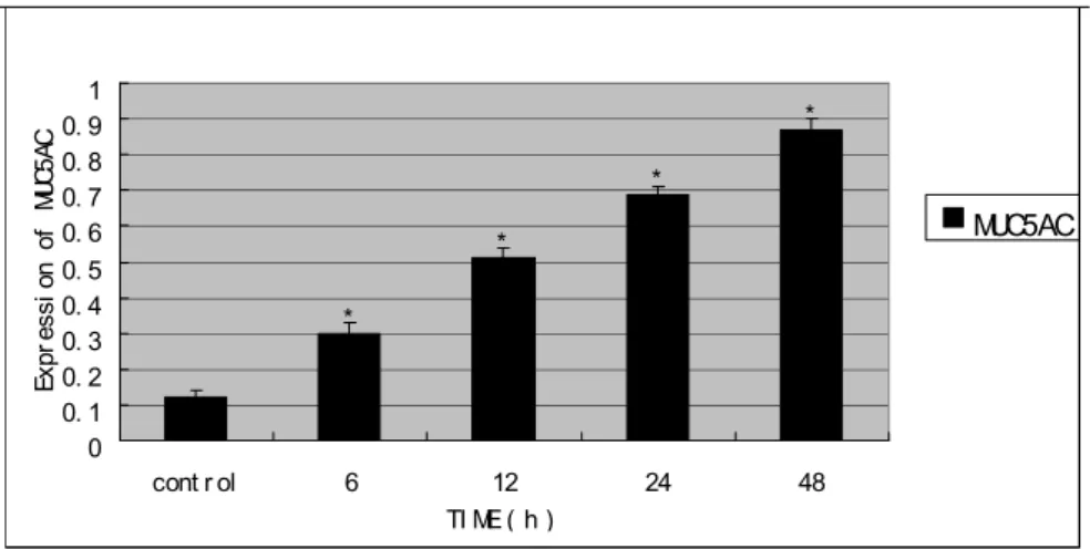
Influence of different times of hypertonic culture on MUC5AC secretion of 16HBE Studies on the effects of hypertonicity on the expression of MUC5AC in the 16HBE cells, as monitored by the ELISA showed that the treatment with different times (0, 6, 12, 24, 48h) of 600 mOsm/L hypertonic medium significantly
improved the secretion of MUC5AC (all P<0.05).
*
*
*
*
0 0. 1 0. 2 0. 3 0. 4 0. 5 0. 6 0. 7 0. 8 0. 91
cont r ol 6 12 24 48
TI ME(h)
Ex
pr
es
si
on
o
f
MU
C5
AC
MUC5AC
Figure 1 - Expression of MUC5AC protein in each group by ELISA *P<0.05 versus control group.
Wnt/β-catenin signaling pathway modulates
expression of MUC5AC induced by
hypertonicity in 16HBE cells
Results for the effects of manipulating Wnt/β
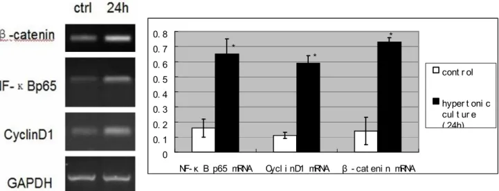
-catenin signaling pathway on the MUC5AC expression stimulated by hypertonicity were statistically significant. The 16HBE cells were cultured in 600 mOsm/L hypertonic conditions for different times (0, 12, 24, 48h). The expression
levels of β-catenin and Cyclin D1 proteins were
evaluated by the western blot analysis. The treatment with 600 mOsm/L hypertonic medium
significantly boosted the expression of β-catenin
and Cyclin D1 proteins (all P<0.05) (Fig.2).
Similar results were obtained when the expression
levels of β-catenin and Cyclin D1 mRNA were
increased markedly by RT-PCR assay (P<0.05)
(Fig.3). The knockdown of endogenous β-catenin
for 24h by si-β-catenin resulted in about 2-fold
reduction in the expression levels of the
MUC5AC, β-catenin and Cyclin D1 proteins
compared to the hypertonicity group (all P<0.05)
(Fig.4). These results suggested that Wnt/β-catenin
signaling pathway might play a vital role in the
upregulation of MUC5AC in hypertonic
conditions.
*
*
*
#
#
#
** **
**
0 0. 1 0. 2 0. 3 0. 4 0. 5 0. 6 0. 7 0. 8 0. 9 1
0 12 24 48
TI ME( h)
Ex
pr
es
si
on
o
f
pr
ot
ei
ns
β - cat eni n Cycl i nD1 NF- κ B p65
Figure 2 - Expression of β-catenin, Cyclin D1 and NF-κB p65 in groups which were trained in
* *
*
0 0. 1 0. 2 0. 3 0. 4 0. 5 0. 6 0. 7 0. 8
NF- κ B p65 mRNA Cycl i nD1 mRNA β - cat eni n mRNA
cont r ol
hyper t oni c cul t ur e ( 24h)
Figure 3 - Expression of β-catenin, Cyclin D1 and NF-κB p65 mRNAs in groups which were trained
in hypertonic atmosphere for different times by RT-PCR. *P<0.05 experimental groups versus control group.
* *
* *
* *
* *
0 0. 2 0. 4 0. 6 0. 8 1 1. 2 1. 4
A B C D
re
la
ti
ve
e
xp
re
ss
io
n
of
p
ro
te
in
s
β - cat eni n Cycl i nD1 NF- κ B p65 MUC5AC
Figure 4 - The cells were divided into four different treatment groups. The cells were transfected with
10 nmol/L β-catenin siRNA or control siRNA. Group A signifies hypertonicity group. Group B signifies hypertonicity with 10nmol/L control siRNA for 24h. Group C signifies hypertonicity with 10nmol/L β-catenin siRNA for 24h. Group D signifies isotonicity group. The cell lysates were analysed by western blotting with β-catenin, Cyclin D1, NF-κB p65 and MUC5AC antibodies. *p<0.05, compared with hypertonicity group (Group A). Data are shown as mean±S.D. of three independent experiments.
NF-κB signaling pathways modulates
expression of MUC5AC induced by
hypertonicity in 16HBE cells
To further confirm the mechanisms in the regulation of MUC5AC by hypertonicity, the
NF-κB signaling pathways was testred. The results
were statistically significant. The16HBE cells were cultured in 600 mOsm/L hypertonic conditions for different times (0, 12, 24, 48h). The
evaluated by the western blotting. The treatment
with 600 mOsm/L hypertonic medium
significantly boosted the expression of NF-κB p65
protein (all P<0.05) (Fig.2). Similar results were
obtained when the level of NF-κB p65 mRNA
expression was increased markedly by RT-PCR
assay (P<0.05) (Fig.3). Knockdown of
endogenous β-catenin by si-β-catenin for 24h
resulted in about 2-fold reduction in the expression
level of NF-κB p65 protein compared to the
hypertonicity group (all P<0.05) (Fig.4). These
results suggested that NF-κB signaling pathway
might play a positive role in the upregulation of MUC5AC in hypertonic conditions.
DISCUSSION
Excessive mucus secretion is one of the most indispensable pathological traits of a variety of chronic airway inflammatory diseases. The expression of MUC5AC is regulated by the multiple signaling pathways, such as ERK, JNK, p38, and signal transducer and activator of transcription pathways (Yu et al. 2011; Lim et al. 2009). Clinically, 3-5% hypertonic sodium chloride solution is often used to induce and get rid of airway mucus. However, little information has been focused on its mechanisms. This study showed that hypertonicity exerted a time-dependent upregulation of the MUC5AC in
16HBE cells. Augmented expression of β-catenin,
Cyclin D1 and NF-κB p65 stimulated the
expression of MUC5AC, while, conversely, the
knockdown of β-catenin resulted in a notable
decrease in Cyclin D1, NF-κB p65 and MUC5AC.
These findings indicated that β-catenin played a
positive role in the regulation of MUC5AC
production. Furthermore, both Wnt/β-catenin and
NF-κB pathways were involved in the action of β
-catenin. These results collectively highlighted a
crucial role for β-catenin in the control of mucus
secretion of 16HBE cells in vitro, and Wnt/β
-catenin and NF-κB signaling pathways could be
one of the important causes for the excessive secretion of MUC5AC in hypertonic atmosphere. Wnt signaling pathway is divided into three types:
the Wnt/β-catenin signaling pathway, Wnt/Ca2+
signaling pathway and the Wnt/PCP signaling
pathway. Wnt/β-catenin signaling pathway is the
most classic one, which is also known as the canonical Wnt signaling pathway, which regulates the differentiation, migration, proliferation and
polarity of the cells (Yao et al. 2011). It also involves in regulating the expressions of many genes. It is reported that the abnormal activation of this pathway takes positive effect in the formation of a variety of tumors (Amado et al. 2011; Bulut et al. 2011; Sethi et al. 2011). The anomalous
activation of β-catenin in the nucleus is regarded
as a sign of abnormal activation of Wnt signaling pathway. The abnormal activation of Wnt ligand leads to proliferation of epithelial cells and fibrosis of lung. Wang et al. (2011) reported that COPD patients with Wnt signaling pathway inhibited had a reduction in the ability to repair the damages and injuries of the lung. Morrisey (2003) showed that
Wnt/β-catenin signaling pathway played a critical
role in the repair process in lung tissue. Huang et al. (2008) found that cigarette somking could
up-regulate the Wnt/β-catenin signaling pathway.
This signaling pathway is also known to have relationships with mucus production and secretion (Konigshoff and Eickelberg 2010). However, previous studies have failed to explain the role of
the Wnt/β-catenin signaling pathway in mucus
hypersecretion of hypertonic conditions.
β-catenin is the core molecule of Wnt/β-catenin
signaling pathway, which has been found as an
adhesion molecule originally. It is a
multifunctional protein, is produced by the CTNNB1 gene (88kD size) and located in 3p22-p21.3 of human chromosome (Morin and
Weeraratna 2003). β-catenin is composed of 781
amino acids and contains 12 Armadillo (arm) regions and special structures of N-terminal and C-terminal. With plenty of Ser/Thr residues,
N-terminal of β-catenin is comprised of 130 amino
acids and controls the stability of β-catenin.
C-terminal of β-catenin is comprised of 100 amino
acids, which regulates the transcription of target genes downstream (Willert and Nusse1998). In
normal conditions, β-catenin is mainly localized in
the cell membrane, with the functions of mediating cell adhesion and regulating expression of the genes. Mucenski et al. (2005) pointed out that the
long-term activation of β-catenin could lead to the
hyperplasia of airway goblet cells. Since it is well-known that airway mucus is secreted by the goblet
cells, β-catenin is expected to be closely related to
the mucus hypersecretion of airway. Farkas et al. (2005) reported that thehypertonic liquid could
induce reversible phosphorylation of β-catenin in
the epithelial cells of the brain. It suggested that
there could be some connections with Wnt/β
pressure. The present findings showed that β -catenin was increased when 16HBE cells were cultured in the hypertonic atmosphere, which
demonstrated that Wnt/β-catenin pathway was
activated.
Encoding 295 amino acids, Cyclin D1 gene, the
key element of Wnt/β-catenin signaling pathway,
is located on 11q1-3 of chromosome. Cyclin D1
gene is one of the target genes of β-catenin/LEF-1
complex (Shtutman et al. 1999). Cyclin D1 gene was originally found in the parathyroid. It can induce the growth of tumors in parathyroid in transgenic mice and adjust the transmission of
Ca2+. It is also the regulator for macrophage
transfer (Imanishi et al. 2001; Neumeister et al. 2003). The overexpression of Cyclin D1 can shorten the G1 phase of the cell cycle and prompt the cells into S phase, which causes the uncontrolled growth and arrests differentiation of cells. Hence, the variation of Cyclin D1 is closely related with the occurrence of a variety of tumors. However, no studies have focused on the
relationships between the airway mucus
hypersecretion in hypertonic conditions and Cyclin D1 gene yet. The present results showed that
Cyclin D1, as a downstream gene of Wnt/β
-catenin pathway, was also increased significantly,
which demonstrated that Wnt/β-catenin pathway
was activated further.
There are binding sites for NF-κB gene in the
promoter of MUC5AC, which indicates that
NF-κB plays a key role in the formation and secretion
of mucus. Although working independently,
NF-κB and Wnt/β-catenin signaling pathways have
close relationships. A series of studies have found that there is a "crosstalk" between the two
signaling pathways, which is mediated by the β
-TrCP. IκB and β-catenin have an identical
structure—DAGX2+nS degradation domains.
Phosphorylated Ser32, Ser36 of IκB and
phosphorylated Ser33, Ser37 of β-catenin can bind
with the WD40 of β-TrCP, and then be degraded.
Hence, the content of NF-κB p65 protein increased
when 16HBE cells were cultured in the hypertonic atmosphere and such change was the same as
Wnt/β-catenin pathway.
CONCLUSIONS
From the results it could be concluded that the
proteins of β-catenin, Cyclin D1 and NF-κB p65
played a positive role in the hypertonicity
stimulated MUC5AC production of 16HBE cells.
The results demonstrated that both Wnt/β-catenin
and NF-κB signaling pathways were involved in
this process. Further studies are ought to focus on
the role of Wnt/β-catenin and NF-κB signaling
pathways in the airway mucus secretion in vivo.
These findings in vitro encourage the explorations
of the therapeutic potentials of manipulating β
-catenin in the management of mucus
overproduction in chronic inflammatory airway diseases.
REFERENCES
Fahy JV. Remodeling of the airway epithelium in
asthma. Am J Respir Crit Care Med. 2001; 164(10 Pt2): S46-51.
Hogg JC, Chu F, Utokaparch S, Woods R, Elliott WM, Buzatu L, et al. The nature of small-airway obstruction in chronic obstructive pulmonary disease.
N Engl J Med. 2004; 350(26): 2645-53.
Shao MX, Nadel JA. Neutrophil elastase induces MUC5AC mucin production in human airway epithelial cells via a cascade involving protein kinase C, Reactive oxygen species, and TNF-α-converting enzyme. J Immunol. 2005; 175(6): 4009-16.
Voynow JA, Rubin BK. Mucins, Mucus, and Sputum.
Chest. 2009; 135(2): 505-12.
Kneidinger N, Yildirim AO, Callegari J, Takenaka S, Stein MM, Dumitrascu R, et al. Activation of the Wnt/β-catenin pathway attenuates experimental emphysema. Am J Respir Crit Care Med. 2011; 183(6): 723-33.
Baarsma HA, Spanjer AI, Haitsma G, Engelbertink LH, Meurs H, Jonker MR, et al. Activation of Wnt/β -catenin signaling in pulmonary fibroblasts by TGF-β is increased in chronic obstructive pulmonary disease.
Plos One. 2011; 6(9): e25450.
Yu HM, Li Q, Kolosov VP, Perelman JM, Zhou XD. Interleukin-13 induces mucin 5AC production involving STAT6/SPDEF in human airway epithelial cells. Cell Commun Adhes. 2010; 17: 83-92.
Lim JH, Kim HJ, Komatsu K, Ha U, Huang Y, Jono H, et al. Differential regulation of Streptococcus pneumoniae induced human MUC5AC mucin expression through distinct MAPK pathways. Am J Transl Res. 2009; 1(3): 300–11.
Yao H, Ashihara E, Maekawa T. Targeting the Wnt/β -cateninsignaling pathway in human cancers. Expert Opin Ther Targets. 2011; 15(7): 873-887.
Bulut G, Fallen S, Beauchamp EM, Drebing LE, Sun J, Berry DL, et al. β-cateninaccelerates human papilloma virus type-16 mediated cervical carcinogenesis in transgenic mice. Plos One. 2011; 6(11): e27243.
Sethi K, Sarkar S, Das S, Rajput S, Mazumder A, Roy B, et al. Expressions of CK-19, NF-κB, E-cadherin, β-cateninand EGFR as diagnostic and prognostic markers by immunohistochemical analysis in thyroid carcinoma. J Exp Ther Oncol. 2011; 9(3): 187-199.
Wang R, Ahmed J, Wang G, Hassan I, Strulovici-Barel Y, Hackett NR, et al. Down-regulation of the canonical Wnt/β-catenin pathway in the airway epithelium of healthy smokers and smokers withCOPD. Plos One. 2011; 6(4): e14793.
Morrisey EE. Wnt signaling and pulmonary fibrosis.
Am J Pathol. 2003; 162(5): 1393-7.
Huang CL, Liu D, Ishikawa S, Nakashima T, Nakashima N, Yokomise H, et al. Wnt1 overexpression promotes tumor progression in non-small cell lung cancer. Eur J Cancer. 2008, 44(17): 2680-8.
Konigshoff M, Eickelberg O. Wnt signaling in lung disease: a failure or a regeneration signal? Am J Respir Cell Mol Biol. 2010; 42(1): 21-31.
Morin PJ, Weeraratna AT. Wnt signaling in human cancer. Cancer Treat Res. 2003; 115(2): 169-87.
Willert K, Nusse R. β-catenin: a key mediator of Wnt signaling. Curr Opin Genet Dev. 1998; 8(1): 95-102. Mucenski ML, Nation JM, Thitoff AR, Besnard V, Xu
Y, Wert SE, et al. β-catenin regulates differentiation of respiratory epithelial cells in vivo. Am J Physiol Lung Cell Mol Physiol. 2005; 289(6): L971-9. Farkas A, Szatmari E, Orbok A, Wilhelm L, Wejksza
K, Nagyosizi P, et al. Hyperosmotic mannitol induces Src kinase-dependent phosphorylation of β-catenin in cerebral endothelial cells. J Neurosci Res. 2005, 80(6): 855-61.
Shtutman M, Zhurinsky J, Simcha I, Albanese C, D’Amico M, Pestell R, et al. The Cyclin D1 gene is a target of the beta-catenin/LEF-1 pathway. Proc Natl Acad Sci USA. 1999; 96(4): 5522-7.
Imanishi Y, Hosokawa Y, Yoshimoto K, Schipani E, Mallya S, Papanikolaou A, et al. Primary hyperparathyroidism caused by parathyroid-targeted overexpression of Cyclin D1 in transgenic mice. J Clin Invest. 2001; 107(9): 1093-102.
Neumeister P, Pixley FJ, Xiong Y, Xie H, Wu K, Ashton A, et al. Cyc1in D1 governs adhesion and motility of macrophages. Mol Biol Cell. 2003; 14(5): 2005-15.